Universidade de Lisboa
Faculdade de Farmácia
Evaluation of the activity of molecules isolated from
Laurencia genus against Acanthamoeba
José Luís Gil Pio
Mestrado Integrado em Ciências Farmacêuticas
Universidade de Lisboa
Faculdade de Farmácia
Evaluation of the activity of molecules isolated from
Laurencia genus against Acanthamoeba
José Luís Gil Pio
Monografia de Mestrado Integrado em Ciências Farmacêuticas
apresentada à Universidade de Lisboa através da Faculdade de Farmácia
Orientador:
Doutor Jacob Lorenzo Morales, Professor Auxiliar
Co-Orientador:
Doutor José E. Piñero Barroso, Professor Auxiliar
Co-Orientador:
Doutora Quirina dos Santos Costa, Professora Auxiliar
Some notes from the author:
This work was carried out in the ambit of the Erasmus + programme in the “Universidade de La Laguna” from 1st of February to 30th of April 2019.
It was presented at “Faculdade de Farmácia da Universidade de Lisboa” as an element of the final evaluation of the “Mestrado Integrado em Ciências Farmacêuticas”.
Nevertheless, the work developed has already been presented and published in: a) “Universidade de La Laguna”, in the ambit of Erasmus + programme, on the 8th of April 2019, and registered/identified as followed:
Universidad de La Laguna Oficina de Sede Electrónica
Entrada Nº registro: 2019/17372, Nº reg. oficina: OF002/2019/16216 Fecha: 03/04/2019 11:46:52 “.
“XXI congresso socepa, Pontevedra, Universidade de Vigo”
Abstract:”Evaluación in vitro de la actividad de compuestos derivados del alga roja Laurencia
jonhstonii sobre el protozoo parásito Acanthamoeba spp.”
www.socepa2019.com
XVIII Internation Meeting on the Biology and Pathogenicity of Free-Living Amoebae, Abstract: In vitro activity evaluation of Laurencia derivatives against Acanthamoeba castellanii Neff
Resumo
Resumo
O género Acanthamoeba é um parasita oportunista e largamente distribuído, classificado como Ameba de Vida Livre com uma crescente importância global, nas últimas décadas, como um patogeno emergente. Este protozoário possui a fase ativa de trofozoíto, responsável pela reprodução e alimentação ou a fase de latência e muito resistente de quisto, quando é submetido a condições desfavoráveis e de stress. Pode provocar uma doença ocular, Queratite por
Acanthamoeba, sobretudo em utilizadores de lentes de contacto e também encefalite amebiana
granulomatosa, uma patologia cerebral, muito fatal, que está normalmente associada a indivíduos imunocomprometidos. As opções de tratamento existentes não são 100% eficazes e apresentam efeitos tóxicos para as células humanas. Além disso, devido sobretudo à forma de quisto, este parasita é muito resistente às opções de tratamento disponíveis. Até há data foram identificados 22 genótipos de Acanthamoeba diferentes, dos quais o T4 tem sido o mais associado aos estados patológicos provocados por esta ameba. Devido a estes fatores, existe uma preocupação em descobrir novos fármacos para tratar as infeções causadas pela
Acanthamoeba. Neste caso, as algas marinhas podem ser um bom fornecedor de potenciais
espécies com valor terapêutico para produzir medicamentos. Assim sendo, o género Laurencia aparece como uma possível fonte de novos agentes terapêuticos contra este parasita, como mencionado num artigo anterior. Neste trabalho, foi testada a atividade de doze moléculas de
Laurencia viridis versus Acanthamoeba castellanii Neff. Das moléculas testadas, o CL3, CL4
e CL12 apresentaram as melhores atividades, apesar de que, o CL3 também foi o mais tóxico. Por conseguinte, prosseguiram-se os estudos com o CL4, que demonstrou atividade quisticida. Através ensaios adicionais mostrou afetar a membrana citoplasmática, bem como a cromatina e também a mitocôndria devido à redução na produção de ATP e às alterações no potencial da membrana mitocondrial. Tendo em conta as observações desta investigação, os compostos de
Laurencia viridis demonstraram potencial face à Acanthamoeba castellanii Neff, sendo que
esta espécie parece uma boa opção para prosseguir com os estudos em relação a este parasita e, se possível, desenvolver novos fármacos e eficazes.
Abstract
Abstract
Acanthamoeba genus is a widely distributed and opportunistic parasite classified as a
Free-Living Amoebae with increasing importance worldwide in the past decades, as an emerging pathogen. This protozoan has an active trophozoite stage, which is responsible for the feeding and reproduction, and a cyst one. The last happens when the parasite is submitted to stress and harsh conditions, with the cyst being dormant and very resistant. It can cause an ocular sight-threatening disease, Acanthamoeba Keratitis that occurs mainly in contact lens wearers. Can also originate Granulomatous Amoebic Encephalitis, a chronic, very fatal brain pathology, which is more associated with immunocompromised people. The options currently available for the treatment are not 100% effective and present toxic effects to human cells, adding the fact that this parasite is also resistant to these molecules, mostly due to the cyst form. To date, there are 22 Acanthamoeba genotypes identified, where T4 has been the most associated with pathological states caused by this amoeba. Thereby, exists a concern to find drugs to treat Acanthamoeba infections. Here, seaweeds can be a rich supplier of potential species with therapeutic value to produce new and active medicine. Therefore, the genus
Laurencia appears to be a possible source of novel therapeutic agents against this parasite, as
mentioned in a previous article. In this study, the amoebicidal activity of twelve Laurencia
viridis molecules was evaluated versus Acanthamoeba castellanii Neff. From the tested
molecules, CL3, CL4 and CL12 presented the best activity values, with CL3 also being the most toxic compound. CL4 was chosen to proceed with the investigation, showing cysticidal activity. Through further assays, CL4 showed to affect the mitochondria. Such fact was due to the reduction in the ATP production and changes in the mitochondrial membrane potential. Besides that, CL4 affected the cytoplasmic membrane and interacted with the chromatin of the cells. Based on the observations from this work, Laurencia viridis molecules showed potential towards Acanthamoeba castellanii Neff. This species appears a good option to keep studying its effect on this parasite and hopefully develop new and effective medication.
Acknowledgements
Acknowledgements
I can’t express the way I’m feeling right now. It’s a mixture of amazing fellings,
and also some sad ones. I feel amazed for seeing that a long and hard-working journey
paid off, but sad to finish a course that gave me so many good memories.
With that said I would like to thank my parents and my family for allowing me to
study in FFUL, and always support me in my choices and be a great motivation from the
very beginning to the end. Also to thank my girlfriend for maintaining my mental sanity
and always be there for me and motivate me to keep going. To my friends, the ones since
I was a little child, and the ones that this house gave me. For the ones of FFUL, and
pardon me my childhood friends, I would like to give a special word, for making a lot
easier these five years.
Last but not least, guys from the “Instituto Universitario de Enfermedades Tropicales y Salud Pública de Canarias, de la Universidad de La Laguna”, “muchas
Table of Contents
Table of Contents
1. Introduction ... 10
1.1. Acanthamoeba ... 10
1.2. Acanthamoeba life cycle ... 11
1.3. Acanthamoeba infections ... 13
1.3.1. Acanthamoeba Keratitis ... 13
1.3.2. Granulomatous Amoebic Encephalitis ... 15
1.4. Acanthamoeba classification ... 17
1.5. Seaweeds ... 17
1.5.1. Laurencia genus ... 18
2. Objectives ... 19
3. Materials and Methods ... 20
3.1 Materials ... 20
3.1.1. Cell Strains ... 20
3.1.2. Chemicals ... 20
3.1.3. Reagents and products ... 21
3.1.4. Equipment ... 21
3.1.5. Software ... 22
3.2. Methods ... 22
3.2.1. In vitro activity against Acanthamoeba castellanii Neff trophozoites ... 22
3.2.2. Cytotoxicity assay ... 22
3.2.3. In vitro activity against Acanthamoeba castellanii Neff cysts ... 23
3.2.4. ATP measurement ... 23
3.2.5. Changes in the mitochondrial membrane potential (ΔΨm) ... 23
3.2.6. Plasma membrane permeability ... 24
3.2.7. Chromatin condensation analysis ... 25
4. Results and Discussion ... 26
4.1. In vitro activity against Acanthamoeba castellanii Neff trophozoites and cytotoxicity assay ... 26
4.2. In vitro activity against Acanthamoeba castellanii Neff cysts ... 28
4.3. Apoptosis-like assays ... 29
4.4. Precipitation problems ... 32
Table of contentes
6. References ... 35 7. Attachments ... 42
List of Figures
List of Figures
Figure 1. Acanthamoeba phylogenetic scheme ... 10
Figure 2. The life cycle of Acanthamoeba spp. in humans ... 12
Figure 3. Corneal damage in AK shown after sodium fluorescein application ... 14
Figure 4. Chemical structure of the compounds evaluated... 21
Figure 5. Activity assays of the Laurencia compounds towards A. castellanii Neff trophozoites. ... 27
Figure 6. Activity assays of Laurencia compounds towards A. castellanii Neff cysts . 28 Figure 7. The effect of CL4 on the mitochondrial potential ... 29
Figure 8. ATP measurement assay with negative control and CL4 compound... 30
Figure 9. The effect of CL4 on the plasma membrane permeability ... 31
Figure 10. The effect of CL4 on chromatin condensation ... 32
Figure 11. Example of precipitate formation in CL7 at a concentration of 25 µg/mL .. 32
Figure 12. Book of Abstracts of the XXI Congreso Socepa www.socepa2019.com .... 42
Figure 13. Abstract: Evaluación in vitro de la actividad de compuestos derivados del alga roja Laurencia jonhstonii sobre el protozoo parásito Acanthamoeba spp. ... 43
Figure 14. Book of Abstracts of the XVIII Internation Meeting on the Biology and Pathogenicity of Free-Living Amoebae ... 43
Figure 15. Abstract: In vitro activity evaluation of Laurencia derivatives against Acanthamoeba castellanii Neff ... 43
List of Tables
List of Tables
Table 1. Activity assay (IC₅₀) of the different Laurencia compounds against
Acanthamoeba castellanii Neff trophozoites and cytotoxicity assay (CC₅₀) in murine
macrophages (J774A.1) ... 26
List of acronyms and abbreviations
List of acronyms and abbreviations
AK - Acanthamoeba Keratitis
ATCC - American Type Culture Collection DMEM - Dulbecco’s Modified Eagle Medium DMSO - Dimethyl sulfoxide
FBS - Fetal Bovine Serum FLA - Free-Living Amoebae
GAE - Granulomatous Amoebic Encephalitis HIV - Human immunodeficiency virus NEM - Neff’s encystment medium PCR - Polymerase chain reaction PHMB - Polyhexamethylene biguanide PI - Propidium iodide
PYG - Peptone Yeast Glucose
Introduction
1. Introduction
1.1. Acanthamoeba
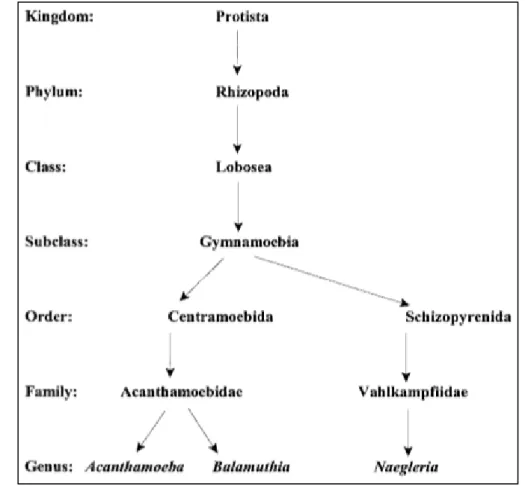
Acanthamoeba spp. from the family Acanthamoebidae and order Centramoebida
(Figure 1), are opportunistic Free-Living Amoebae. This species is ubiquitous in nature and widely distributed in different environments as seawater, soil, water lakes, in the air, contact lenses, air-conditioning units, bottled water, swimming pools, as well as hospitals (1–3).
The genus Acanthamoeba was established in 1931 by Volkonsky, in which the first amoeba isolated from dust, in 1913, is included. Firstly was designated as Amoeba
polyphagus, but was later renamed Acanthamoeba polyphaga (4).
Balamuthia mandrillaris, Naegleria fowleri, Sappinia diploidea, Vahlkampfia
spp., and Vermamoeba vermiformis, along with Acanthamoeba spp. are opportunistic pathogens to humans and other animals that can lead to death (5,6). When first identified,
Acanthamoeba was not considered a human pathogen, however, in the ’50s, it was
reported that they were able to lyse in monkey kidney cells. Also, in the ’70s, the two first
Introduction
infection cases by this parasite were reported on a person with cerebral haemorrhage and another with Hodgkin's disease going through chemotherapy, respectively (7).
The importance of this protozoan has developed in the last decades, not only because of their ability to cause pathologies but also by virtue of their interaction with bacteria’s (8). Acanthamoeba, in a poor nutrient environment, has the capacity to act as a bacterial predator, with non-pathogenic bacteria acting like prey. In this case, the bacteria are taken through phagocytosis and then lysed in the parasite phagolysosomes, having significant importance in the regulation of bacterial population. Additionally, they may behave as a “bacterial Trojan Horse”. Here, pathogenic bacteria invade and preserve themselves inside Acanthamoeba to survive adverse conditions, like during transmissions to hosts susceptible to infection (9,10). Thus, the bacterias can resist the host defences and stay viable, yet they are not able to multiply. Acanthamoeba can also be used as a bacterial reservoir and normally, those bacteria are human pathogens of the greatest importance (Helicobacter pylori, for example). In this situation, they not only survive but also multiply themselves inside the amoeba. Therefore, under favourable conditions, bacteria cause the lysis of their host, allowing them to induce disease or infect another parasite (10).
This amoeba, in addition, is also acknowledged as a “Trojan Horse” for viruses and with so, is responsible for accentuating the virulence of these and protecting them against hostile environments. Adenoviruses, enteroviruses and different members of giant virus families, are examples, that have been isolated from cultures of this parasite (11).
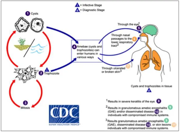
1.2. Acanthamoeba life cycle
Regarding the life cycle of this parasite, it is divided into two stages (Figure 2): 1) A trophozoite form which measures among 25 to 40 µm, is vegetative, feeds on yeasts, bacteria, amid others and reproduce through binary fission. It forms pseudopods to feed itself and has a hyaline pseudopodium to allow the locomotion. This stage holds distinctive characteristics like acanthopodia that are fine, spiny prominences on the surface, a noticeable contractile vacuole that regulates the water of the trophozoite in the cytoplasm, and one big nucleolus in a central nucleus. It also has food vacuoles, Golgi complex, ribosomes, many mitochondria and a trilaminar plasmatic membrane (1,12–14).
Introduction
2) A cyst (13 to 20 µm) that is dormant and resistant to antibiotics, biocides and cold. It is formed out of the trophozoite stage in a process called encystment, which occurs due to a response by Acanthamoeba to stress and hostile conditions like toxins, temperature, pH modifications and absence of nutrients. It is a process that leads to significant modifications, not only of cellular constituents and organelles composition and function but of morphological features too. Also, different proteases, such as serine and cysteine proteases, are implicated in the encystment (1,15). At the beginning of this process, an amorphous and irregular layer named ectocyst, consisting of a mixture of proteins and polysaccharides, is formed. It is also formed an inner layer called the endocyst, a much more solid one, composed of polysaccharides, mainly cellulose. The endocyst can have different appearances, it can be spherical, polygonal, stellate, or oval. Together, the ecto and endocyst form the double-walled cyst. With the encystation, the trophozoites uptake a great amount of food and shut themselves, having almost no metabolic activity with the cyst formation. The cysts, like the trophozoites, possess one nucleus with a dense nucleolus in the centre. Carbohydrates, proteins and unidentified material are the major constituents of cyst walls and, in less quantity, by lipids and ash (12,14,16,17).
Introduction
1.3. Acanthamoeba infections
Worldwide are being continually described new cases of infections caused by FLA. Even though the morbidity of these infections is low, the mortality rate, on the contrary, is reasonably high. Acanthamoeba species, apart from having the capacity to originate systemic diseases, can also lead to widespread infections (18). It can cause an ocular infection known as Acanthamoeba Keratitis and Granulomatous Amoebic Encephalitis, a systemic infection that affects the central nervous system. Moreover, Acanthamoeba is also the causative agent of nasopharyngeal and cutaneous infections (19).
1.3.1. Acanthamoeba Keratitis
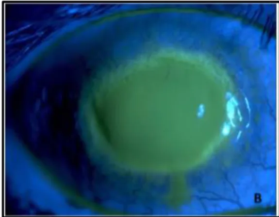
AK is a sight-threatening infection that affects the cornea (Figure 3). The parasite adheres to the corneal surface, binding to glycoproteins on the epithelium. In this process, the acanthopodia of the amoeba surface are very important to the extent that they are strictly related to the adhesion rate. This factor allows the release of enzymes and toxins, like phospholipases and proteases, in which serine proteases have a higher degradative capability and play a major role in the encystment and excystment, as mentioned before. Cytopathic factors are also released, including mannose-induced protein that will bind to mannose-rich glycoproteins on the corneal surface, leading to corneal epithelial destruction (20–22). Thus, Acanthamoeba is able to invade the stroma and secrete collagenolytic factors, dissolving the stromal matrix, which results in an inflammatory response that leads to corneal cell death and keratitis (23). It can provoke epithelial defect, lid oedema, pain with photophobia and may even lead to blindness. Although the biggest risk to develop AK is by wearing contact lens (more than 80% of the cases), since it facilitates adhesion to the corneal surface, people who do not wear them can also suffer from this infection. Likewise, the diagnostic of AK is complicated, in the means that could be easily misdiagnosed as Herpes Simplex keratitis (24,25).
The first occurrence described of this infection was in 1974 in the United Kingdom, and in 1984 was reported the first case by a contact lenses wearer, by wearing them in a hot tub (26,27). In Portugal, a 16-year-old male had been diagnosed with AK by the department of ophthalmology of the Braga Hospital, although in the beginning, he was misdiagnosed with Herpes Simplex keratitis. The boy was a regular user of contact lenses (28). There were also six cases of patients who were diagnosed with AK, in the Central Lisbon Hospital Center, from 2007 to 2012. They had ages comprehended
Introduction
between 18 and 41 years old and all of them had in common the fact of wearing contact lenses (29).
With respect to the diagnose, the cultivation of the parasite from the contact lenses or from the corneal biopsy is still the gold standard method, although PCR and in vivo confocal microscopy, corroborated by laboratory-based tests are emerging in this field (3). In terms of the culture, it’s more economical and simple, yet the results can take a few days to draw any conclusion and therefore a delay in starting appropriate treatment. Apart from this, is considered to have low sensitivity, with values described within 7 and 51%. On the contrary, PCR has a higher sensitivity, is faster and has the capacity to detect less than five trophozoites (24,30). Regarding confocal microscopy, it can be advantageous since it allows to identify modifications on the morphological characteristics of the cornea. Nonetheless, for this, it’s necessary a skilled analyst, acquainted with the characteristics of Acanthamoeba in order to detect the disease. Also, in the diagnostic field, proton nuclear magnetic resonance spectroscopy is seen as an optimistic technique (2,31).
Focusing on treatment, the commonly used includes a biguanide, PHMB (polyhexamethylene biguanide) or chlorhexidine digluconate along with a diamidine like propamidine isethionate or hexamidine (22). During the first 48 or 72 hours, these drugs are instilled hourly day and night. After this period, hourly just daytime, for some weeks and later, four times per day. Thereby, this is a treatment that could last for months and the patients need to be strictly followed until there is a gradual decrease in the parasite burden. This is due to the potential chemical toxicity of these biocides in the cornea. Furthermore, the cyst stage further complicates the treatment of infections due to
Figure 3. Corneal damage in AK shown after sodium fluorescein
Introduction
Acanthamoeba. So, in order to avoid these resistance issues, monotherapy is not
recommended, even though biguanides can be employed individually (31).
In the case of suspected or coinfections with bacteria, antibiotics like neomycin or chloramphenicol should be added to the regimen, not merely to prevent bacterial infection but also to eliminate the food source of the parasite. If the treatment with the drugs is not effective, as a last resort, penetrating keratoplasty can be suggested, with topical treatment after surgery throughout a year, due to the possibility of cyst survival (2).
1.3.2. Granulomatous Amoebic Encephalitis
GAE is a chronic infection, persisting between six months and two years, which has a mortality rate higher than 90% and generally occurs in immunocompromised patients, like those who have HIV or doing chemotherapeutic procedures. Nevertheless, some cases have been reported in people with no clear signs of being immunocompromised. Common symptoms and signs of GAE consist of headache, lethargy, seizures, nausea, vomiting, loss of consciousness, cerebral oedema and coma. In fact, the cerebral hemispheres, tend to be the most affected tissue of the central nervous system. Different Acanthamoeba species like A. castellani and A. polyphaga have been identified as causative agents of the disease (12,32,33).
The ways of entry for Acanthamoeba is either by the skin injuries, allowing it to go straight to the blood circulation, or through the respiratory tract. In the second way, the parasite enters the alveolar blood vessels reaching the central nervous system by crossing the blood-brain barrier and consequently, causes granulomatous amoebic encephalitis (18). In order to cross the barrier, it is believed to involve not only properties from the parasite, like adhesins and proteases but also from the host, like interleukin alpha and gamma interferon. Those factors culminate in an increase of permeability and/or apoptosis of the brain endothelial cells, allowing Acanthamoeba to invade the central nervous system (13).
Tavares et al. (34) reported the first Portuguese case of GAE in an 8 year-old-boy that turned out to be fatal. Initially, it was misdiagnosed as a tumour, but histopathologic examination exhibited evidence of amoebic infection, that was later corroborated through PCR. The parasite responsible for the infection was another FLA (Balamuthia
mandrillaris), whereas to date there are no confirmed cases in Portugal caused by Acanthamoeba. Moreover, globally, a large number of this infection cases have probably
Introduction
burden of GAE should be related to the number of cases of this infection in patients with HIV. Moreover, the low number of detected cases may be linked with the lack of reported occurrences in developing countries.
In relation to GAE diagnose, still prevails a challenge and keeps being identified after autopsy in most cases. Microscope detection by histopathological examination or immunohistochemistry of the morphological forms of the parasite has been the common and conventional method (35).
With respect to microscope detection, cerebrospinal fluid, brain and skin tissue have been the used samples, with the histopathological examination having the capacity to detect both cysts and trophozoites. Unfortunately, these are usually confused as macrophages or necrosed keratinocytes. This problem is overcome by immunohistochemistry, where specific antibodies are used to target the Acanthamoeba antigens. Molecular diagnosis, through PCR, is also rising as an up-and-coming method due to the high sensitivity and fast result, being the most promising test in this field. Serological diagnose, even though being noninvasive and relatively easy to perform, just gives a hypothetical diagnosis (36).
Concerning GAE therapy, it is vital to have an early diagnose and use a combination of two different agents so the chances of a successful therapy increase. In this disease, the drugs used are rifampicin, amphotericin B, cotrimoxazole, miltefosine, pyrimethamine and others. Although there is no standard therapy to treat this infection, of these described drugs, the most commonly used is amphotericin B. The issue, is that even so, the prognosis still remains bad, with a mortality rate of more than 90%. This is a direct result of the difficulty to cross the hematoencephalic barrier and also because of the toxicity and adverse effects of these compounds (37,38).
When GAE started gaining attention, therapeutics with corticosteroids were instituted due to the common symptoms of cerebral oedema and inflammation, however, it turned out that they worsen the infection and with so, steroids should be avoided (1).
There are reported GAE survivors, but some of them showed sequels of neurocognitive disorders due to cerebral oedema. These disorders display the need to handle very gently this infection, to dodge this kind of complications in those who are successfully treated (36).
Introduction
1.4. Acanthamoeba classification
In terms of Acanthamoeba classification, it was initially carried out based on the size and shape of cysts, dividing them into three groups, group I, II and III. However, this method is quite misleading by virtue of the discrepancy of cysts characteristics (39). With that said, currently, the genotyping of the species is done by analysing the diagnostic fragment 3 (DF3) region of the nuclear small subunit 18s ribosomal RNA gene, and so far 22 different genotypes (T1-T22) have been identified (40–42). Among these 22 genotypes, T2, T4, T5, T10, for example, are linked with AK and GAE infections. For instance, T4 is the one who is responsible for the majority of Acanthamoeba infections and about 90% of AK cases occur due to this genotype (43,44). The T4 genotype looks to adhere more firmly to the cells and consequently have a more negative effect on the host than other genotypes. This is probably due to a greater expression of the mannose-binding protein, which is strongly connected to Acanthamoeba’s harmful activity, as previously mentioned (45).
1.5. Seaweeds
Seaweeds are rich in minerals, vitamins, essential amino acids and specific bioactive compounds, which have health-promoting properties. They are spread all over the globe, in the different climatic zones and are divided in red (Rhodophyceae), brown (Phaeophyceae) and green (Chlorophyceae) algae based on their characteristic pigments. The green and red ones have primary endosymbiosis, belonging to the Plantae kingdom, and the brown has secondary endosymbiosis, being part of the Chromista kingdom (46). Green algae do not have any pigments to cloak the chlorophyll, responsible for the green colour. Red algae, besides the chlorophyll, possess the phycocyanin and phycoerythrin pigments that give the red colouration. Brown algae, in turn, has, apart from chlorophyll, golden and brown pigments to mask the green colour, with fucoxanthin being the main pigment found in this algae (47).
Their consumption has been linked with a lower risk of chronic diseases, including cardiovascular disease, cancer (namely breast cancer), metabolic syndrome, gut problems, helping to regulate insulin in type 2 diabetes and even on weight control (48). Also, multiple marine polysaccharides had shown therapeutic potential, like antiviral activity, owing to their biological properties. Due to these reasons, seaweeds are seen as an unlimited resource of bioactive compounds for research and development of novel and
Introduction
Once, natural products have shown antiparasitic activity and were applied in the treatment of these infections. Hence, marine sources, such as seaweeds, became an object of study to evaluate their antiparasitic activities (50).
1.5.1. Laurencia genus
Marine organisms’ metabolites have been linked with pharmaceutical and cosmeceutical (a combination of both pharmaceuticals and cosmetics) areas. Furthermore, they have been used for generations as traditional medicines in East Asia (51,52). Amid them, red algae are the richer producers of these, with the genus Laurencia, part of order Ceramiales and family Rhodomelaceae being one of the richest sources, among red algae. The most of Laurencia metabolites belong to sesquiterpenes, diterpenes, triterpenes and C15 acetogenins classes and demonstrate a wide biological activity. They
showed cytotoxic activity, especially against cancer, along with antifungal, antibacterial activity and antiviral activity (HSV-1, EBV, VZV, among others). In addition, had evidenced anti-inflammatory and antiparasitic activity, more specifically insecticidal and nematicidal activity contra Culex pipiens pallens and Caenorhabditis elegans (53).
Laurencia also presented activity against Trypanosoma cruzi as shown by Veiga-Santos et al. (51) and Leishmania amazonensis as demonstrated by Veiga-Santos et al. (54).
Thus, with the activity against Acanthamoeba spp. also evidenced by Laurencia
johnstonii in a previous study by García-Davis et al. (50), the genus Laurencia has the
Objectives
2. Objectives
Acanthamoeba has gain notability in the past decades due to its interaction with other
microorganisms and pathogenicity. Accordingly, the need to develop novel and truly effective anti-amoebic agents against both trophozoites and cysts, in order to treat this parasite infections, is also indispensable. Having this in mind, the “Instituto Universitario de Enfermedades Tropicales y Salud Pública de Canarias, de la Universidad de La Laguna”, has been testing countless molecules, with the intention of discovering new, active, non-toxic drugs that could be used for treatment.
Laurencia genus has demonstrated to possess antiparasitic activity, including against
Acanthamoeba by Laurencia johnstonii, as mentioned earlier. On account of this, it was
decided to elaborate this study with molecules isolated from Laurencia viridis, hoping to have promising antiparasitic results as well.
Hence, the aims of this investigation were to determine the in vitro activity of compounds from Laurencia genus against Acanthamoeba castellanii Neff trophozoites and assess the cytotoxicity effect of them in a murine macrophage cell line (J774A.1). It was also decided to appraise the in vitro activity of the molecules under evaluation towards Acanthamoeba castellanii Neff cysts and evaluate the apoptotic action mechanisms of the active Laurencia compounds against Acanthamoeba castellanii Neff trophozoites.
Materials and Methods
3. Materials and Methods
The elaboration of this fieldwork was based on the investigation developed at the “Instituto Universitario de Enfermedades Tropicales y Salud Pública de Canarias, de la Universidad de La Laguna”. Was also established by the analysis, interpretation and different article synthesis, as well as web pages, published between 1974 and 2019.
To obtain the electronic bibliography the sources used were: PubMed (www.ncbi.nlm.nih.gov/pubmed/); CDC (www.cdc.com).
This research was conducted in the period between February 1st and August 30th of 2019.
3.1 Materials
3.1.1. Cell Strains
The anti-Acanthamoeba activity of the compounds and their apoptotic effects were evaluated versus the Acanthamoeba castellanii Neff (ATCC 30010) type strain from the American Type Culture Collection. This strain was axenically incubated in PYG medium (0.75% (w/v) proteose peptone, 0.75% (w/v) yeast extract and 1.5% (w/v) glucose) supplemented with 10 µg/mL gentamicin (Sigma-Aldrich). As for the cytotoxicity assays, murine macrophages J774A.1 (ATCC TIB-67) were used, cultivated in DMEM medium (Gibco Life Technologies) with 10% supplement of FBS, 10 µg/mL gentamicin in 5% and 37ºC CO₂ incubator.
3.1.2. Chemicals
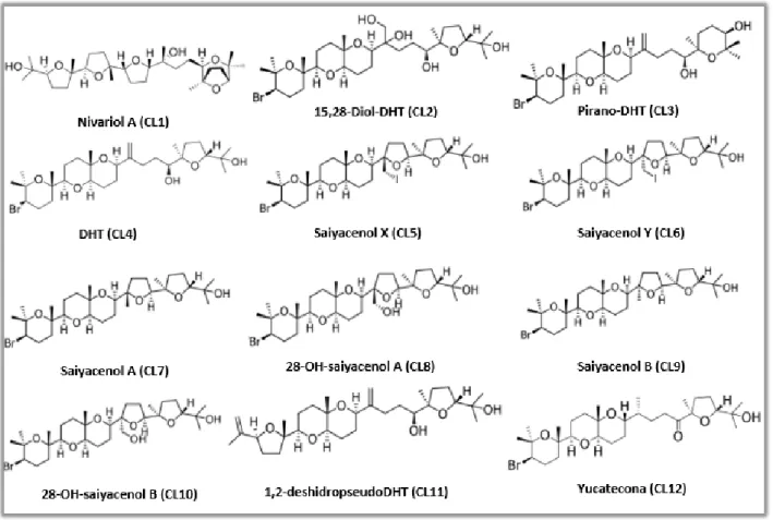
In this research, twelve molecules; 8 isolated from Laurencia viridis and 4
semi-synthetic derivatives, were evaluated in vitro against Acanthamoeba castellanii Neff.
These molecules were kindly provided by the Marine Natural Products Laboratory, Instituto Universitario de Bio-Orgánica Antonio González (IUBO-AG), ULL. The natural compounds evaluated were Nivariol A (CL1), Pirano-DHT (CL3), DHT (CL4), Saiyacenol A (CL7), 28-OH-saiyacenol A (CL8), Saiyacenol B (CL9), 1,2-deshidropseudoDHT (CL11) and Yucatecona (CL12), with the semi-synthetic molecules synthesized from DHT (CL4), being 15,28-Diol-DHT (CL2), Saiyacenol X (CL5), Saiyacenol Y (CL6), 28-OH-saiyacenol B (CL10) (55). These molecules are represented in Figure 4.
Materials and Methods
3.1.3. Reagents and products
alamarBlue™ (Life Technologies), DMSO (Merck KGaA), Trypan Blue stain 0,4% (Invitrogen) and Tween 20% (acofarma).
3.1.4. Equipment
Allegra™ 25R Centrifuge (Beckman Coulter), Cell counting chamber slides (Invitrogen), Countess II FL (Invitrogen), Electronic Multichannel Pipette (Eppendorf), EnSpire Multimode Plate Reader (Perkin Elmer), EVOS FL Cell Imaging System AMF4300 (Life Technologies), Inverted microscope Leica DMIL (Leica), Orbital Shaker (Comecta), Vertical Laminar Flow Bench (Telstar AV-100), 26ºC Incubator (Heraeus), 37ºC CO₂ Incubator (ThermoFisher Scientific), 96-well microtiter plate (ThermoFisher Scientific), white-wall 96-well microtiter plate (ThermoFisher Scientific) and 96 well deep well plate (VWR).
Figure 4. Chemical structure of the compounds evaluated. Molecular weight: CL1 = 524 g/mol; CL2 =
621 g/mol; CL3 = 587 g/mol; CL4 = 587 g/mol; CL5 = 712 g/mol; CL6 = 715 g/mol; CL7 = 587 g/mol; CL8 = 603 g/mol; CL9 = 587 g/mol; CL10 = 603 g/mol; CL11 = 506 g/mol; CL12 = 587 g/mol
Materials and Methods 3.1.5. Software
Sigma Plot 12.0 (Systat Software Inc.) and Excel.
3.2. Methods
3.2.1. In vitro activity against Acanthamoeba castellanii Neff trophozoites
The in vitro activity of the different compounds of Laurencia was evaluated using a previously developed colorimetric assay based on the alamarBlue™ Cell Viability Reagent assay (56,57). Briefly, trophozoites were counted and seeded onto a 96-well microtiter plate, adding 50 µL to each well from a stock solution of 5·10⁴ cells/mL. Then, we let Acanthamoeba adhere to the bottom of the plate for 15 minutes, and after that, the adherence was confirmed by the use of the inverted microscope Leica DMIL. Furthermore, 50 µL of serial dilutions of each compound was added to the 96-well microtiter plate. Finally, alamarBlue™ was placed into each well at 10% of the final volume and incubated at 26ºC with slight agitation. After 96 hours the fluorescence was determined with EnSpire Multimode Plate Reader, at a wavelength of 570/585 nm. As a negative control was used the A. castellanii Neff (ATCC 30010) culture incubated with PYG medium. The inhibitory concentration to inhibit the growth of 50% of the parasites (IC₅₀) was calculated through non-linear regression analysis with 95% confidence limits using the software Sigma Plot 12.0 and Excel. The experiments were performed three times each, allowing to have the standard deviation.
3.2.2. Cytotoxicity assay
The cytotoxicity of the tested compounds was determined in vitro with murine macrophages (J774A.1). For this assay, the DMEM culture medium was changed for RPMI medium with L-Glutamine, supplemented with 10% (v/v) inactivated FBS and with 10 µg/mL gentamicin. In this assay the macrophages were counted and seeded into a 96-well microtiter plate, adding 50 µL to each well from a stock solution of 2·10⁵ cells/mL. Cells were then let to adhere during 15 minutes to the bottom of the plate, checking them under an inverted microscope Leica DMIL. After that, 50 µL of a serial dilution of the compounds were added to the 96-well microtiter plate. Finally, alamarBlue™ was added into each well at an amount equal to 10% of the final volume and incubated at 37ºC in the presence of CO₂ at 5% for 24 hours. After this period, as in
Materials and Methods
the in vitro activity against Acanthamoeba castellanii Neff trophozoite assay, the fluorescence was determined with EnSpire Multimode Plate Reader at a wavelength of 570/585 nm. As a negative control was used the murine macrophages (J774A.1) culture with RPMI medium. The cytotoxicity concentration to inhibit the growth of 50% of murine macrophages (CC₅₀) was calculated through non-linear regression analysis with 95% confidence limits using the software Sigma Plot 12.0 and Excel, with the experiments being performed three times each, allowing to have the standard deviation.
3.2.3. In vitro activity against Acanthamoeba castellanii Neff cysts
For this assay, the A. castellanii Neff cysts were prepared as described by Lorenzo-Morales et al. (58). The attached trophozoites were transferred from PYG medium to NEM medium (0.1 M KCL, 8 mM MgSO₄·7H₂O, 0.4 mM CaCl₂·2H₂O, 1 mM NaHCO₃, 20 mM ammediol [2-amino-2-methyl-1,3-propanediol; Sigma Aldrich Chemistry], pH 8.8, at 25ºC). In order to have mature cysts, the trophozoites were kept under slight agitation on the orbital shaker for 7 days. At that point, the cysts were harvested and washed with PYG medium. Afterwards, we performed the serial dilution of the compound under evaluation, with PYG medium, in a 96-well microtiter plate. Then we add the mature cysts from a stock solution of 10⁵ cells/mL obtaining a volume of 100 µL in each well. The plate was then incubated at 26ºC for 7 days, and after this time the plate was centrifuged for 15 minutes at 3000 rpm, with the supernatant being removed and 100 µL of new PYG medium added. Lastly, 10 µL of alamarBlue™ was placed into each well and the plates were incubated for 7 days at 26ºC, again. The IC₅₀ was calculated through non-linear regression analysis with 95% confidence limits using the software Sigma Plot 12.0 and Excel.
3.2.4. ATP measurement
In order to evaluate the levels of ATP in amoebae, the cells were incubated with the IC₉₀ of the compound under evaluation (50,2 ± 2,6 µg/mL (value obtain with previous experiences realized by this group of researchers, Lorenzo-Morales J, Sifaoui I, Reyes-Battle M, López-Arencibia A)) for 24 hours. The ATP level was measured using a Cell Titer-Glo® Luminescent Cell Viability Assay (Promega) that requires adding a single reagent (CellTiter-Glo® Reagent), which relies on the properties of a proprietary thermostable luciferase, generating a stable proportional luminescent signal to the amount
Materials and Methods
of ATP present, which is directly proportional to the number of cells present in culture. After the incubation, treated amoebas were mixed with the kit reagent into a white-wall 96-well microtiter plate following the manufacturer’s instructions for measurement of the luminescence on a PerkinElmer spectrophotometer (59–61). As a negative control was used the A. castellanii Neff (ATCC 30010) culture incubated with PYG medium. The reduction percentage of ATP production was obtained using the software Sigma Plot 12.0 and Excel.
3.2.5. Changes in the mitochondrial membrane potential (ΔΨm)
The JC-1 Mitochondrial Membrane Potential Assay Kit (Cayman Chemical) has been used to evaluate the ΔΨm, as described before (60–62). According to the membrane
potential, the lipophilic cationic probe JC-1 accumulates in the mitochondrial matrix. In healthy cells the ΔΨm usually is high, JC-1 spontaneously forms complexes known as J-aggregates, showing intense red fluorescence (emission at 595 nm). However, in apoptotic or unhealthy cells with a low ΔΨm, JC-1 remains in its monomeric cytosolic form and shows only green fluorescence (emission at 535 nm). Thus, the trophozoites were incubated with the IC₉₀ of the compound under evaluation for 24 hours, then they were harvested and washed with buffer. Finally, the cells were incubated at 26°C for 30 min with JC-1 dye, with the fluorescence images being obtained using the EVOS FL Cell Imaging System AMF4300 (63). The negative control used was the A. castellanii Neff (ATCC 30010) culture incubated with PYG medium.
3.2.6. Plasma membrane permeability
The SYTOX® Green Nucleic Acid Stain (Molecular Probes) was performed to detect the Acanthamoeba’s membrane permeability alterations. It is a high-affinity nucleic acid stain that does not cross the plasma membranes of live cells but easily penetrates the ones with compromised membranes. The dead cells nucleic acids, after incubation, exhibits a green fluorescence enhancement of greater than 500-fold due to the nucleic acid-binding. This SYTOX® Green/DNA complex has excitation and emission of 504 and 523 nm, respectively. Acanthamoeba trophozoites were incubated at 26ºC at a final concentration of 10⁵ cells/mL in the presence of the IC₉₀ of the compound under evaluation for 24 hours. After this period, the SYTOX® Green, at a final concentration of 1 µM, was added and the plate was incubated in the dark for 30 minutes (64).
Materials and Methods
Afterwards, the fluorescence images were obtained using the EVOS FL Cell Imaging System AMF4300, and as a negative control was used the A. castellanii Neff (ATCC 30010) culture incubated with PYG medium.
3.2.7. Chromatin condensation analysis
The Chromatin Condensation/Dead Cell Apoptosis Kit with Hoechst 33342 and propidium iodide (GenScript) allows the detection of the compacted chromatin in apoptotic cells, based on the fluorescence. In one hand, Hoechst 33342 is a blue-fluorescent dye, with an excitation and emission of 350 and 461 nm, respectively, that stains more brightly the condensed chromatin of apoptotic cells than the chromatin of the normal ones. On the other hand, PI is red-fluorescent dye (excitation/emission of 535/617 nm), which only penetrates dead cells and therefore it is possible to differentiate from dead, normal and apoptotic cell populations. We start by incubating the trophozoites, at a final concentration of 10⁵ cells/mL with the IC₉₀ of the compound under evaluation for 24 hours. Next, the Hoechst 33342 and PI at a concentration of 5 µg/mL and 1 µg/mL, respectively were added and the plate was incubated at room temperature for 30 minutes (65). Then, the fluorescence images were obtained using the EVOS FL Cell Imaging System AMF4300. The negative control used was the A. castellanii Neff (ATCC 30010) culture incubated with PYG medium.
Results and Discussion
4. Results and Discussion
4.1. In vitro activity against Acanthamoeba castellanii Neff trophozoites and cytotoxicity assay
The results obtained for the in vitro activity against Acanthamoeba castellanii Neff trophozoites and also for the cytotoxicity assay in murine macrophages are shown in Table 1.
As we can see in the obtained results, all the twelve tested compounds of Laurencia (CL1 to CL12) have shown activity against A. castellanii Neff trophozoites. CL3, with an IC₅₀ of 3,11 ± 1,78 µg/mL, was the one with the highest activity. CL4 had the second highest one, with a value of IC₅₀ of 7,53 ± 0,81 µg/mL, followed by CL12 with a 23,05 ± 4,67 µg/mL IC₅₀ value. On the contrary, CL2 presented the worst IC₅₀ of the tested molecules, with an activity value of 69,96 ± 17,42 µg/mL.
In Figure 5, we have images from CL3, CL4 and CL12 activity towards Acanthamoeba trophozoites at a concentration of 25 µg/mL. It is possible to observe that these three molecules
Compound IC₅₀ (µg/mL) A. castellanii Neff Trophozoites CC₅₀ (ug/mL) Murine Macrophages CL1 53,29 ± 7,09 >50 CL2 69,96 ± 17,42 >50 CL3 3,11 ± 1,78 4,53 ± 0,13 CL4 7,53 ± 0,81 16,89 ± 1,82 CL5 36,08 ± 2,18 29,45 ± 0,20 CL6 38,85 ± 4,22 >50 CL7 32,54 ± 3,85 35,17 ± 4,99 CL8 40,13 ± 2,31 >50 CL9 45,72 ± 1,94 >50 CL10 36,25 ± 6,09 >50 CL11 53,01 ± 0,87 >50 CL12 23,05 ± 4,67 >50 Chlorhexidine* 1,53 ± 0,45 6,64 ± 0,35 Amphotericin B** 36 ---
Table 1. Activity assay (IC₅₀) of the different Laurencia compounds against Acanthamoeba castellanii Neff trophozoites
and cytotoxicity assay (CC₅₀) in murine macrophages (J774A.1)
*Reference compound (50)
**Value obtain with previous experiences realized by this group of researchers, Lorenzo-Morales J, Sifaoui I, Reyes-Battle M, López-Arencibia A
Results and Discussion
had inhibited the growth of the trophozoites. In fact, many of them are dead, and others have lost their particular cellular form, in comparison with the negative control, highlighting their trophocidal activity.
In terms of the cytotoxicity assay, of those who showed greater activity (CL3, CL4 and CL12), CL3 turned out to be the most toxic with a CC₅₀ of 4,53 ± 0,13 µg/mL. CL4 and CL12 revealed themselves less toxics, with the last one having a CC₅₀ higher than 50 µg/mL, while CL4 had a CC₅₀ value of 16,89 ± 1,82 µg/mL. Regarding the other molecules, except for CL5 with a CC₅₀ value of 29,45 ± 0,20 µg/mL and CL7 with a value of 35,17 ± 4,99 µg/mL, showed higher CC₅₀ values than 50 µg/mL.
In this way, CL3, due to its cytotoxicity levels, was excluded for further analyses. Given these first analyses, between CL4 and CL12, although both of them seem to be good candidates, CL4 was chosen to proceed with the studies regarding Acanthamoeba castellanii Neff, due to its higher activity values.
Figure 5. Activity assays of the Laurencia compounds towards A. castellanii Neff trophozoites (Magnification 200x)
Negative Control (A), CL3 with a 25 µg/mL concentration (B), CL4 with a 25 µg/mL concentration (C) and CL12 with a 25 µg/mL concentration (D) (pictures provided by Ines Sifaoui)
This figure does not reflect the activity values obtain, since the pictures were taken with the objective to understand if the compound was effective and not to compare them.
Results and Discussion 4.2. In vitro activity against Acanthamoeba castellanii Neff cysts
Acanthamoeba cysts are very resistant, allowing it to survive with slight metabolic
activity in adverse conditions, but still being viable for more than 20 years (66,67). Thus, it is also important to evaluate the activity of new molecules towards this stage of Acanthamoeba life cycle. For this reason, an in vitro activity assay was performed, in order to assess CL4 activity against the cysts. So in table 2, we are able to see the results obtained for the in vitro activity of CL4 against Acanthamoeba castellanii Neff cysts.
Watching table 2, it is possible to verify that CL4 demonstrated activity towards A.
castellanii Neff cysts, with an IC₅₀ value of 23,05 ± 0,09 µg/mL.
In Figure 6 we have pictures of CL4 activity towards Acanthamoeba castellanii Neff cysts at two different concentrations (100 µg/mL and 50 µg/mL), pointing its cysticidal activity.
Compound IC₅₀ (µg/mL) A. castellanii Neff Cysts
CL4 23,05 ± 0,09
Table 2. Activity assay (IC₅₀) of CL4 against Acanthamoeba castellanii Neff cysts
Figure 6. Activity assays of Laurencia compounds towards A. castellanii Neff cysts (Magnification 200x)
CL4 with a 100 µg/mL concentration (A), CL4 with a 50 µg/mL concentration (B) and negative control (C) (picutres provided by Ines Sifaoui)
Results and Discussion 4.3. Apoptosis-like assays
The search for drugs that do not produce necrotic cell death has made the latest research focused on new therapies. Thus, events part of the programmed cell death or apoptosis-like processes are subject of study and include several morphological events as chromatin condensation, nuclear DNA fragmentation or a decrease of cellular ATP level, among others (68).
At this stage, the objective was to understand the mechanism of action of CL4. For that, we performed assays that evaluate changes in mitochondrial membrane potential, in the ATP production, membrane permeability alterations and also in the condensation of the chromatin.
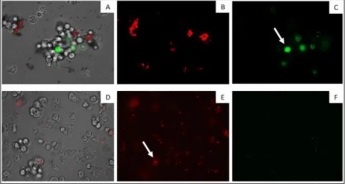
Figure 7 shows a set of images where it denotes the changes in the mitochondrial membrane potential. It can be seen that cells treated with CL4, emitted a higher green fluorescence (C) than the negative control (F). This fluorescence indicates the presence of JC-1 monomers, which reflects the decrease of the mitochondrial membrane potential. It is also possible to observe that, in treated cells, there is a lower red fluorescence (B), than in control (E). In this case, mitochondria potential did not suffer alterations, allowing JC-1 to aggregate.
Figure 7. The effect of CL4 on the mitochondrial potential (Magnification 200X).
In cells treated with the IC₉₀ of CL4 for 24 h ((A), (B) and (C)) and due to the collapse of mitochondrial potential, the JC-1 dye remained in the cytoplasm in its monomeric form, green fluorescence (C). Negative control ((D), (E) and (F)) - JC-1
dye accumulates in the mitochondria of healthy cells as aggregates (red fluorescence) (E) (Images are representative of the population of treated amoeba; Pictures provided by Ines Sifaoui)
Results and Discussion
The ATP measurement assay is documented in Figure 8, where the results demonstrated that the ATP level of A. castellanii Neff, treated with the IC₉₀ of CL4 for 24 hours, has decreased. This reduction, was more precisely in 80,48%, in comparison to the negative control, meaning that the cells only maintained a 19,52% of ATP level after the treatment with CL4.
Figure 9 exhibits the results of the plasma membrane permeability assay. It shows that the parasites treated with the IC₉₀of CL4 displayed a green fluorescence (A and B). This happens because of the formation of SYTOX® Green and DNA complex, as an outcome of the compromised cytoplasmic membrane of the treated cells, emitting the green fluorescence. Oppositely, in the negative control, the plasma membrane did not portray any damage and in consequence, did not allow the penetration of the SYTOX® Green and subsequent complex formation (C and D).
Results and Discussion
The results regarding the chromatin condensation analysis are pointed out in Figure 10. In these, it is possible to observe that the treated cells with CL4 presented a bright blue coloration (C) and in parallel, there was not a blue color in the cells of the negative control (F). This bright blue coloration is due to the condensed chromatin of apoptotic cells stained with the Hoescht blue-fluorescent dye. In the treated cells, we can also see a red coloration (D) while in the negative control, there is no coloration, again (G). The red color is due to the PI, a red-fluorescent dye that only has the capacity to penetrate dead cells. Therefore, the presence of a red coloration means that those are necrotic cells.
Figure 9. The effect of CL4 on the plasma membrane permeability (Magnification 400X).
(A and B) Cells treated with the IC₉₀ of CL4 for 24 h. (A) Overlay image; (B) Dead cells with the SYTOX® Green/DNA complex; (C and D) Negative control
Results and Discussion
4.4. Precipitation problems
Figure 11 shows precipitate formation in CL7 during the in vitro activity assay against
Acanthamoeba castellanii Neff trophozoites. In the course of the investigation, it was detected
that the different Laurencia viridis compounds led to the formation of a precipitate. This fact was noticed with both of the two solvents used to dissolve the Laurencia viridis molecules, DMSO and Tween 20%.
Figure 10. The effect of CL4 on chromatin condensation (Magnification 200X).
(A to D) Cells treated with the IC₉₀ of CL4 for 24 h. ; (E to G) Negative control; (B) Overlayed C and D images; (C) Apoptotic cells with condensed chromatin stained with Hoechst; (D) Necrotic cells stained with PI
(Images are representative of the population of treated amoeba; Pictures provided by Ines Sifaoui)
Figure 11. Example of precipitate formation in CL7 at a concentration of 25 µg/mL
Conclusions
5. Conclusions
With the objective to find new, safe and active drugs versus Acanthamoeba
castellanii, twelve compounds of Laurencia virids (CL1 to CL2) have been assessed to
find their activity towards this parasite.
Thanks to the in vitro activity assay, it was discovered that all compounds possessed activity against Acanthamoeba castellanii Neff trophozoites. Among these, CL3 was the most active, while CL2 was the less one. It is important to mention that although these molecules displayed good activity values, none of them tested presented better IC₅₀ value than chlorhexidine, which is the common drug used to treat AK. But, CL3, CL4, CL7 and CL12, presented better activity values than amphotericin B, that is another typical molecule used against this pathology. Another conclusion drawn was that CL3 revealed himself as the most toxic compound as evidenced by the results of the cytotoxicity assay in J774A.1 murine macrophages.
Given these data, the CL4 molecule proved to be worthy for more comprehensive insight and additional tests. In fact, this compound showed activity versus A. castellanii Neff cysts in the in vitro assay. Yet, the IC₅₀ value of CL4 activity facing the cysts was higher than its cytotoxicity value in murine macrophages, meaning that it is only effective towards the cysts at a toxic concentration for the host. Unluckily, this result is in line with what has been observed in relation to cysticidal activity of the majority of the different drugs currently available. The use of the JC-1 Mitochondrial Membrane Potential Assay Kit and the Cell Titer-Glo® Luminescent Cell Viability Assay pointed to damage in the mitochondria induced by CL4. This interpretation was due to the reduction in the mitochondrial membrane potential and in ATP levels illustrated in these assays. With the application of the SYTOX® Green Nucleic Acid Stain, it also led to believe that CL4 has the capacity to cause permeability in the cytoplasmic membrane. The Chromatin Condensation/Dead Cell Apoptosis Kit allowed us to conclude that CL4, although it triggered cell apoptosis, unfortunately, it also has shown to cause necrosis.
Another conclusion reached was that this Laurencia compounds precipitated in the solvents used to dissolve them, DMSO and Tween 20%. Moreover, there is a possibility that this factor has affected the outcome of the assays. Therefore, the solubility optimization of these molecules should be the first step to carrying on with the studies.
Despite the necessary optimization of the solubility and the unfavourable results of the cysticidal activity and chromatin condensation analysis, Laurencia viridis remains as
Conclusions
a viable option to continue investigating and hopefully develop a novel and truly effective antiparasitic drug against Acanthamoeba.
References
6. References
1. Cabral G, Marciano-Cabral F. Acanthamoeba spp. as Agents of Disease in Humans. Clin Microbiol Rev [Internet]. 2003;16(2):273–307. Available from: http://www.pubmedcentral.nih.gov/articlerender.fcgi?artid=153146
2. Khan NA. Acanthamoeba: Biology and increasing importance in human health. FEMS Microbiol Rev. 2006;30(4):564–95.
3. Siddiqui R, Khan NA. Biology and pathogenesis of Acanthamoeba. Parasites and Vectors [Internet]. 2012;5(1):6. Available from: http://www.parasitesandvectors.com/content/5/1/6
4. Visvesvara GS. Infections with free-living amebae [Internet]. 1st ed. Vol. 114, Handbook of Clinical Neurology. © 2013, Elsevier B.V. All rights reserved.; 2013. 153–168 p. Available from: http://dx.doi.org/10.1016/B978-0-444-53490-3.00010-8
5. Abd El Wahab WM, El-Badry AA, Hamdy DA. Molecular characterization and phylogenetic analysis of Acanthamoeba isolates in tap water of Beni-Suef, Egypt. Acta Parasitol. 2018;63(4):826–34.
6. Reyes-Batlle M, Hernández-Piñero I, Rizo-Liendo A, López-Arencibia A, Sifaoui I, Bethencourt-Estrella CJ, et al. Isolation and molecular identification of free-living amoebae from dishcloths in Tenerife, Canary Islands, Spain. Parasitol Res. 2019;
7. Marciano-cabral F, Puffenbarger R, Cabral GUYA. the Increasing Importance of Hospitals. JAMA J Am Med Assoc. 2011;96(13):1087.
8. Rojas M del C, Rodríguez Fermepín M, Gracia Martínez F, Costamagna SR. Presencia de Acanthamoeba spp. en agua para consumo ganadero en la provincia de La Pampa, Argentina. Rev Argent Microbiol [Internet]. 2017;49(3):227–34. Available from: http://dx.doi.org/10.1016/j.ram.2016.12.003
9. Siddiqui R, Khan NA. War of the microbial worlds: Who is the beneficiary in Acanthamoeba-bacterial interactions? Exp Parasitol [Internet]. 2012;130(4):311– 3. Available from: http://dx.doi.org/10.1016/j.exppara.2012.01.021
References
11. Guimaraes AJ, Gomes KX, Cortines JR, Peralta JM, Peralta RHS. Acanthamoeba spp. as a universal host for pathogenic microorganisms: One bridge from environment to host virulence. Microbiol Res [Internet]. 2016;193:30–8. Available from: http://dx.doi.org/10.1016/j.micres.2016.08.001
12. Visvesvara GS, Moura H, Schuster FL. Pathogenic and opportunistic free-living amoebae: Acanthamoeba spp., Balamuthia mandrillaris , Naegleria fowleri , and
Sappinia diploidea. FEMS Immunol Med Microbiol [Internet]. 2007;50(1):1–26.
Available from: https://academic.oup.com/femspd/article-lookup/doi/10.1111/j.1574-695X.2007.00232.x
13. Khan NA. Acanthamoeba and the blood-brain barrier: The breakthrough. J Med Microbiol. 2008;57(9):1051–7.
14. CDC. CDC - Acanthamoeba Infection - Biology [Internet]. https://www.cdc.gov/. 2019 [cited 2019 Feb 20]. Available from: https://www.cdc.gov/parasites/acanthamoeba/pathogen.html
15. Silva LK dos S, Boratto PVM, La Scola B, Bonjardim CA, Abrahão JS. Acanthamoeba and mimivirus interactions: The role of amoebal encystment and the expansion of the “Cheshire Cat” theory. Curr Opin Microbiol. 2016;31:9–15.
16. Anwar A, Khan NA, Siddiqui R. Combating Acanthamoeba spp. cysts: What are the options? Parasites and Vectors. 2018;11(1):4–6.
17. Anwar A, Khan NA, Siddiqui R. Galactose as novel target against Acanthamoeba cysts. PLoS Negl Trop Dis. 2019;13(7):e0007385.
18. Król-Turmińska K, Olender A. Human infections caused by free-living amoebae. Ann Agric Environ Med. 2017;24(2):254–60.
19. Schuster FL, Visvesvara GS. Free-living amoebae as opportunistic and non-opportunistic pathogens of humans and animals. Int J Parasitol. 2004;34(9):1001– 27.
20. Lorenzo-Morales J, Martín-Navarro CM, López-Arencibia A, Arnalich-Montiel F, Piñero JE, Valladares B. Acanthamoeba keratitis: An emerging disease gathering importance worldwide? Trends Parasitol. 2013;29(4):181–7.
21. Dudley R, Alsam S, Khan NA. The role of proteases in the differentiation of Acanthamoeba castellanii. FEMS Microbiol Lett. 2008;286(1):9–15.
References
22. Maycock NJR, Jayaswal R. Update on Acanthamoeba Keratitis: Diagnosis, Treatment, and Outcomes. Cornea. 2016;35(5):713–20.
23. Alkharashi M, Lindsley K, Sikder S. Medical interventions for acanthamoeba keratitis. Cochrane Database Syst Rev. 2013;2013(10).
24. Lorenzo-Morales J, Khan NA, Walochnik J. An update on Acanthamoeba keratitis: diagnosis, pathogenesis and treatment . Parasite. 2015;22:10.
25. Neelam S, Niederkorn JY. Pathobiology and immunobiology of Acanthamoeba keratitis: Insights from animal models. Yale J Biol Med. 2017;90(2):261–8. 26. Nagington J, Watson PG, Playfair TJ, Mcgill J, Jones BR, Steele ADMG. Amoebic
Infection of the Eye. Lancet. 1974;304(7896):1537–40.
27. Samples JR, Binder PS, Luibel FJ, Font RL, Visvesvara GS, Peter CR. Acanthamoeba Keratitis Possibly Acquired from a Hot Tub. Arch Ophthalmol. 1984;102(5):707–10.
28. Ferreira Mendes J, Monteiro T. Ulcère pseudodendritique. J Fr Ophtalmol [Internet]. 2017;40(4):341–2. Available from: http://dx.doi.org/10.1016/j.jfo.2016.12.015
29. Anjos R, Vicente A, Vieira L, Maduro V, Alves N, Feijão J, et al. Queratite por Acantamoeba - Revisão de 6 Casos Clínicos. Centro Hospitalar Lisboa Central. Oftalmologia. 2013;37:283–90.
30. Costa AO, Furst C, Rocha LO, Cirelli C, Cardoso CN, Neiva FS, et al. Molecular diagnosis of Acanthamoeba keratitis: evaluation in rat model and application in suspected human cases. Parasitol Res. 2017;116(4):1339–44.
31. Carrijo-Carvalho LC, Sant’ana VP, Foronda AS, de Freitas D, de Souza Carvalho FR. Therapeutic agents and biocides for ocular infections by free-living amoebae of Acanthamoeba genus. Surv Ophthalmol [Internet]. 2017;62(2):203–18. Available from: http://dx.doi.org/10.1016/j.survophthal.2016.10.009
32. Visvesvara GS. Infections with free-living amebae [Internet]. 1st ed. Vol. 114, Neuroparasitology and Tropical Neurology. © 2013, Elsevier B.V. All rights reserved.; 2013. 153–168 p. Available from: http://www.sciencedirect.com/science/article/pii/B9780444534903000108
References
33. Baig AM, Khan NA. A proposed cascade of vascular events leading to granulomatous amoebic encephalitis. Microb Pathog [Internet]. 2015;88:48–51. Available from: http://dx.doi.org/10.1016/j.micpath.2015.08.005
34. Tavares M, Da Costa JMC, Carpenter SS, Santos LA, Afonso C, Aguiar Á, et al. Diagnosis of first case of Balamuthia amoebic encephalitis in Portugal by immunofluorescence and PCR. J Clin Microbiol. 2006;44(7):2660–3.
35. Theel ES, Pritt BS. Parasites. 2016;
36. Parija S, Venugopal H, KP D. Management of granulomatous amebic encephalitis: Laboratory diagnosis and treatment. Trop Parasitol. 2015;5(1):23.
37. Kulsoom H, Baig AM, Siddiqui R, Khan NA. Combined drug therapy in the management of granulomatous amoebic encephalitis due to Acanthamoeba spp., and Balamuthia mandrillaris. Exp Parasitol [Internet]. 2014;145(S):S115–20. Available from: http://dx.doi.org/10.1016/j.exppara.2014.03.025
38. Taravaud A, Loiseau PM, Pomel S. In vitro evaluation of antimicrobial agents on Acanthamoeba sp. and evidence of a natural resilience to amphotericin B. Int J Parasitol Drugs Drug Resist. 2017;7(3):328–36.
39. Chan LL, Mak JW, Ambu S, Chong PY. Identification and ultrastructural characterization of acanthamoeba bacterial endocytobionts belonging to the alphaproteobacteria class. PLoS One. 2018;13(10):1–21.
40. Tice AK, Shadwick LL, Fiore-Donno AM, Geisen S, Kang S, Schuler GA, et al. Expansion of the molecular and morphological diversity of Acanthamoebidae (Centramoebida, Amoebozoa) and identification of a novel life cycle type within the group. Biol Direct [Internet]. 2016;11(1). Available from: http://dx.doi.org/10.1186/s13062-016-0171-0
41. Taher EE, Méabed EMH, Abdallah I, Wahed WYA. Genotyping and risk factors , a study from Cairo , Egypt. J Infect Public Health [Internet]. 2017;7–13. Available from: https://doi.org/10.1016/j.jiph.2017.09.013
42. Kot K, Łanocha-arendarczyk NA, Kosik-bogacka DI. Original papers Amoebas from the genera Acanthamoeba and their. 2018;64(December):299–308.
43. Haniloo A, Pezeshki A, Mahmmodzadeh A, Kadkhodamohammadi E. Genotyping of Acanthamoeba spp. from water sources from Northwestern Iran. Acta Parasitol.
References
2017;62(4):790–5.
44. Castro-Artavia E, Retana-Moreira L, Lorenzo-Morales J, Abrahams-Sandí E. Potentially pathogenic Acanthamoeba genotype T4 isolated from dental units and emergency combination showers. Mem Inst Oswaldo Cruz. 2017;112(12):817–21. 45. Carnt N, Stapleton F. Strategies for the prevention of contact lens-related
Acanthamoeba keratitis: A review. Ophthalmic Physiol Opt. 2016;36(2):77–92. 46. Gutiérrez-Rodríguez AG, Juárez-Portilla C, Olivares-Bañuelos T, Zepeda RC.
Anticancer activity of seaweeds. Drug Discov Today [Internet]. 2018;23(2):434– 47. Available from: http://dx.doi.org/10.1016/j.drudis.2017.10.019
47. Murugan AC, Karim MR, Yusoff MBM, Tan SH, Asras MFBF, Rashid SS. New insights into seaweed polyphenols on glucose homeostasis. Pharm Biol [Internet]. 2015;53(8):1087–97. Available from: http://dx.doi.org/10.3109/13880209.2014.959615
48. Brown EM, Allsopp PJ, Magee PJ, Gill CI, Nitecki S, Strain CR, et al. Seaweed and human health. Nutr Rev. 2014;72(3):205–16.
49. Shi Q, Wang A, Lu Z, Qin C, Hu J, Yin J. Overview on the antiviral activities and mechanisms of marine polysaccharides from seaweeds. Carbohydr Res [Internet]. 2017;453–454:1–9. Available from: https://doi.org/10.1016/j.carres.2017.10.020 50. García-Davis S, Reyes-Batlle M, Viveros-Valdez E, Díaz-Marrero A, Fernández
J, Sifaoui I, et al. Anti-Acanthamoeba Activity of Brominated Sesquiterpenes from Laurencia johnstonii. Mar Drugs. 2018;16(11):443.
51. Veiga-Santos P, Pelizzaro-Rocha KJ, Santos AO, Ueda-Nakamura T, Filho BPD, Silva SO, et al. In vitro anti-trypanosomal activity of elatol isolated from red seaweed Laurencia dendroidea. Parasitology. 2010;137(11):1661–70.
52. Martins A, Vieira H, Gaspar H, Santos S. Marketed marine natural products in the pharmaceutical and cosmeceutical industries: Tips for success. Mar Drugs. 2014;12(2):1066–101.
53. Harizani M, Ioannou E, Roussis V. The Laurencia Paradox: An Endless Source of Chemodiversity. Vol. 102, Progress in the chemistry of organic natural products. 2016. 91–252 p.
References
54. Veiga-santos P, Prado B, Filho D, Sudatti DB, Pereira RC, Nakamura CV, et al. Effect of Elatol , Isolated from Red Seaweed. 2010;2733–43.
55. IUBO LM. Laurencia compounds [Internet]. .ull.es/institutos/instituto-bio-organica/. 2019 [cited 2019 Feb 19]. Available from: https://www.ull.es/institutos/instituto-bio-organica/
56. Martín-Navarro CM, Lorenzo-Morales J, Cabrera-Serra MG, Rancel F, Coronado-Álvarez NM, Piñero JE, et al. The potential pathogenicity of chlorhexidine-sensitive Acanthamoeba strains isolated from contact lens cases from asymptomatic individuals in Tenerife, Canary Islands, Spain. J Med Microbiol. 2008;57(11):1399–404.
57. McBride J, Ingram PR, Henriquez FL, Roberts CW. Development of colorimetric microtiter plate assay for assessment of antimicrobials against Acanthamoeba. J Clin Microbiol. 2005;43(2):629–34.
58. Lorenzo-Morales J, Kliescikova J, Martinez-Carretero E, De Pablos LM, Profotova B, Nohynkova E, et al. Glycogen Phosphorylase in Acanthamoeba spp.: Determining the Role of the Enzyme during the Encystment Process Using RNA Interference . Eukaryot Cell. 2008;7(3):509–17.
59. Promega. CellTiter-Glo® Luminescent Cell Viability Assay [Internet]. worldwide.promega.com. 2016 [cited 2019 Mar 20]. Available from: https://worldwide.promega.com/products/cell-health-assays/cell-viability-and-
cytotoxicity-assays/celltiter_glo-luminescent-cell-viability-assay/?catNum=G7570
60. Sifaoui I, López-Arencibia A, Martín-Navarro CM, Reyes-Batlle M, Wagner C, Chiboub O, et al. Programmed cell death in Acanthamoeba castellanii Neff induced by several molecules present in olive leaf extracts. PLoS One. 2017;12(8):1–12.
61. López-Arencibia A, Reyes-Batlle M, Freijo MB, McNaughton-Smith G, Martín-Rodríguez P, Fernández-Pérez L, et al. In vitro activity of 1H-phenalen-1-one derivatives against Acanthamoeba castellanii Neff and their mechanisms of cell death. Exp Parasitol [Internet]. 2017;183:218–23. Available from: http://dx.doi.org/10.1016/j.exppara.2017.09.012