1. Department of Physical Medicine and Rehabilitation, Erenkoy Hospital, Istanbul
2. Faculty of Medicine, Department of Ophthalmology, Celal Bayar University, Manisa
3. Faculty of Medicine, Department of Physical Medicine and Rehabilitation, Balikesir University, Balikesir
4. Faculty of Medicine, Department of Dermatology, Inonu University, Malatya
5. Department of Physical Medicine and Rehabilitation, Malatya Hospital, Malatya
6. Department of Physical Medicine and Rehabilitation, Ersoy Hospital, Istanbul
7. Faculty of Medicine, Department of Physical Medicine and Rehabilitation, Inonu University, Malatya
muscle strengths are lower in BD. We therefore recom-mend careful monitoring of patients with BD in terms of muscle strength.
Keywords: Behçet’s disease; Knee; Muscle strength; Isokinetic; Peak torque.
IntroductIon
Behçet’s disease (BD) is a chronic, multisystemic in-flammatory disease, characterized by recurrent oro-genital aphthous ulcerations, uveitis and skin lesions. The disease may affect other systems such as locomo-tor, neurological, gastrointestinal, nephrologic and vas-cular systems1,2.
Arthritis has been found present at the time of diagno sis in about 70% of patients, and 9% of patients have had arthritis only as an initial manifestation in BD. The most frequently involved joints are the knees, an-kles, wrists, and elbows3. The arthritis of BD is usually
mono-oligo or polyarticular, and it characteristically manifests a nondestructive course4.
Isokinetic muscle strength (IMS) is a measure of the maximal dynamic muscle strength throughout the range of motion in a joint. Considered to be a valid and reliable measure5,6, IMS is often used to evaluate sports
performance and the efficacy of exercise training in sports medicine, as well as to evaluate the efficacy of surgical intervention and rehabilitation in orthopedic and rehabilitation clinics7-9. In recent years it has also
been used for the evaluation of muscle strength in rheumatological diseases associated with functional disabilities10,11. In a recent study, quadriceps muscle
strength was measured with isokinetic dynamometer in rheumatoid arthritis (RA) and ankylosing spondyli-tis (AS) patients who had low disease severity and suf-fered little knee pain during the walking exercise11.
The involvement of the knee joint in BD is
well-Isokinetic evaluation of knee extensor/flexor
muscle strength in Behcet’s patients
Durmus B1, Emre S2, Sahin N3, Karincaoglu Y4, Dogan E5, Baysal O6, Ersoy Y7, Altay Z7
ACTA REUMATOL PORT. 2015;40:348-354
AbstrAct
Background: Behçet’s disease (BD) is an idiopathic, multisystemic, progressive disease. The purpose of this study is to compare the knee flexor and extensor isoki-netic muscle strengths of Behcet’s patients with that of healthy subjects.
Methods: Twenty-five (13 male and 12 female) patients with BD and 25 (15 male and 10 female) healthy indi-viduals were included in the study. Velocities of 90°/sec, 120°/sec, and 150°/sec were used for the isokinetic muscle strength testing. Patients with active inflamma-tory knee arthritis were excluded. Peak torque (Nm) and peak torque adjusted to body weight (%) were ta -ken into consideration for comparison between study groups.
Results: Compared to healthy controls, there was a sta-tistically significant decrease in both the bilateral knee extensor and flexor muscle isokinetic peak torques (Nm) as well as the peak torques adjusted to body weight (%) at velocities of 90°/sec, 120°/sec and 150°/sec in patients with BD (p < 0.05). However, there was no significant difference in the agonist-antagonist ratio of the isokinetic peak torques of knee muscles be-tween the two groups.
Conclusion: In light of these findings, we have con-cluded that both knee flexor and extensor isokinetic
known; however, to our knowledge, until now there have not been any studies in the literature which relate BD to the strength of the knee muscles. Motivated by this, our study was conducted in order to measure knee flexor and extensor muscle strength in patients with BD by using an isokinetic dynamometer, and to compare the results of the measurements with those obtai ned for an age- and sex-matched healthy control group. In that respect, this study is the first controlled study to evaluate knee flexor and extensor isokinetic muscle strength in patients with BD.
MAtErIAls And MEthods
The study was designed as a crosssectional, controll -ed, clinical study. We included 25 (13 male and 12 fe-male) consecutive patients presenting at the Physical Medicine and Rehabilitation and Dermatology De-partments, and diagnosed with BD according to the International Study Group for BD criteria12. The same
number of healthy disease-free individuals (15 male and 10 female) were included to serve as controls. Each participant provided written informed consent to par-ticipate in this study, which was also approved by the local ethics committee.
The patients were evaluated from the systemic, rheumatologic, dermatologic, ophthalmologic, and neurologic points of view. The patients were screened for age, sex, weight, height, and the durations of disea -se. We excluded from the study all patients and healthy subject’ s with any known neurologic, orthopedic, car-diovascular, pulmonary disease, diabetes mellitus, or any thyroid disorders that may have an effect on mus-cle strength. Moreover, we excluded those patients with active inflammatory knee joint involvement. None of the patients had neurologic involvement and active uveitis. None of the individuals had engaged in a regular exercise program for the last 6 months. The overall incidence of joint symptoms and the involved joints were recorded. None of the patients were using medications that affect muscle strength either posi-tively or negaposi-tively. Arthralgia was defined by physical examination as pain in the joint without demonstrable inflammation. The healthy volunteers were selected among relatives of the patients admitted to our clinic. Before the formal testing, each subject performed a warm up on a bicycle ergo meter with a load of 1 W/kg and with stretching exercises of the lower limbs for 5 minutes. We used a calibrated isokinetic testing
dynamometer (Biodex System 3 Pro, Biodex Corp., Shir -ley, NY) for isokinetic muscle strength testing of the trunk. The reliability of the isokinetic testing is esti-mated to be r=0.97 for the quadriceps femoris muscles and r=0.85 for the hamstring muscles6. The reliability
and reproducibility of the device used, is well esta -blished13.
The procedure to measure the knee flexor and ex-tensor isokinetic muscle strength of each subject was as follows: Firstly, the subject was seated upright on the Biodex chair with the axis of the dynamometer aligned to the knee joint axis. Once positioned properly, the subjects shoulder, waist, thigh, and lower right leg proximal to the ankle were secured with straps. The subject was then instructed to perform concentric knee exercise at a range of motion (ROM) from 0° to 90° of flexion. Five warm-up and practice repetitions were performed before each testing repetition. The isoki-netic testing protocol consisted of tests at three angu-lar velocities of 90°, 120° and 150°/sec, with 10 repe-titions. The initial five repetitions at each velocity were performed at sub maximal effort, and the last five were performed at maximum effort. There was a 5 minutes of rest interval between successive repetitions (Figure 1). All tests were performed on both extremities and performed for concentric muscle strength. Peak torque (PT), peak torque/body weight (PT/BW) ratio (%) and the agonist/antagonist (flexor/extensor) ratio (%) (AG/AN) were recorded. PT is the highest torque va -lue obtained from the measurements in all joint action distinction. There are multiple parameters in the iso-metric measurements evaluating muscle strength and
FIGurE 1.Isokinetic knee muscle strength testing with Biodex System 3 Pro dynamometer
(Figure 2). All extensor PT/BW ratios (%) of the knee strengths at velocities of 90°/sec, 120°/sec and 150°/sec were significantly lower (P < 0.001 for all comparisons) in the BD group. Behçet's patients group exhibited statis tically significantly lower PT/BW ratio (%) at ve -lo cities of 90°, 120° and 150°/sec both for the right (p < 0.001, = 0.003 and = 0.005 respectively) and the left (p = 0.005, 0.016 and 0.012 respectively) knee fle -xors (Figure 3). However, the difference between bi-lateral knee agonist-antagonist ratios (%) of isokinetic peak torques of the two groups was not significant at any of the velocities (Figu re 4).
dIscussIon
In this study, we aimed to investigate bilateral knee fle -xor and extensor muscle strength at velocities of 90º/sec, 120º/sec and 150º/sec, by using isokinetic methods, in patients with BD. We included patients who had never had active inflammatory arthritis up to the time of the examinations. We compared the pa-tient’s results with age- and sex-matched controls. The isokinetic measurements at the three velocities revealed that both PT and PT/BW were significantly lower in Be-hcet’s patients. However, no significant difference, was observed between the two groups in the ago nist--antagonist ratios of the knee muscles, which suggest that in BD there is a relationship between the muscle strength loss in the anterior and posterior muscu lature. In diseases associated with joint involvement both muscle function. Among those parameters, PT
isomet-ric test parameter, whose unit is newtonmeter (Nm), is the most suitable and is highly recommended14. PT is
affected by age, gender, dominant extremity, BMI, body fat ratio, and waist thickness. PT/BW is a parameter that is used for commenting on the isokinetic evalua-tion and making the measurements subjective and unique to the person15. AG/AN ratio eva luates the
bal-ance of trunk flexor and extensor16.
The software SPSS 16.0 (SPSS, Chicago, IL, USA) was used for statistical evaluation. Whenever variables were normally distributed, as assessed using the Kol-mogorov-Smirnov test, we performed parametric test statistics; such as independent sample t-test. For the categorical variables, we used the Chi-square test. The data were expressed as mean ± standard deviation for parametric variables and as frequency (count or per-cent) for categorical variables. All p values less than 0.05 were considered to indicate significance.
rEsults
The mean age of the patients was 34.72 ± 9.21 (range 22-52), and that of the controls was 33.24 ± 9.01 (range 23-57) years. Therefore, there was no statisti-cally significant difference between the two groups in age. Likewise, no statistically significant difference was detected in weight, height and BMI either. The mean du-ration of disease was 8.25 ± 6.38 (range 0.5-25) years for the BD group. Descriptive characteristics of the patient and control groups are presented in Table I.
Up to the examination date, only 6 patients had ne -ver experienced any joint complaints. Among the re-maining 19 patients, 9 had had inflammatory arthritis anamnesis. On the date of examination, none of the patients had active inflammatory knee arthritis. How-ever, 2 patients had active joint inflammation in the el-bow. In the BD group, 11 subjects had anterior or pos-terior uveitis; among these subjects, 2 patients had bi-lateral involvement. For all patients with uveitis, the disease was under control with medical treatment.
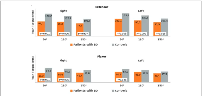
The control group exhibited statistically significan -tly greater strength at velocities of 90°/sec, 120°/sec and 150°/sec both for the right (p = 0.001, 0.006 and 0.007 respectively) and left (P = 0.006, 0.009 and 0.016 res -pectively) knee extensors. Isokinetic knee flexor strength was significantly lower at 90°/sec and 120°/sec for the right knee (p < 0.001, = 0.025 respectively) and at 90°/sec for the left knee (p = 0.046) in the BD group
tAblE I. dEMoGrAphIc chArActErIstIcs oF pAtIEnts wIth bEhcEt’s dIsEAsE And controls
BD patients Controls n = 25 n = 25 P Age (years) 34.72±9.21 33.24±9.01 0.568 Gender (M/F) (n) 13/12 15/10 0.569* Weight (kg) 71.96±14.07 68.48±8.93 0.302 Height (cm) 168.20±8.92 167.80±7.26 0.877 BMI (kg/m2) 25.30±3.60 24.27±2.29 0.238 Dominant side 22/3 24/1 0.297* (R/L) Disease duration 8.25±6.38 (years)
Data presented as mean ± S.D. *Chi-Square test
immobilization and functional disabilities, as well as inflammation in joints and periarticular tissues, may lead to a decrease in muscle strength10,17. In prospective
studies, the incidence of arthralgia or arthritis in BD is observed in more than half of patients18,19. In Behcet’s
patients, lower extremity joint involvement, particu-larly knee involvement, is common and is seen with remissions and activations4. In a recent study,
involve-ment of the knee was reported to be found in 40% of patients with BD20. In our study, arthralgia and/or
arthritis anamnesis was present in 13 (52%) BD pa-tients, but none of those patients had active inflamma-tory knee arthritis.
BD is a multisystemic disease, thus many factors may lead to functional disability in patients. Uveitis is a com-mon and important clinical finding during the follow--up of BD. The frequency of ocular involvement in patients with BD is around 70%. The typical form of ocu -lar involvement is a relapsing remitting uveitis. Less frequently, it may present in the form of conjunctivitis, conjunctival ulcers, keratitis, episcleritis, scleritis, and extra ocular muscle paralysis from neurologic involve-ment of BD. Intraocular inflammation may involve the anterior or posterior segment or, more commonly, both segments21. In a study involving patients with AS, the
presence of acute anterior uveitis (AAU) in AS patients was reported to lead to a significant decrease in the pa-tients’ functionali ty in daily activities. It was suggested that the presence of AAU in AS patients may be
associated with higher disease activity, poor functional abi -lity and advanced physical impairment22. In our study,
none of the patients has active intraocular inflamma-tion; however, 11 (44%) patients have experienced uveitis attacks in their medical anamnesis and uveal in-flammation was clinically under control with medica-tion.
In BD, other probable causes of muscle weakness could be nervous system involvement and muscular tissue inflammation. Although central nervous system involvement is well described in BD; in recent studies, an axonal type of distal sensory or sensorimotor po -lyneuropathy predominantly involving the lower ex-tremities has been commonly reported in BD pa-tients23,24. Silent neurologic involvement may occur in
BD, and there is a possibility of subclinical involvement without neurologic signs and symptoms in some ca -ses23. In this study, despite the lack of electromyogram
(EMG) results, we performed neurologic examinations thoroughly. We could not detect any signs of periphe -ral or cent-ral nervous system involvement in any of the patients. Subclinical muscle involvement and necro-tizing myositis with pain, swelling and tenderness or only myalgia have rarely been reported in BD25,26. In
addition to these, myopathy and neuropathy cases re-lated to agents used in the treatment of BD such as colchicine and cyclosporine A have been reported27,28.
In our study, colchicine was used alone or as a combi-nation by 17 patients, as was cyclosporine A by 7 pa-90º130,2117,1101,685,274,386,7120º150º90º120º150ºRightLeftExtensor
Peak Torque (Nm)Patients with BDControlsP=0.001P=0.006P=0.007133,8120,5103,481,993,1102,1P=0.006P=0.009P=0.016 90º63,259,152,844,941,440,2120º150º90º120º150ºRightLeftFlexor Peak Torque (Nm)Patients with BDControlsP<0.001P=0.02557,652,347,339,145,945,3P=0.046
90º 130,2 117,1 101,6 85,2 74,3 86,7 120º 150º 90º 120º 150º Right Left Extensor Pe ak T or qu e (N m )
Patients with BD Controls
P=0.001 P=0.006 P=0.007 133,8 120,5 103,4 81,9 93,1 102,1 P=0.006 P=0.009 P=0.016 90º 63,2 59,1 52,8 44,9 41,4 40,2 120º 150º 90º 120º 150º Right Left Flexor Pe ak T or qu e (N m )
Patients with BD Controls
P<0.001 P=0.025 57,6 52,3 47,3 39,1 45,9 45,3 P=0.046
tients. All of this information showed that, muscle strength and functional status could be affected in BD patients even without causing disability.
BD is characterized by exacerbations and remissions. The duration of attacks ranges from a few days to a few weeks and sequelae can be found after attacks25. These
attacks could lead to immobilization of patients, as well as result in loss of muscle strength and functional disa -bility. In a study which was based on a comparison of the functional disabilities between BD and rheumatoid arthritis, Behcet’s patients were found to have higher functional disability and fatigue than RA patients. In that study, the functional disability scores of RA pa-tients were similar to Behçet’s papa-tients without arthri-tis29. In RA patients it has been shown that, knee
isoki-netic muscle strengths are correlated with Health As-sessment Questionnaire (HAQ) scores that are used for the measurement of functional disabilities. In 36 RA patients, it was found that knee isokinetic muscle strength was correlated with HAQ, which is a patient reported outcome measure for functional disability in RA17. Current tools for assessing BD do not include
pa-tient-reported outcomes for assessment of functional disability such as HAQ29. In one study, it was reported
that knee extensor strength reduced by 25% in patients without a clinically defined affected knee in RA and with an affected knee joint, while knee-extensor strength decreased up to 78% compared to a control group10. In another study, muscle strength of 67 RA
patients with low and moderate disease activity was eva -luated with isokinetic and isometric methods. Knee in-volvement was detected in just 16 of the patients. Com-parison with healthy controls revealed a significant de-crease in both isometric and isokinetic muscle strengths and endurance in RA patients30. Systemic lupus erythe
-matosus (SLE) is also another multisystemic rheuma-tologic disease and usually does not cause destructive arthritis as BD does. In a study involving 43 SLE pa-tients where, quadriceps muscle strength and exercise capacity with a bicycle ergo meter were compared with sedentary controls, it was found that both parameters were nearly 21% lower compared to those measured for the controls31. In another study, which investigated
the aerobic capacity of Behcet’s patients, it was shown that exercise capacity with bicycle ergo meters was less than that of the controls32.
In a recent study, possible involvement of the quadri-ceps muscle was shown in BD patients. Quadriquadri-ceps and 90º187,8168,1146,1116,2101,3119,1120º150º90º120º150ºRightLeftExtensor
PT/BW (%)Patients with BDControlsP<0.001P<0.001P<0.001193,5174,1149,8111,4126,8140,3P<0.001P<0.001P<0.001 90º91,984,576,560,455,654,5120º150º90º120º150ºRightLeftFlexor PT/BW (%)Patients with BDControlsP<0.001P=0.003P=0.00583,779,168,752,561,661,7P=0.005P=0.016P<0.012
90º 187,8 168,1 146,1 116,2 101,3 119,1 120º 150º 90º 120º 150º
Right Extensor Left
PT /B W ( % )
Patients with BD Controls
P<0.001 P<0.001 P<0.001 193,5 174,1 149,8 111,4 126,8 140,3 P<0.001 P<0.001 P<0.001 90º 91,9 84,5 76,5 60,4 55,6 54,5 120º 150º 90º 120º 150º
Right Flexor Left
PT /B W ( % )
Patients with BD Controls
P<0.001 P=0.003 P=0.005 83,7 79,1 68,7 52,5 61,6 61,7 P=0.005 P=0.016 P<0.012
FIGurE 3.Peak torque/body weight ratios (%) of flexor and extensor knee isokinetic muscle strength in patients with BD and healthy subjects (controls)
Achilles tendon thicknesses were measured with ul-trasonography (US) in BD patients, with the results demonstrating that quadriceps tendon thickness was significantly higher compared to the controls. An im-portant point of that study is that tendon thickness was also higher in patients without arthritis33. This could be
a sign of subclinical periarticular inflammation. In a similar study, hand and foot tendon thicknesses were measured in 33 BD patients with US, and in both re-gions tendon thickness was found to be significantly higher compared to controls. In the same study, weak hand grip strength was observed in BD patients, which was not significantly obvious in the controls34.
In clinical practice for assessment of joints; joint ten-derness and swelling as well as, erythrocyte sedimen-tation rate (ESR) and C-reactive protein (CRP) are the clinical and laboratory parameters more commonly used than other parameters available. In diseases asso-ciated with arthritis, it is not practical to use generic or disease specific patient reported outcome measures such as HAQ for the evaluation of functional disabili-ty. Manual muscle testing (MMT) is the most com-monly used non-quantitative test for clinical assess-ment of muscle strength. The success of MMT is de-pendent on personal experience and abilities35, since it
is very difficult to detect minor alterations in muscle strength with MMT. Supporting this general claim, in a recent study, a comparison of the isokinetic torque with the MMT values for the knee flexors and extensors showed that the isokinetic measurement was more sen-sitive than MMT, even in cases of severe weakness36.
Therefore, in diseases with subclinical muscle weak-ness, it is better to use an isokinetic dynamometer for a more precise evaluation.
In our study, mild and moderate angular velocities were used for isokinetic muscle strength testing. Since the purpose of this study was to evaluate muscle strength, high angular velocities, which are mainly used for sports training were not preferred14,37.
To summarize our findings; in BD patients without active inflammatory knee arthritis, both knee flexor and extensor muscle strength were reduced. Muscle weak-ness in BD could be related to arthropathy; whereas fac-tors such as uveitis attacks and neuropathy can also cause muscle weakness due to functional impairment. Because there was a relationship between muscle strength loss in the anterior and posterior musculature, muscle strengthening should be not only in the exten-sor musculature but global. Since there has not been any research related to isokinetic knee muscle strength in Behcet’s patients so far, our results may lead to spec-ulations and therefore should be supported with new studies and with long-term follow-up. Furthermore, we would like to point out remind that the limi ted sample of patients, which is a drawback of our study, might be insufficient to make a definite conclusion.
In conclusion, isokinetic knee flexor and extensor muscle strength could be decreased in patients with BD. In clinical evaluation, regular muscle strength measure-ment could provide useful data related to functio nal im-pairment of Behcet’s patients. Our first and preliminary findings pertaining to decreased bilateral knee extensor and flexor isokinetic muscle strength of BD patients should be complemented with future clinical studies.
corrEspondEncE to
Nilay Sahin
Balikesir University, Cagıs Yerleskesi, 10145 Balıkesir E-mail: [email protected]
90º
50,651,152,651,955,145,6120º150º90º120º150º RightLeft
Agonist/Antagonist (%)
Patients with BDControls42,542,545,648,549,244,7
90º 50,6 51,9 51,1 55,1 52,6 45,6 120º 150º 90º 120º 150º Right Left A go n is t/ A n ta go n is t (% )
Patients with BD Controls
42,5 42,5 48,5 45,6 49,2
44,7
FIGurE 4.Agonist/antagonist (flexor/extensor) ratios (%) for right and left knee at 90°, 120° and 150°/sec velocities in patients with BD and healty subjects (controls)
rEFErEncEs
1. Duzgun N, Ates A. Erosive arthritis in a patient with Behcet’s di-sease. Rheumatol Int 2003;23:265-267.
2. Sakane T, Takeno M, Suzuki N, et al. Behcet’s disease. N Engl J Med 1999;341:1284-1291.
3. Gur A, Sarac AJ, Burkan YK, et al. Arthropathy, quality of life, depression, and anxiety in Behcet’s disease: relationship bet-ween arthritis and these factors. Clin Rheumatol 2006;25:524--531.
4. Calguneri M, Kiraz S, Ertenli I, et al. Characteristics of peripheral arthritis in Behcet’s disease. N Z Med J 1997;110:80-81. 5. Giles B, Henke P, Edmonds J, McNeil D. Reproducibility of
iso-kinetic muscle strength measurements in normal and arthritic individuals. Scand J Rehabil Med 1990;22:93-99.
6. Hewett TE, Myer GD, Ford KR. Decrease in neuromuscular con-trol about the knee with maturation in female athletes. J Bone Joint Surg Am 2004;86-A:1601-1608.
7. Osternig LR. Isokinetic dynamometry: implications for muscle testing and rehabilitation. Exerc Sport Sci Rev 1986;14:45-80. 8. Pua YH, Bryant AL, Steele JR, et al. Isokinetic dynamometry in anterior cruciate ligament injury and reconstruction. Ann Acad Med Singapore 2008;37:330-340.
9. Silva RT, Gracitelli GC, Saccol MF, et al. Shoulder strength profile in elite junior tennis players: horizontal adduction and abdu -ction isokinetic evaluation. Br J Sports Med 2006; 40: 513-517; discussion 517.
10. Hsieh LF, Didenko B, Schumacher HR, Jr., et al. Isokinetic and isometric testing of knee musculature in patients with rheuma-toid arthritis with mild knee involvement. Arch Phys Med Re-habil 1987;68:294-297.
11. Mengshoel AM, Jokstad K, Bjerkhoel F. Associations between walking time, quadriceps muscle strength and cardiovascular capacity in patients with rheumatoid arthritis and ankylosing spondylitis. Clin Rheumatol 2004;23:299-305.
12. Criteria for diagnosis of Behcet’s disease. International Study Group for Behcet’s Disease. Lancet 1990;335:1078-1080. 13. Drouin JM, Valovich-mcLeod TC, Shultz SJ, et al. Reliability
and validity of the Biodex system 3 pro isokinetic dynamome-ter velocity, torque and position measurements. Eur J Appl Phy-siol 2004;91:22-29.
14. Kannus P. Isokinetic evaluation of muscular performance: im-plications for muscle testing and rehabilitation. Int J Sports Med 1994; 15 Suppl 1:S11-18.
15. Borges O. Isometric and isokinetic knee extension and flexion torque in men and women aged 20-70. Scand J Rehabil Med 1989;21:45-53.
16. Jacoby SM. Isokinetics in rehabilitation. In: Techniques in Mus-culoskeletal Rehabilitation, edited by W.E. Prentice, M.L. Voigth. NewYork, McGraw-Hill Medical Publishing Division, 2001: 154-160.
17. Schiottz-Christensen B, Lyngberg K, Keiding N, et al. Use of iso-kinetic muscle strength as a measure of severity of rheumatoid arthritis: a comparison of this assessment method for RA with other assessment methods for the disease. Clin Rheumatol 2001;20:423-427.
18. Kim HA, Choi KW, Song YW. Arthropathy in Behcet’s disease. Scand J Rheumatol 1997;26:125-129.
19. Yurdakul S, Yazici H, Tuzun Y, et al. The arthritis of Behcet’s di-sease: a prospective study. Ann Rheum Dis 1983;42:505-515. 20. Alekberova ZS, Elonakov AV, Goloeva RG, et al. Behcet’s
disea-se and joint affection. Ter Arkh 2008;80:56-58.
21. Sungur G, Hazirolan D, Hekimoglu E, et al. Late-onset Behcet’s disease: demographic, clinical, and ocular features. Graefes Arch Clin Exp Ophthalmol 2010; 248:1325-30
22. Chen CH, Lin KC, Chen HA, et al. Association of acute anterior uveitis with disease activity, functional ability and physical mo-bility in patients with ankylosing spondylitis: a cross-sectional study of Chinese patients in Taiwan. Clin Rheumatol 2007;26: 953-957.
23. Atasoy HT, Tunc TO, Unal AE, et al. Peripheral nervous system involvement in patients with Behcet disease. Neurologist 2007; 13:225-230.
24. Birol A, Ulkatan S, Kocak M, et al. Peripheral neuropathy in Behcet’s disease. J Dermatol 2004; 31:455-459.
25. Borhani Haghighi A, Pourmand R, Nikseresht AR. Neuro-Beh-cet disease. A review. Neurologist 2005;11:80-89.
26. Sarui H, Maruyama T, Ito I, et al. Necrotising myositis in Beh-cet’s disease: characteristic features on magnetic resonance ima-ging and a review of the literature. Ann Rheum Dis 2002;61: 751-752.
27. Fujii Y, Arimura Y, Takahashi N, et al. A case of Behcet’s disea-se associated with neuromyopathy induced by combination the-rapy with colchicine and cyclosporin. Ryumachi 2003;43:44--50.
28. Saleh FG, Seidman RJ. Druginduced myopathy and neuropa -thy. J Clin Neuromuscul Dis 2003; 5:81-92.
29. Moses Alder N, Fisher M, Yazici Y. Behcet’s syndrome patients have high levels of functional disability, fatigue and pain as mea-sured by a Multi-dimensional Health Assessment Questionnai-re (MDHAQ). Clin Exp Rheumatol 2008;26:S110-113. 30. Ekdahl C, Broman G. Muscle strength, endurance, and aerobic
capacity in rheumatoid arthritis: a comparative study with healt-hy subjects. Ann Rheum Dis 1992;51:35-40.
31. Tench C, Bentley D, Vleck V, et al. Aerobic fitness, fatigue, and physical disability in systemic lupus erythematosus. J Rheuma-tol 2002;29:474-481.
32. Gokoglu F, Yorgancioglu ZR, Ustun N, et al. Evaluation of pul-monary function and bicycle ergometry tests in patients with Behcet’s disease. Clin Rheumatol 2007;26:1421-1425. 33. Ozcakar L, Onat AM, Ureten K, et al. Sonographic evaluation
of the tendons in familial Mediterranean fever and Behcet’s di-sease. Joint Bone Spine 2006;73:514-517.
34. Gokoglu F, Ceceli E, Ramadan SU, et al. Ultrasonographic eva-luation of hand and foot tendons in Behcet’s disease. Arch Med Res 2008;39:709-713.
35. Schmitt WH, Jr., Cuthbert SC. Common errors and clinical gui-delines for manual muscle testing: “the arm test” and other inac-curate procedures. Chiropr Osteopat 2008;16:16.
36. Tiffreau V, Ledoux I, Eymard B, et al. Isokinetic muscle testing for weak patients suffering from neuromuscular disorders: a re-liability study. Neuromuscul Disord 2007;17:524-531. 37. Dvir Z. Isokinetics: Muscle Testing, Interpretation, and Clinical
Applications, 2nd Ed; London; Churchill Livingstone, 2004: 49-74.