2017
UNIVERSIDADE DE LISBOA
FACULDADE DE CIÊNCIAS
DEPARTAMENTO DE FÍSICA
Development and Characterization of Lithium Formate EPR
Dosimetry for Proton Radiation Therapy
Tatiana Pedro Cordeiro Costa
Mestrado Integrado em Engenharia Biomédica e Biofísica
Perfil em Biofísica Médica e Fisiologia dos Sistemas
Dissertação orientada por:
Prof. Doutor Luís Peralta
Nothing in life is to be feared, it is only to be understood.
Now is the time to understand more, so that we may fear less.
i
ACKNOWLEDGMENTS
As my five-year journey as a MSc student ends, it is time to thank to everyone who guided and supported me throughout this process.
To my supervisors in Linköping, without who this work would not have been possible. Eva Lund, thank you for always supporting and believing in me. You always showed such a big care, interest and high expectations for this work and your enthusiasm for EPR was certainly contagious and a great motivation! You are an inspiration as a person and as a professional for all the work that you have done and still do regarding the EPR research.
Emelie Adolfsson, you transmitted such a big confidence and feeling of easiness when you taught me all the first practical things about EPR. Thank you for always having your door open for my “basic” questions and though you haven’t accompanied this work until the end I hope you are proud of it and of our results.
I hope I have represented well the EPR team and that this research is continued!
A big thank you to Skandion Clinic, especially to Marcus Fager and Alexandru Dasu. Your contribution was great and I cannot express my appreciation for the help and interest you showed in these experiments and for showing and explaining me all the practical details about proton therapy.
Também, não posso deixar de agradecer ao professor Luís Peralta por estar sempre disponível para me ajudar e responder às minhas dúvidas, mesmo não dominando por completo este tema. Sem qualquer dúvida, a exigência e rigor que o caracterizam ajudaram bastante a melhorar e a chegar a este meu trabalho final.
Ainda, um agradecimento à Reitoria da Universidade de Lisboa pelo apoio financeiro que, no âmbito do programa ERASMUS+, ajudaram à realização deste estágio.
Also, as this journey was much more than work, many other have to be mentioned.
To all my Linköping friends and to Karin Dahlgren for being my second family when I missed home the most. I don’t think I could have gone through the cold and dark Swedish winter without you and all the good and fun moments we shared!
A todos os amigos que, mais longe ou mais perto, nunca me deixaram sentir só. Às minhas meninas de Leiria que já me acompanham há muitos anos e me conhecem melhor que ninguém e a aos amigos que ganhei na Universidade que verdadeiramente fizeram este caminho muito mais fácil.
Por último, não posso deixar de agradecer às pessoas mais importantes da minha vida. Pai, mãe e mano, obrigada por todos os votos de confiança que me deram - às vezes mesmo sem perceberem bem no que eu me estava a meter -, por me aturarem nos melhores e piores dias e, acima de tudo, por me apoiarem incondicionalmente em tudo. É a vocês que devo tudo e, por isso, um grande OBRIGADA!
iii
RESUMO
A técnica de Ressonância Paramagnética Eletrónica, RPE (EPR do inglês Electron Paramagnetic Resonance) utiliza métodos de espectroscopia para a deteção de eletrões livres - ou radicais livres - em moléculas cuja estrutura cristalina permite a retenção e estabilização destes radicais. Os radicais livres formam-se, entre outras causas, devido à exposição das moléculas a radiação eletromagnética de energia considerável sendo a quantidade de radicais formados e retidos na molécula proporcional à dose de radiação absorvida por esta. Esta relação é o principio básico da RPE e diz-nos que através da contabilização dos radicais formados, por espectroscopia, é possível inferir acerca da dose radiativa absorvida por uma molécula, sendo este o principal objetivo da dosimetria.
Apesar da utilização da RPE para fins de dosimetria em radioterapia ser uma aplicação relativamente recente, trata-se de uma técnica já muito bem estabelecida em outras áreas. Em particular, a técnica RPE com dosímetros de alanina é mesmo recomendada pela Agência Internacional de Energia Atómica (IAEA do inglês International Atomic Energy Agency) não só para fins de dosimetria retrospetiva como também para a medição de doses de radiação superiores a 6 Gy.
As vantagens da utilização de alanina como material dosimétrico recaem na estabilidade dos seus radicais, na resposta linear num intervalo alargado de dose e no facto de ser um material cujas características de dispersão e absorção de radiação ionizante serem muito semelhantes às dos tecidos corporais. No entanto, apesar de todas as vantagens, a baixa sensibilidade da alanina não permite a sua utilização para a medição e verificação de doses inferiores a 6Gy, comummente utilizadas em tratamentos radioterapêuticos. Deste modo, foram estudados diversos materiais para encontrar o dosímetro mais adequado que, mantendo as características vantajosas da alanina, colmatasse o problema encontrado.
A melhor alternativa encontrada foi o formato de lítio que apresenta uma sensibilidade quase sete vezes superior à da alanina e uma maior equivalência à água no que respeita os coeficientes de absorção massa-energia em dose de radiação relevantes em tratamentos de radioterapia. Por conseguinte, o sistema de RPE com dosímetros de formato de lítio começou a ser considerado como uma melhor opção e diversos testes de caracterização têm vindo a ser realizados para aferir acerca da qualidade, estabilidade e adequabilidade desde novo sistema a várias aplicações em radioterapia.
O sistema em questão foi aplicado, com sucesso, para verificações dosimétricas em tratamentos de Radioterapia Externa como radioterapia de intensidade modulada (IMRT) ou radioterapia conformacional tridimensional (3D-CRT), em que doses de cerca de 2 Gy por dia – dependendo do plano de tratamento - são depositadas no tumor do paciente por um feixe de fotões produzidos num acelerador linear, e em tratamentos de Braquiterapia, em que radioisótopos são colocadas dentro ou próximo do alvo do tratamento. Em ambos os casos o sistema dosimétrico de formato de lítio mostrou
iv ser robusto, ter uma resposta linear às diferentes doses absorvidas e permitir determinações das doses com uma incerteza inferior a 2.5 %. Assim, mesmo não sendo uma técnica considerada para uso diário em clínicas, devido ao moroso processo de leitura do sinal de RPE, o sistema de dosimetria RPE de formato de lítio torna-se uma das mais vantajosas alternativas aos sistemas utilizados atualmente em aplicações em que a obtenção de medições de alta precisão é priorizada relativamente à eficácia de tempo, tendo já sido considerado um ótimo candidato para auditorias de dosimetria nas técnicas estudadas.
No entanto, apesar das elevadas expectativas, nenhuma investigação tinha sido ainda realizada relativamente ao funcionamento do sistema de formato de lítio após irradiação com partículas mais pesadas como protões, cujas interações com a matéria são consideravelmente diferentes das dos fotões. Apesar do número de clínicas especializadas em terapia de protões ser relativamente reduzido devido aos elevados custos associados e, por conseguinte, a oferta deste tratamento ser ainda limitada, trata-se de uma alternativa à radioterapia convencional, com claras vantagens no que diz respeito à precisão e eficácia do tratamento que advêm principalmente da deposição de energia característica dos protões e do associado pico de Bragg, cuja profundidade de ocorrência é definida pela energia inicial dada às partículas. Os protões entram no novo meio com uma energia muito elevada e uma taxa de depositação de energia reduzida mas, com o aumento da profundidade e a diminuição da energia da partícula, esta taxa de deposição aumenta gradualmente, atingindo o seu máximo na profundidade do pico de Bragg, onde toda a restante energia das partículas é depositada e as partículas ficam em repouso, não depositando mais energia. Assim, controlando a profundidade do pico de Bragg, a maior dose pode ser depositada na área tumoral e os órgãos e tecidos envolventes podem ser poupados, evitando-se os danos colaterais associados à radioterapia convencional.
Deste modo, é de extrema importância a existência de um sistema de dosimetria de elevada qualidade que permita, com elevada precisão, a verificação das doses de radiação entregues pelos feixes de protões utilizados, devendo o sistema RPE ser considerado para esta aplicação.
O trabalho de investigação apresentado nesta dissertação foi realizado com o objetivo de aferir acerca das características do sistema RPE com formato de lítio após irradiações de protões e da influência que a diferente deposição de energia e interações destas partículas com a matéria podem ter no sistema em questão.
A investigação e consequente caracterização foi feita relativamente a dois tópicos principais: a taxa de resposta do sinal de RPE obtido pelos dosímetros aquando submetidos a doses de radiação entre os 0 Gy e os 9 Gy, através da obtenção e análise da curva Dose-Resposta do sistema, e do estudo do desvanecimento do sinal durante um período de um mês. Adicionalmente foi realizado um “teste cego” a fim de verificar a exatidão conseguida na estimação da dose absorvida por um dosímetro somente através do seu sinal RPE e da curva Dose-Resposta. Este trabalho de investigação foi realizado na Universidade de Linköping, na Suécia, em parceria com a recente clínica de terapia de protões Skandionkliniken, em Uppsala (Suécia), onde todas as irradiações de protões foram realizadas.
Os resultados confirmaram a elevada qualidade e precisão do sistema para verificações dosimétricas em terapia de protões.
A curva Dose-resposta mostrou uma relação linear entre o sinal de RPE dos dosímetros e a dose absorvida por estes pelo que um maior sinal corresponde a uma maior dose absorvida pelo respetivo dosímetro. A elevado valor do coeficiente de correlação de Pearson entre as variáveis foi interpretada como um forte indicador da qualidade das estimações que se obteriam através da relação encontrada e isto foi confirmado no teste às cegas em que, utilizando apenas dois grupos de dosímetros para determinar a curva de Dose-Resposta, a dose desconhecida fornecida a um terceiro grupo de dosímetros foi estimada com um erro de somente 1 %, apenas com base nos seus sinais de RPE e na regressão obtida.
v Relativamente ao estudo do desvanecimento do sinal, cada grupo de dosímetros foi irradiado em semanas consecutivas durante um mês, tendo o sinal de RPE de todos os dosímetros sido medido no final desse mês, no mesmo dia. Os resultados foram evidentes: os sinais de RPE dos dosímetros cujas irradiações decorreram nas primeiras semanas da experiência sofreram um desvanecimento maior, tendo sido registado um desvanecimento máximo na ordem dos 6.50 % durante o período de tempo estudado. Este fenómeno, que não havia sido verificado nos sinais de dosímetros sujeitos a radiação de fotões, constitui assim um dos mais importantes fatores a ter em conta aquando da utilização do sistema para verificações dosimétricas neste tipo de irradiações com feixes de protões.
Após os testes efetuados, é possível afirmar que o sistema de dosimetria RPE com formato de lítio mostrou ser robusto e bastante eficaz em verificações dosimétricas também nesta aplicação. No entanto, e apesar de uma análise mais extensa deste fenómeno ser necessária, foi comprovado o desvanecimento do sinal de RPE com o tempo pelo que o intervalo de tempo decorrido entre a irradiação e a leitura do sinal do dosímetro se torna num elemento preponderante e determinante na medição de doses absorvidas com a maior precisão possível.
Assim, apesar de poderem ser necessárias algumas correções aos sinais obtidos no espectrómetro, quando a leitura destes sinais não é imediata à irradiação, o potencial deste sistema é inquestionável, devendo o sistema RPE com formato de lítio ser visto como um forte candidato a ser usado, por exemplo, em auditorias de dosimetria em terapia de protões.
Palavras-chave: Ressonância Paramagnética Eletrónica Formato de Lítio
Terapia de Protões
Curvas Dose-Resposta
vii
ABSTRACT
Electron Paramagnetic Resonance (EPR) dosimetry using lithium formate dosimeters started to be studied as an alternative to the well-established dosimetry method that uses alanine as dosimeter material, so that a higher precision and accuracy in the measurements of low absorbed doses commonly used in radiation therapy could be achieved.
Lithium formate has shown to be a material with properties very similar to alanine and thus very suitable for EPR dosimetry, but with the advantage of being up to seven times more sensitive to smaller radiation doses. The proposed dosimetry system was tested in both external beam therapy (photon therapy) and in brachytherapy and the system showed to be very robust, allowed dose determinations with a standard uncertainty lower than 2.5 % and was considered a good candidate for dosimetry audits in both radiotherapy modalities.
The next step and the aim of the present dissertation work is the study and characterization of the lithium formate system under proton beam irradiations. The use of heavy charged particles is associated to very different interactions of the energy with matter and, consequently, to different dose deposition processes that may influence the dosimetry system performance. So, despite the suitability of the system for the other clinical applications, no assumptions can be made about the system quality for dose measurements in proton therapy.
In this way, the system was studied regarding two main characteristics: the dose response and the phenomenon of signal fading. This research work was mainly based in Linköping University (Sweden) but it involved a partnership with Skandionkliniken in Uppsala (Sweden) to perform the necessary proton irradiations.
The first test studied the relation between the EPR signals and the absorbed dose and the results showed that not only the obtained regression that characterizes that relation is, as expected, linear but also allowed absorbed dose to water estimations with an average estimation uncertainty below the 2 %. To complement this test and to verify what accuracy could be reached with a linear regression estimated with only two groups of dosimeters, a group of four dosimeters was irradiated with an unknown dose that was further estimated with an error of 1 %, confirming the great determinations that can be achieved with the present dosimetry system.
The second study indicated that, after proton irradiations, the EPR signal stored in the lithium formate dosimeters decreases with time and, for a period of 31 days, a maximum fading of 6.50 % was discovered. This phenomenon is not unexpected in irradiations with heavy particle beams and needs to be considered if the irradiation and the EPR signal measurement are not done in the same day because
viii if a smaller signal than the one associated to the real absorbed dose is considered, erroneous conclusions will be consequently taken regarding the absorbed dose by the dosimeter.
Overall, though more studies need to be done, especially regarding the fading associated to the dosimeters EPR signals, the lithium formate dosimetry system is considered a very promising tool for dose verifications in proton beam irradiations. The system not only presents a linear behaviour as it allows dose estimations with uncertainties lower than the 4 % uncertainty limit accepted in dose delivery processes. Therefore, the lithium formate dosimetry system might actually be a good candidate for audits in proton radiation therapy.
Key-words: Electron Paramagnetic Resonance (EPR) dosimetry Lithium Formate
Proton therapy
Dose Response
TABLE OF CONTENTS
L IS T O F FIG URE S ...xi
L IS T O F T AB LE S ... xiii ACRO N Y MS ... xv IN T RO D UC T IO N ... 1 1.1. AIM ... 3 1.2. OUTLINE ... 4 L ITE RA T URE RE V IE W ... 5 RAD IA T IO N IN R AD IO TH E R APY ... 9 3 . 1 . PH O T O N RAD I AT I O N ... 9 3 . 2 . PAR T I C L E RAD I AT I O N ... 13 3 . 2 . 1 . PR O TO N TH E R AP Y ... 14
RAD IA T IO N D O SIME TRY ... 19
4 . 1 . DO S I M E T R Y QU AN T I T I E S ... 19 4.1.1. FL UE N C E ... 19 4.1.2. EN E R GY IM P AR TE D ... 20 4.1.3. KE R M A ... 20 4.1.4. CE M A ... 20 4.1.5. AB S O R B E D DO S E ... 21 4 . 2 . RAD I AT I O N PR O T E C T I O N QU AN T I T I E S ... 21 4.2.1. OR G AN DO S E ... 21 4.2.1. EQ U IV AL E N T DO S E ... 21 4.2.2. EF FE C T IV E DO S E ... 22
x
4 . 3 . RAD I AT I O N DO S I M E T E R S ... 22
4.3.1. IONIZATION CHAMBER DOSIMETRY ... 24
4.3.2. THERMO LUMINESCENT DOSIMETRY ... 24
4.3.3. EL E C TR O N PAR AM A GN E T IC RE S O N AN C E DO S IM E TR Y ... 25 4.3.3.1. Ba sic Pr i n ci pl es ... 25 4.3.3.2. EPR Spectroscopy ... 28 4.3.3.3. A d va n ta g es of lit hi u m f or ma te E PR S p e ctr o sc o p y ... 30 MA TE R IA L S AN D ME TH O D S ... 33 5 . 1 . BAT C H PR O D U C T I O N AN D AC C E P T AN C E ... 33 5 . 1 . 1 . DO S IM E TE R PR O D UC T IO N ... 33 5 . 1 . 2 . HO M O GE N E I TY TE S T ... 35 5 . 2 . EPR RE AD O U T S ... 36 5 . 3 . LI T HI UM FO RM ATE DOS IM E TR Y SY S TEM: CH AR AC T ER IZ AT I ON ... 37 5 . 3 . 1 . LIN E AR I TY TE S T ... 39 5 . 3 . 2 . BL IN D TE S T ... 39 5 . 3 . 3 . FAD IN G TE S T ... 41 RE SU L T S AN D D I SC U SS IO N ... 43 6 . 1 . LI N E AR I T Y TE S T ... 43
6.1.1.BATCH HOMOGENEITY ANALYSIS ... 43
6.1.2.LINEARITY TEST ANALYSIS ... 45
6.1.3. DIS C US S IO N ... 51
6 . 2 . BL I N D TE S T ... 53
6.2.1.BATCH HOMOGENEITY ANALYSIS ... 53
6.2.2.BLIND TEST ANALYSIS ... 54
6.2.3.DISCUSSION ... 58
6 . 3 . FAD I N G TE S T ... 59
6.3.1.BATCH HOMOGENEITY ANALYSIS ... 59
6.3.2.FADING ANALYSIS ... 61
6.3.2.DIS C US S IO N ... 66
CO N CL US IO N S AN D F UT U RE W O RK ... 69
xi
LIST OF FIGURES
FIGURE 3.1 - CONTRIBUTION OF EACH TYPE OF INTERACTION TO THE TOTAL PHOTON BEAM
ATTENUATION IN WATER. ... 10
FIGURE 3.2 - DOSE DEPOSITION AS A FUNCTION OF DEPTH WITHIN THE BODY: COMPARISON
BETWEEN PHOTON BEAMS AND A PROTON BEAMS. ... 12
FIGURE 3.3 – MONTE CARLO SIMULATION OF THE PLANAR INTEGRATED DOSE DISTRIBUTION
OF A PROTON BEAM IN WATER. ... 16
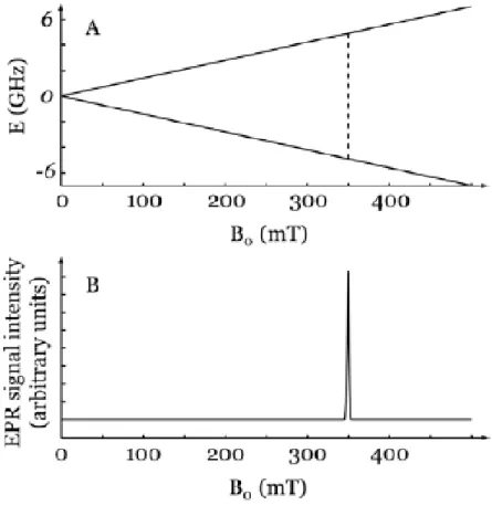
FIGURE 4.1-ILLUSTRATION OF THE ZEEMAN EFFECT IN ONE FREE ELECTRON... 26
FIGURE 4.2-A:REPRESENTATION OF THE INCREASE IN THE ENERGY LEVELS OF AN ELECTRON
SPIN AS THE MAGNETIC FIELD APPLIED INCREASES. B: SIMULATED EPR ABSORPTION
SPECTRUM FOR THE ALLOWED TRANSITION. ... 27
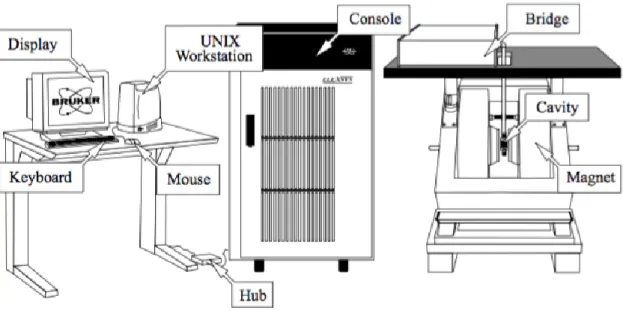
FIGURE 4.3-MAIN COMPONENTS OF AN EPRELEXSYS E500 SPECTROMETER. ... 28
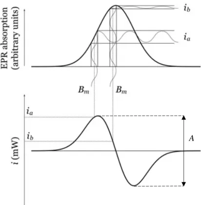
FIGURE 4.4 - ILLUSTRATION OF THE EPR ABSORPTION LINE AND THE RESPECTIVE FIRST
DERIVATIVE, AS THE EPR SIGNAL IS PRESENTED IN THE EPR SPECTROSCOPY TECHNIQUE.
... 30
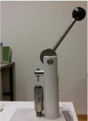
FIGURE 5.1-TABLE-TOP PELLET-PRESS USED FOR PRODUCING THE DOSIMETERS. ... 34
FIGURE 5.2–FIVE DOSIMETERS LABELLED ACCORDING TO THE SYSTEM CHOSEN. ... 35
FIGURE 5.3 - SCHEMATIC REPRESENTATION OF THE PMMA PHANTOM USED FOR IN
xii
FIGURE 5.4- SCHEMATIC REPRESENTATION OF THE PMMA PHANTOM USED FOR THE PROTON
BEAM IRRADIATIONS. ... 38
FIGURE 5.5 – SCHEMATIC REPRESENTATION OF THE EXPERIMENTS SET-UP FOR THE PROTON
BEAM IRRADIATIONS. ... 38
FIGURE 6.1 - HOMOGENEITY ANALYSIS OF THE BATCH OF DOSIMETERS USED IN THE
LINEARITY TEST.. ... 44
FIGURE 6.2 - RELATION BETWEEN THE ABSORBED DOSE FROM THE PROTON BEAM BY EACH
DOSIMETER AND THE RESPECTIVE EPR SIGNAL. ... 47
FIGURE 6.3 – HOMOGENEITY ANALYSIS OF THE BATCH OF DOSIMETERS PRODUCED FOR THE
BLIND TEST. ... 53
FIGURE 6.4 - RELATION BETWEEN THE ABSORBED DOSE BY GROUP 1 AND GROUP 3
DOSIMETERS IRRADIATED IN THE BLIND TEST AND THEIR RESPECTIVE EPR SIGNAL... 56
FIGURE 6.5 –HOMOGENEITY ANALYSIS OF THE BATCH OF DOSIMETERS USED IN THE FADING
TEST. ... 60
FIGURE 6.6 - RELATION BETWEEN THE RELATIVE EPR SIGNAL OF THE DOSIMETERS AND THE
xiii
LIST OF TABLES
TABLE 4.1-FIELDS OF RESONANCE FOR THE TYPICAL MICRO WAVE BANDS AVAILABLE IN EPR
SPECTROMETERS. ... 28
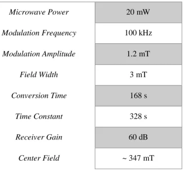
TABLE 5.1–SPECTROMETER SETTINGS FOR EPR MEASUREMENTS. ... 37
TABLE 5.2–ABSORBED DOSES BY EACH SUB-GROUP OF DOSIMETERS IN THE LINEARITY TEST.
... 39
TABLE 5.3–ABSORBED DOSES BY EACH SUB-GROUP OF DOSIMETERS IN THE BLIND TEST. . 40
TABLE 5.4-REGISTER OF THE ABSORBED DOSES BY EACH OF THE GROUPS IRRADIATED. .... 41
TABLE 6.1-OVERALL ANALYSIS OF THE BATCH TO BE USED IN THE LINEARITY TEST. ... 45
TABLE 6.2-SUMMARY OF THE EPR SIGNALS MEASURED TO EACH OF THE IRRADIATED GROUPS
IN THE LINEARITY TES T. ... 46
TABLE 6.3- SUMMARY OF THE RESULTS REGARDING THE ERROR OF EACH SIGNAL ESTIMATION
TO EACH INDIVIDUAL DOSIMETER O F THE BATCH. ... 48
TABLE 6.4 - ESTIMATION OF THE 95 % AND 99 % CONFIDENCE INTERVALS ASSOCIATED TO
THE EPR SIGNALS OBTAINED FOR EACH IRRAD IATED GROUP. ... 49
TABLE 6.5 – ESTIMATION OF THE ABSORBED DOSE WITH A 95 % AND 99 % CONFIDENCE FOR
FIVE GROUPS IRRADIATED WITH THE PROTON BEAM CONSIDERIN G ONLY THEIR AVERAGE
xiv
TABLE 6.6 - PERCENT ERRORS ASSOCIATED TO EVERY DOSIME TER IRRADIATED WITH THE
PROTON BEAM DOSE ESTIMATIONS. ... 51
TABLE 6.7-OVERALL ANALYSIS OF THE BATCH TO BE USED IN THE BLIND TEST.. ... 54
TABLE 6.8-SUMMARY OF THE EPR SIGNALS MEASURED TO THE IRRADIATED DOSIMETERS IN
THE BLIND TEST. ... 55
TABLE 6.9-ESTIMATION OF THE UNKNOWN ABSORBED DOSE BY GROUP B DOSIMETERS.. .. 57
TABLE 6.10 – DOSE ESTIMATIONS AND ASSOCIATE ERRORS OF THE FOUR DOSIMETERS OF
GROUP B. ... 58
TABLE 6.11–OVERALL ANALYSIS OF THE BATCH TO BE USED IN THE LINEARITY TEST. ... 61
TABLE 6.12 - SUMMARY OF THE MEASURED EPR SIGNALS TO EACH OF THE GROUPS
IRRADIATED OVER THE WEEKS IN THE FADING TEST... 61
TABLE 6.13 - SUMMARY OF THE RESULTS REGARDING THE ERROR OF THE RELATIVE SIGNALS
ESTIMATION FOR EVERY DOSIMETER... 64
TABLE 6.14 - FADING ESTIMATIONS, IN PERCENTAGE, ASSOCIATED TO THE D AYS STUDIED IN
THE FADING TEST WITH THE CALCULATION OF THE RESPECTIVE CONFIDENCE INTERVALS.
xv
A
CRO
NYMS
3 D - C R T Three-dimensional Conformal Radiotherapy
C T Computer Tomography
EPR Electron Paramagnetic Resonance
IA EA International Atomic Energy Agency
IC F Individual Calibration Factor
IC R U International Commission on Radiation Units and Measurements
IM R T Intensity Modulated Radiation Therapy
ISS Istituto Superiore de Sanità
LET Linear Energy Transfer
LIU Linköping University
OA R Organ at Risk
PM M A Polymethyl Methacrylate
SSD Source-Surface Distance
TLD Thermoluminescence Dosimeter
VM A T Volumetric Modulated Arc Therapy
WHO World Health Organization
1
Chapter 1
INTRODUCTION
Cancer is one of the leading causes of morbidity and mortality worldwide. This group of diseases is characterized by an uncontrolled growth and spread of cells, which can invade various parts of the body and other organs, and affects around 14 million people every year, being responsible for about 8 million deaths per year according to the World Cancer Report [1] from the World Health Organization (WHO).
Each of the over one hundred different types of cancer require unique diagnosis and specific treatments that must be planned by specialized oncologic teams. Radiation therapy, or radiotherapy, together with surgery and chemotherapy are the three main modalities used for cancer treatment.
Radiotherapy as the procedure of using ionising radiation for cancer treatment can be divided into two main groups: external beam radiotherapy and brachytherapy. In the first, the radiation source is at a certain distance from the patient and the target is irradiated with an external radiation beam while in brachytherapy the radiation sources are placed close or even inside the target volume [2].
The use of ionising radiation for medical purposes has been a topic of study and concern for many years now. The effectiveness of this treatment modality is related with the sensitivity that cells have to radiation and how this radiation can damage the genes, interrupting the cell cycle and, consequently, leading to the death of cells. The main problem is that, regardless of its efficacy in killing the cancerous cells, the ionising radiation also affects normal tissues and damage them, which originates the severe side effects associated to most radiotherapy treatments like sore skin, tiredness or hair loss, among many others. Consequently, all over the world, specialists and researchers work on the development of new methods and solutions to reach the best balance between destroying cancer cells and minimizing damages to the surrounding healthy tissues.
When not considering the influence of the type of cancer and its degree of development, the effectiveness of a treatment depends mainly on two factors: the accuracy of the treatment planning and the quality of the equipment and technologies of the radiation systems available.
Concerning the treatment planning process, the dose distributions of target volumes and critical organs are assessed for each patient and the optimal technique among the treatment modalities available is chosen by qualified professionals. The planning process starts with the obtainment of a 3D image of the patient, most commonly a Computed Tomography (CT), so that the most accurate and realistic data regarding the anatomy of the patient and the tumour characteristics (location and volume) are used. This data is then used as the input for the planning software that contours the different volumes encountered.
CH A PTE R 1 | IN T R O D U C TI O N
2 Accounting this, a specialized medical physicist identifies both the tumour volumes and the surrounding tissues and Organs at Risk (OAR) and defines the necessary dose distributions.
A team of physicists together with radiotherapy doctors work to set the final details of the plan and, in the end, if every step of the planning process is successfully and accurately done, the best and less harmful treatment for each individual situation is obtained.
Besides having a precise treatment plan with all the structures well-defined and bordered it is of the greatest importance that the dose delivery system is also working as expected so that the delivered dose matches the planned one.
Medical physicists working with radiotherapy have the responsibility to verify that the correct dose is being delivered in the right direction towards the patient by performing regular quality controls and, with this, make sure that the tinny margin that separates the treatment efficacy and the occurrence of severe side effects is not exceeded.
The ideal dose is defined as [3]:
“The ideal radiation with which to treat cancer is one that delivers a defined dose distribution within a target volume.”
All radiotherapy procedures as susceptible to errors and even the smallest errors during the treatment planning, the dose delivery process or in the dosimetry verifications may cause substantial negative consequences for the patient. Considering this, it has been argued that the accuracy of the dose delivery to the patient in a radiotherapy treatment should be better than 3-4 % [4], considering all the possible inaccuracies associated to radiotherapy beam characteristics, patient data, treatment planning dose calculations and absorbed dose determinations.
In order to accomplish the accuracy and precision needed, radiation dosimetry or, more specifically, medical radiation dosimetry, becomes an essential part of the treatment control.
Medical radiation dosimetry consists on the evaluation, through measurement or calculation, of the absorbed dose received by a patient that is exposed to ionising radiation as a consequence of therapeutic or diagnostic procedures [5]. Nowadays, there are several methods and equipment available for dose verification measurements and each has different characteristics so, according to the aim of the measurement, the most suitable and convenient method should be used.
Radiation detectors may be divided in two main groups according to how the signal read is displayed. Using a direct reading detector, such as ionization chambers or semiconducting diodes, the signal is displayed instantaneously whereas using passive detectors like thermoluminescent detectors (TLD) or Electron Paramagnetic Resonance (EPR) detectors the effects caused by ionizing radiation result in data that has to be processed to obtain the signal information regarding the radiation [6].
The first group of detectors are, therefore, preferred for clinical applications because they can show the result much fast way. In fact, historically, ion chambers have been the main choice for the measurement of the absorbed dose in beams of ionising radiation and most dosimetry protocols are based on the use of ionization chamber since it is one of the most accurate and well-known method for dose determinations [7]. Though, passive detectors are usually characterized by a lower dependence on the direction of the beam, do not require connecting cables – a factor that sometimes makes impossible the use of ionization chambers -, and are associated to very precise measurements so, in some circumstances, passive detectors can become the most suitable equipment to be used.
The present thesis is focused on the EPR technique. Its use in radiation therapy is a relatively new advance but EPR spectroscopy is already a well-established technique for other dosimetry applications. In fact, the EPR spectroscopy technique using alanine as the dosimeter material has been accepted and recommended by the International Atomic Energy Agency (IAEA) mostly for retrospective dosimetry [8] and, concerning radiotherapeutic purposes, for the measurement of doses of
CH A PTE R 1 | IN T R O D U C TI O N
3 radiation over the 6 Gy [9]. However, the low sensitivity of most EPR materials used so far made it very difficult to achieve the precision recommended by the International Commission on Radiation Units and Measurements (ICRU) at the dose levels administrated in radiotherapy.
The dose determination using the EPR technique is rather time consuming so it is not the most suitable technique to be used on the daily basis in clinics and, additionally, requires the purchase and use of an expensive high precision spectrometer. Nonetheless, this technique gains relevance due to the accuracy that might be reached if more sensitive dosimeter materials are used and because of the possibility of repeating the measurements several times without influencing the radiation signal, allowing the increase of the results certainty.
So, in applications that prioritize the achievement of a high precision and accuracy and do not require a time efficiency, EPR dosimetry with more suitable and sensitive dosimetry materials, become a very realistic and advantageous alternative to other dosimetry systems.
Research was made to find more appropriate materials for this purpose and one of the most promising materials is polycrystalline lithium formate monohydrate, commonly referred as lithium formate. The EPR dosimetry system with this material has already been tested for dosimetry purposes in some radiotherapy techniques like external beam therapy and brachytherapy and the conclusions were very pleasing.
Under high LET (Linear Energy Transfer) radiation, however, the behaviour of the system was still not studied and for the technique to gain status and consideration among the other dosimetry techniques used in radiotherapy, the full characterization of the lithium formate EPR dosimetry system under every type of radiation is needed. Particularly, due to the promising benefits of proton therapy upcoming its high precision dose delivery, the search for accurate and well-known systems for dose verifications is becoming more and more important.
1.1. A
IM
The present project is focused on the continuation of the characterization of the promising lithium formate EPR system for dose measurements under clinical proton beams.
The alternative of using lithium formate instead of alanine is thought to be considerably more beneficial for dose verifications because it is argued to have such a great accuracy that it can measure absorbed doses down to 0.2 Gy, accordingly to previous research. In this way, when low uncertainty dosimetry methods are needed and the high precision is preferred over the time efficiency, the EPR dosimetry method may be considered as the most suitable one in comparison with ionization chamber dosimetry or thermoluminescence dosimetry, for example.
The lithium formate EPR system has been developed, tested and optimised for photon beams and was considered a good candidate for dosimetry audits in both external beam therapy and brachytherapy. However, the different characteristics of photon beam therapy and charged particle therapy result in different radiation-matter interactions and in different dose distribution over the target. Consequently, the behaviour of a dosimetry system is expected to have some differences.
Considering these differences, the greater need to have accurate dosimetry systems for dose verifications in proton beams and the promising results obtained for photon beam dose verifications, the lithium formate EPR dosimetry must also be fully characterized for proton beam irradiations. This is where the present project is inserted.
CH A PTE R 1 | IN T R O D U C TI O N
4 A lithium formate EPR dosimetry system was developed and tested for proton beams with the aim of being characterized regarding the linearity of its response to different doses and also study the stability of the signal read on the dosimeters over a period of one month.
All the process of the dosimeters production, calibrations, EPR signal readouts and respective analysis were performed in Linköping University (LiU). The proton irradiations needed for the study of the lithium formate dosimeters response were done in the new Swedish proton therapy facility in Uppsala, Skandionkliniken.
1.2. O
UTLINE
Chapter 2 provides a literature review over the EPR technique and the present state of the use of this
dosimetry system for radiation therapy dosimetry controls.
Chapter 3 describes the types of radiation used for radiotherapy purposes: photon therapy used in the
conventional radiotherapy and particle therapy where the proton therapy is inserted. The main interactions of the ionising particles with matter are clarified and the main characteristics of the proton radiation therapy are enhanced.
Chapter 4 clarifies the main concepts of radiation dosimetry. The most important quantities in
dosimetry used for the quantification of radiation are explained as well as the most important terms used in Radiation Protection and in Medical Dosimetry. Also, a section regarding radiation dosimeters contains the description and principal characteristics of the most commonly used systems with special focus on the Electron Paramagnetic Resonance technique.
Chapter 5 presents the materials and methodology used in each experiment. This section includes the
description of the manufacture of the lithium formate dosimeters, the homogeneity test and standards needed for a batch to be considered homogeneous, the EPR readout procedure and the methodology followed for the Linearity Test, Blind Test and Fading Test.
Chapter 6 presents the results obtained in each experiment. The analysis is firstly focused on the
acceptance of the batch as homogenous or non-homogeneous and, secondly, focused on the results of the main tests where statistical analysis is also presented. Finally, the obtained results are discussed and compared to previous research made regarding the EPR dosimetry and the use of lithium formate dosimeters.
Chapter 7 gives general conclusions regarding all the tests made and the lithium formate dosimetry
5
Chapter 2
LITERATURE REVIEW
Since the early 20th century, radiation therapy has been used for the treatment of cancer.
However, medical dosimetry is a relatively new field and, particularly over the past 40 years, the concept as changed a lot and, nowadays, is seen as an essential part of quality assurance in medicine all around the world [10].
The medical physics main goal to increase the accuracy and precision of the dose delivery justifies a current research in several fields related to medical dosimetry and, for this reason, a big focus is on the passive dosimeters since most of them are characterized by long term stability, wide dosimetric range, small dimensions and the lack of cables or power supplies [11].
To understand the EPR technique principles is important to go back to the 1920s, where scientists began to apply quantum mechanics principles to describe atoms and molecules. From the Stern-Gerlach experiments was possible to conclude that an electron magnetic moment in an atom can only take discrete orientations under the influence of a magnetic field and that atoms can absorb energy and orientate themselves according to the frequency of the radiation they are exposed to. As a result of this and many other quantum physics discoveries, the first observation of an EPR peak was registered in 1945 by Zavoisky when he detected a radiofrequency absorption line from a CuCl22H2O sample.
In 1962, Bradshaw [12] demonstrated that EPR was a satisfactory technique for the measurement of radiation doses since the amplitude of the obtained absorption line was directly related with the dose itself. He suggested the use of a polycrystalline sample of the amino acid α-alanine as detector material (dosimeter) due several of this material features like:
- high sensitivity to radiation damage: large amounts of free radicals are created in the alanine structure after irradiation
- stability: the radical population formed is stable in time, making the dosimeter signal virtually independent of the time of irradiation
- similarity to biological systems in absorbing radiation: the atomic composition of alanine is tissue or water equivalent to that of human tissue
In the consequent years, EPR dosimetry has developed in a way that allowed it to be accepted as a competitive method at higher dose ranges (10 to 105 Gy) to other dosimetry methods [13].
CH A PTE R 2 | LI TE R A T U R E RE V I E W
6 Moreover, in the beginning of the 1980s, IAEA chose alanine as the standard dosimetric material for high dose measurements due to its particular and well-known characteristics [14].
At the Istituto Superiore de Sanità (ISS) in Italy, the method of manufacturing solid state alanine dosimeters was set up in 1983 and the same method was patented in 1985. There, the physicists made a big analysis regarding the response of the dosimeters to gamma rays and the main results were coherent with previous experiments: dosimeters with very precise and with reproducible results, no substantial energy or dose rate dependence neither influence of the angle in which the dosimeters detect the radiation beam [15].
After this, alanine EPR was established as a precise and accurate dosimetric method likely to be applied not only in industrial radiation processing but in radiation therapy as well.
Despite the advantages associated to alanine dosimeters like their tissue-equivalence and requiring no energy correction within the range of typical therapeutic beams, their sensitivity was not good enough for them to be used in clinical applications that require determinations of lower doses, ranging from 0.2 to 5 Gy [16]. So, aiming to make EPR a competitive dosimetry method for medical applications, research was made to find more sensitive materials for EPR dosimeters.
In 2002, polycrystalline lithium formate monohydrate, commonly known as lithium formate, was confirmed to have a sensitivity up to seven times higher than alanine [17] and so, dosimeters made from this material started being analysed with the thought that they could have some clear and practical advantages over alanine dosimeters. The advantages of using lithium formate as dosimeter materials were enhanced in a later study [18] where was shown that these dosimeters allow the measurement of absorbed doses down to 0.2 Gy. Consequently, they were considerable suitable to measure the lower dose ranges that could not be measured by the alanine dosimeters and, additionally, their linear response to all ranges indicated promising results for the measurements of doses both in the target of radiotherapy procedures as in the surrounding areas.
For intensity-modulated radiation therapy (IMRT), three-dimensional conformal radiotherapy (3D-CRT) and volumetric modulated arc therapy (VMAT), the lithium formate EPR dosimetry was tested and compared with the ionization chamber [19]. The determined doses agreed very well with planned doses and with the doses measured by ionization chambers confirming the potential of this dosimetry technique in terms of accuracy and the reproducibility of the measurements in external beam therapy with photons. Concerning dose measurements around brachytherapy sources with photon energies relevant to this technique, the referred EPR system also showed very accurate results [20, 21]. For irradiations with proton beams, the radical formation in lithium formate dosimeters was studied and reported [22]. In this research, the doses given were between 5 and 20 Gy and conclusions showed that the EPR reading signal increased linearly with the dose given and the precision of the reading was high and comparable with the precision obtained in previous studies of lithium formate using photon and electron beams.
At the same time, as the LET increased the relative effectiveness of the irradiation - defined as the dosimeter signal per absorbed dose in the detector - decreased and this decrease was interpreted as the result of several interactions and recombination within the tracks of the high LET particles. With this study the indication that the radical population trapped in the lithium formate structure after irradiation with densely ionising radiation is less stable than irradiation after low LET beams was clear and the importance of a study regarding the whole lithium formate dosimeter characterization especially with high LET particles was more enhanced. The lower stability of radicals after high LET radiation exposures had been previously referred in a publication from 1989 [23] where the signal from alanine radicals faded over the time.
Though the research regarding lithium formate EPR is quite promising, many studies are still focusing in alanine EPR technique since it is the standard dosimetry method. In 2015, research showed
CH A PTE R 2 | LI TE R A T U R E RE V I E W
7 the suitability to use alanine EPR technique for dosimetry characterization of proton beams with range between 10 Gy and 70 Gy [24]. Yet, not much more has been done regarding the characterization of the system, particularly to lower radiation doses justifying the need and adequacy of the present project. The viability of any dosimetry system using for dose verifications in therapy is primarily verified with dosimetry audits where the same technique is tested in several clinics [25]. The great performance of an audit for clinical trials is what assures the dosimetry accuracy among the different participating clinics and is what warrants reproducible treatment protocols.
In the present days, EPR dosimetry using alanine has already been used for audits in the United Kingdom, Italy and Belgium. These audits enhanced the possibility to use alanine detectors to verify the dosimetry accuracy of conventional photon and electron beams [26]. Additionally, an end-to-end test using alanine dosimetry was designed and tested showing that alanine dosimeters are also suitable detectors for audits in heavy particle therapies. However, in this same work some of the weaknesses of the alanine dosimeters were exposed, in particular the lower sensitivity to lower doses.
Considering lithium EPR dosimetry, in Linköping University (Sweden), an audit system for high energy external beam therapy using lithium formate dosimeters was also developed [27] with the aim of evaluating the behaviour of these dosimeters during the whole treatment chain (end-to-end test) including CT imaging, planning and dose delivery. To get more realistic results, a head to neck phantom made of Polymethyl Methacrylate (PMMA) with heterogeneities was designed and built and the system was tested in four clinics in Sweden. Results showed that the developed audit system was suitable for end-to-end audits since all the measured dose values agreed quite well with the calculated doses enhancing, once again, the great characteristics of the used dosimetry system.
9
Chapter 3
RADIATION IN RADIOTHERAPY
Radiation therapy consists on the use of ionizing radiation to treat a disease, most likely cancer. The concept of ionizing radiation includes any electromagnetic or particle radiation with enough energy to ionize common molecules and create electrically charged particles – ions – with the liberation of one or several electrons [28].
The ionizations can occur directly once the particle radiation interacts with the matter or the interaction might initiate nuclear or elementary-particle transformations that, by their turn, result in ionization. So, ionizing radiation can be divided in directly and indirectly ionizing radiation.
Directly ionizing radiation includes several types of charged particles such as electrons, protons, alpha particles or heavy ions and means that these particles transfer most of their kinetic energy directly to the medium resulting in absorbed dose by the medium. Indirectly ionizing radiation, on the other hand, means that the energy is imparted to matter in two steps: firstly, the indirectly ionizing radiation transfers energy as kinetic energy to secondary particles and only then these charged particles transfer most of their kinetic energy to the medium, resulting in the absorbed dose. In this last type of radiation are included neutral particles like photons and neutrons.
For medical purposes is also possible to distinguish two types of radiation used in radiotherapy treatments: photon radiation and particle radiation, which will be described next. The basis of radiotherapy, whatever type is considered, is the way the ionizing radiation and matter interact with each other and the expected effects of radiation in tumorous cells are always a consequence of the transfer of energy of the charged particles moving through a medium to the atoms of the body cells.
3.1.
P
HOTON
R
ADIATION
Under the modern theory of light, presented by Albert Einstein in the beginning of the 20th
century, a photon is a quantum of electromagnetic radiation. In radiotherapy, the most common beams used are high energy x-rays and gamma rays, generically named as photon beams. However, the electromagnetic spectrum also includes radio waves, infrared rays, visible light and ultraviolet rays and
CH A PTE R 3 | RA D I A TI O N I N RA D I O TH E R A PY
10 all these consist of photons so when referring to photon beams is important to specify what type of radiation is being used.
What differs among the several radiation types is the photons energy (E) that is known to be proportional to the photons frequency 𝑓.
𝐸 = ℎ𝑓 (Equation 3.1)
The relation between the variables is given by Equation 3.1 where ℎ is the Planck’s constant1.
𝑓 =𝑐
𝜆 (Equation 3.2)
Since the frequency of the electromagnetic radiation and its wavelength 𝜆 can be related by Equation 3.2, where 𝑐 represents the speed of electromagnetic-field propagation, Equation 3.1 can be rewritten so that the photon energy can be calculated only considering the wavelength 𝜆 of the radiation.
𝐸 =ℎ𝑐
𝜆 (Equation 3.3)
Therefore, the energy of each photon, given by Equation 3.3, does not depend on the intensity of the radiation since they all contain the exact same amount of energy. The intensity of a beam is a function of the number of photons striking a given surface area per unit time.
Most external radiation therapy is delivered through linear accelerators – linac - where electrons are accelerated to very high velocities and led to strike a heavy metal target to create a beam of photons. When moving through materials they are likely to interact with the matter in different ways, producing different ionizing photon radiations.
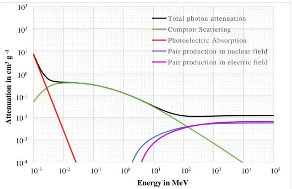
The most important interactions [6] to consider in radiotherapy include: - Photoelectric effect
This effect is the consequence of the interaction between the electromagnetic radiation and the atoms from an attenuator, usually a metal. When the energy of the photons is higher than the electrons’ binding energy, all their energy is transferred to the orbital electron that is ejected from the attenuator with a kinetic energy equivalent to the photon energy minus the binding energy. The empty spot left by the ejected electron is then filled by an electron from an upper orbital, resulting in the emission of characteristic X-rays or in the ejection of Auger electrons.
The probability of this effect depends on several factors and among those factors are the photons energy and the atomic number of the material that absorbs the radiation. For example, for materials with high atomic number, the photoelectric is considerable for higher energies but for materials or compounds with low atomic number (like water) the photoelectric effect is not likely to occur at energies over 200 keV.
- Compton scattering
Represents the interaction of an energetic photon with a quasi-free orbital electron. The incident photon energy has to be considerably higher than the binding energy of the orbital electron and, with the interaction, the photon transfers part of its energy to the electron and is scattered in a different direction with a higher wavelength.
1 Constant that relates de energy in one quantum (photon) of electromagnetic radiation to the frequency of that
CH A PTE R 3 | RA D I A TI O N I N RA D I O TH E R A PY
11 The energy given to the electron is, of course, dependant on the initial photon energy and the angle of scattering: a scattering angle closer to 180 results in a larger amount of energy to the electron which is scattered approximately in the photons original path. On the other hand, a small scattering angle corresponds to a smaller amount of energy given to the electron. After the photon-electron interaction, the photon with a lower energy may continue undergoing additional interactions.
The angular distribution of the photons scattered is given by the Klein-Nishina cross section that is independent of the atomic number of the material and that is why it is quite difficulty to distinguish tissues in medical images when these images are obtained with energies where the Compton scattering dominates: between 25 keV and 25 MeV. In this range are included the energy levels used in most radiation treatments and so, the Compton scattering is one of the most important interactions in radiotherapy with photon radiation.
- Pair production
Originated by the interaction of a photon with the nucleus of an atom and not their orbital electrons, the pair production refers to the creation of an electron-positron pair. In this context, the photon disappears and an electron-positron pair is produced in the nuclear Coulomb field.
The new particles move away with the remaining energy of the photon converted to kinetic energy. Since mass is produced out of the photon energy, the pair production effect has an energy threshold of 2𝑚𝑒𝑐2= 1.02 𝑀𝑒𝑉. The two will typically be annihilated once they lose their kinetic
energy, originating annihilation radiation (characteristically photons with 0.511 keV).
The probability for it to happen is proportional to the logarithm of the incoming photon energy and is dependent on the atomic number of the material it goes through. The energy in which pair production dominates is above the 25 MeV in water.
All the above interactions influence the total attenuation of the photon beam in a medium and have to be considered to obtain a more exact quantification of the photon absorption and scattering. The contribution of each interaction to the beam attenuation in water is represented in Figure 3.1.
A tte n u ati o n i n c m 3g -1 Energy in MeV
Total photon attenuation Compton Scattering Photoelectric Absorption Pair production in nuclear field Pair production in electric field
103 102 101 100 10-1 10-2 10-3 10-4 10-3 10-2 10-1 100 101 102 103 104 105 Figure 3.1 - Contribution of each type of interaction to the total photon beam attenuation in water. All interactions must be considered in order to calculate accurately, in the treatment planning, the radiation dose that the body will receive and therefore the dose that will be absorbed by the different body tissues.
CH A PTE R 3 | RA D I A TI O N I N RA D I O TH E R A PY
12 In a radiotherapy treatment, the photons used produce a cascade of interactions before the energy is absorbed as dose, both in the treatment machine and in the patient body. These interactions, that might be either one of the above mentioned, where the energy is transferred from the photons to (secondary) electrons and positrons occur as soon as the photons interact with the material atoms. So, characteristically, the peak dose of photon beams is reached within a few millimetres from the entrance point – “build-up effect” -, at a depth correspondent to the range of the first secondary electrons produced. The new energetic particles will then ionize and excite other atoms until the initial energy transferred to them is lost or the particles exit the body. The beam energy is controlled so that the peak dose occurs after a few millimetres from the entrance point and the skin is saved from the radiation.
Another important characteristic is the low-LET that X-rays and Gamma rays are characterized for. LET, the acronym for Linear Energy Transfer, is a term applied to quantify energy transferred per unit length of the track. So, being classified as low-LET means that the radiation penetrates tissues very easily but deposits its energy very infrequently.
Each accelerated photon causes only one ionization that releases one electron but the secondary electrons produced cause many more ionisations and produce other energetic electrons along their track. The LET from gamma radiation depends on the energy of those secondary electrons.
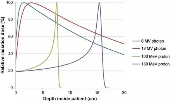
As less energy is transferred from the photon to the target and more is conserved in the photons, they can travel further and the probability of interactions with other particles from the medium (including healthy surrounding tissues) increases. The typical dose deposition from two different photon beams is represented in Figure 3.2 by the blue and red lines.
To control the dose that is deposited in each tissue, the solution passes by the use of several radiation beams delivered from different angles. Nowadays a commonly used technique for this is intensity modulated radiotherapy (IMRT) because it also allows a better dose conformation. This high precision 3D technique uses a computer-driven machine that moves around the patient and, in each of the pre-defined angles, delivers precise radiation doses, that can vary from angle to angle. With
Figure 3.2 - Dose deposition as a function of depth within the body: comparison between photon beams and a proton beams. Photons deposit radiation dose as they penetrate through the body what results in photon beams irradiating healthy tissues in the whole path. On the other hand, with a proton beam, the largest amount of energy is deposited at a defined depth called Bragg Peak and in the rest of the path tissues only absorb relatively small doses of radiation. [52]
CH A PTE R 3 | RA D I A TI O N I N RA D I O TH E R A PY
13 knowledge of the tumour shape and its characteristics and after the medical team identifies the area to treat and the maximum dose that can be absorbed by the organs at risk, the best dose intensity pattern that best conforms to the tumour is determined by a medical physicist.
The combinations of various intensity-modulated fields from different directions allows the delivery of a higher dose to the tumour and, at the same time, limits the amount of radiation that is deposited in the healthy tissues that are on the beam path.
Despite all the improvements in treatment technologies such as multi-leaf collimators, IMRT or image-guided radiotherapy (IGRT), photon beam therapy is always be responsible for healthy tissues to receive considerable radiation doses resulting in harmful side effects for the patient undergoing the treatment. Like so, alternative treatments in which these effects are smaller must be considered.
3.2.
P
ARTICLE
R
ADIATION
This group includes not only hadrons as protons and heavy ions but also neutrons and electrons, all considered ionizing particles for radiotherapy purposes.
The interest in using charged hadrons is related with the existence of the denominated Bragg peak which increases the ability to optimise the spatial distribution of the absorbed dose within a tissue and makes particle radiation the best and more advantageous choice for many cancer treatments.
The Bragg peak indicates the point of greatest penetration in the tissue and it occurs because the stopping power of the particles increases right before they come to rest. It represents the maximum depth the particles reach and this depth is determined by the amount of energy that is given initially to the particles by the accelerator. So, the Bragg peak depth can be controlled by the physicist aiming to a very precise targeting of the tumour tissue. The particles enter the new medium with a very high energy and begin to deposit very small radiation doses that increase gradually with the depth increase and the particle energy reduction. When the Bragg peak depth is reached, the particles deposit their remaining energy and stop irradiating immediately after.
The Bragg curve is a graph of the energy loss rate of ionizing radiation as a function of the distance through a stopping medium. This curve is also represented in Figure 3.2 where the dose deposition of protons is compared with the dose deposition of photon radiation and the differences between both techniques are enhanced.
Contrarily to what happens with photon radiation, where the dose deposition peak is reached within a few millimetres from the entrance point of the photon beam, in the particle radiation case, most energy is deposited at the depth of the Bragg peak, after which the dose rapidly decreases to zero and all particles come to rest if no nuclear reactions occur and secondary neutral particles are created.
This property reduces significantly the exposition of healthy surrounding tissues to radiation so, in theory, this reduction of toxicity will also reduce the probability of developing secondary malignant tumours induced by the radiation that appear some years after the radiotherapy treatments, a risk that appears to be higher for survivors of childhood cancers [29]. In this way, the tolerance of the patients to the treatment is higher as well as their life quality during and after the treatment.
Besides the advantages related to the Bragg peak that optimise the absorbed dose delivered to the target, there are other reasons behind the great interest in the use of particle radiation in radiotherapy. These reasons are based both on the physics of the dose transport and deposition and on the radiobiological effects they have.
The large mass of charged hadrons results a minimal degree of multiple scatter allowing a well-defined lateral definition of the beam.
CH A PTE R 3 | RA D I A TI O N I N RA D I O TH E R A PY
14 Another advantage of hadrons is their high LET when the interaction with the tissues occur. By the definition of LET, high LET radiation causes more molecular damages per unit of exposure because large amounts of energy are deposited in small distances. So, the higher localised deposition of energy results in a greater relative biological effectiveness (RBE) associated to the use of hadrons in comparison with the photon radiation whose secondary electrons are characterised by a low LET. Also, since tumours are characterised for being hypoxic2 which makes them more resistant to radiation, the
oxygen enhancement ratio (OER) of hadrons becomes a considerably important attribute of hadrons. Due to all the potential advantages of particle radiation in radiotherapy that is associated with a more effective treatment and less harmful consequences for the patient, a higher attention is being given to the use of proton and other charged particles like helium or carbon ions.
In the present context, a special focus is given to proton therapy [5].
3.2.1.
P
ROTONT
HERAPYProton therapy is an emerging treatment modality to treat cancer and its concept arises from 1946 [30] after Robert Wilson described some physical properties of protons and hypothesised that high energetic proton beams could be used to increase the radiation doses to tumours and, at the same time, minimize radiation to nearby healthy tissues.
The first treatments using heavy ion beams with protons were performed in 1954 [31] at the Harvard Cyclotron Laboratory and due to the lack of many clinical conditions (treatment planning, beam shaping technology,) were only suited for radiosurgery like pituitary ablation.
In Europe, the first synchrocyclotron was developed in Uppsala University (Sweden) around 1950 by Borje Larsson and there, in 1957, it started to be used for cranial radiosurgery and fractionated radiotherapy [32].
Regardless of the excellent records about the treatment safety and efficacy, the widespread and adoption of this technique is being quite slow mostly because of its technical difficulty, much higher costs of implementation and the lack of evidence of cost-competitiveness. Yet, this technique has grown quite a lot and in 2015 there were 51 active proton therapy centres all over the world, 16 of those in Europe [33]. So, in the future is expected a big spread of proton therapy facilities which will make this technique a more approachable alternative to conventional radiotherapy.
The technique, as the name indicates is based on the use of protons that are accelerated to really high velocities and can be used for therapeutic purposes. Protons are positively charged and heavy particles (in comparison with electrons) and their biggest advantage is the way the destructive power is delivered to the tissues associated due to the characteristically high LET. Also, protons travel fast through tissues with few interactions and without changing much the original trajectory until they reach a defined depth where the Bragg peak occurs and the dose is deposited.
The high dose distribution given by the proton beam to the tumour, with minimal or no exit dose, allows the tumour to be treated with unmatched accuracy, security and efficiency. By sparing healthy surrounding tissues, the characteristic side effects of radiotherapy are significantly reduced and the life quality of the patient during and after the treatment is much better.
For a realistic treatment plan and, thus, a safe treatment is important to be aware of the interactions that protons may have with the matter since these interactions is what determine how the dose will be distributed, how much will be absorbed by tissues and allow to predict how much energy losses during the beam path.
CH A PTE R 3 | RA D I A TI O N I N RA D I O TH E R A PY
15 Protons interact with matter and deposit their energy in mostly in three different ways: energy loss by ionization, multiple Coulomb scattering and nuclear interactions. The first two interactions concern the electromagnetic interaction between the charge of a proton and the charge of atomic electrons - or the Coulomb field of the nucleus - whereas the third regards the interaction with the material nucleus.
- Energy Loss by Ionization
At the energy levels used clinically, the collisions of protons with atomic electrons, named inelastic Coulomb interactions [34], have a considerable importance.
As the protons mass is about 1800 times greater than the mass of electrons [35], each interaction can only reduce the proton energy a little but since the number of interactions is high, these interactions are the main reason for the slowdown of protons in matter and eventually their stopping after the proton range is reached.
The quantum theory of slowing down and stopping process was developed by Bethe and Bloch in the 1930s and predicts the rate of energy loss as a function of the type and energy of the incident particle and of the composition of the stopping material [36].
The energy loss per unit distance is called stopping power (S) [37] and is defined in Equation 3.4, where 𝐸 is the energy and 𝑥 refers to the distance protons go through. The unit for S is 𝑀𝑒𝑉/𝑐𝑚.
𝑆 = −𝑑𝐸𝑑𝑥 (Equation 3.4) Since the stopping power is proportional to the density of the absorbing material, this rate is usually expressed as a quantity independent of its mass density 𝜌 (𝑔 𝑐𝑚⁄ 3), being called mass stopping power. This quantity is expressed in 𝑀𝑒𝑉. 𝑐𝑚2/𝑔 and given by Equation 3.5.
𝑆 𝜌= − 1 𝜌 𝑑𝐸 𝑑𝑥 (Equation 3.5)
Apart from the interaction of charged particles with atoms and molecules being a statistical process, the energy loss occurs as a finite number of individual interactions so it has a statistical error associated. This means that protons with the same initial energy will probably not stop at the exact same depth due to the accumulation of several small variations during the energy loss - range straggling [35]. Though this process is only responsible for a fluctuation of about 1 % in the protons range [36], it can have quite an influence in the Bragg curve shape so it has to be considered if a precise clinical calculation is needed. This precision is of vital interest in radiotherapy because the main advantage of using charged particles is their precise dose deposition in a specific point.
- Multiple Coulomb Scattering
In addition to the possible interactions with atomic electrons that result in some energy loss, when passing near an atomic nucleus protons experience an elastic Coulomb scattering caused by the charge of the nucleus and are deviated through small angles from their original trajectory. Elastic and inelastic Coulomb interactions are designated as Multiple Coulomb Scattering [34].
The energy loss can be neglected in this process but the changes in the trajectory of protons, even the smaller ones, may be of the utmost importance and must be considered in the optimization of dose delivery.