UNIVERSIDADE FEDERAL DO CEARÁ
FACULDADE DE FARMÁCIA, ODONTOLOGIA E ENFERMAGEM
PROGRAMA DE PÓS-GRADUAÇÃO EM ODONTOLOGIA
JORGIANA SILVA DE ASSIS
ANÁLISE ANTIMICROBIANA DO FLAVONÓIDE EPIGALOCATEQUINA-3-GALATO COMO AGENTE DE LIMPEZA CAVITÁRIA EM DENTINA
ARTIFICIALMENTE CARIADA.
FORTALEZA
Dados Internacionais de Catalogação na Publicação Universidade Federal do Ceará
Biblioteca de Ciências da Saúde A865a Assis, Jorgiana Silva de.
Análise antimicrobiana do flavonoide epigalocatequina-3-galato como agente de limpeza cavitária em dentina artificialmente cariada / Jorgiana Silva de Assis. – 2013.
43f. : il. color., enc. ; 30 cm.
Dissertação (Mestrado) – Universidade Federal do Ceará, Programa de Pós-Graduação em Odontologia, Fortaleza, 2013.
Orientação: Prof. Dr. Sérgio Lima Santiago.
1. Catequina. 2. Dentina. 3. Streptococcus Mutans. I. Título.
JORGIANA SILVA DE ASSIS
ANÁLISE ANTIMICROBIANA DO FLAVONÓIDE EPIGALOCATEQUINA-3-GALATO COMO AGENTE DE LIMPEZA CAVITÁRIA EM DENTINA
ARTIFICIALMENTE CARIADA.
Dissertação submetida ao Programa de Pós-graduação em Odontologia da Faculdade de Farmácia, Odontologia e Enfermagem da Universidade Federal do Ceará, como um dos requisitos para obtenção do título de mestre em Odontologia. Área de concentração: Clínica Odontológica
Orientador: Prof. Dr. Sérgio Lima Santiago
FORTALEZA
JORGIANA SILVA DE ASSIS
ANÁLISE ANTIMICROBIANA DO FLAVONÓIDE EPIGALOCATEQUINA-3-GALATO COMO AGENTE DE LIMPEZA CAVITÁRIA EM DENTINA
ARTIFICIALMENTE CARIADA.
Dissertação submetida à Coordenação do
Programa de Pós-graduação em
Odontologia da Universidade Federal do Ceará como requisito parcial para a obtenção do Título de Mestre em Odontologia. Área de concentração: Clínica Odontológica.
Aprovada em: __/__/__
BANCA EXAMINADORA
________________________________________________
Prof. Dr. Sérgio Lima Santiago (orientador)
Universidade Federal do Ceará - UFC
_________________________________________________
Profa. Dra. Cristiane Sá Roriz Fonteles
Universidade Federal do Ceará - UFC
_________________________________________________
Prof. Dr. Cláudio Maniglia Ferreira
À minha mãe ( in memoriam ), que me ensinou a graça que é viver e que, de
onde estiver, certamente está
comemorando mais esta vitória.
“...mas você partiu sem mim. Eu sei que
AGRADECIMENTOS ESPECIAIS
A Deus, pelo dom da vida! Por colocar as pessoas certas na hora certa. Por ter me mantido paciente e perseverante nos momentos de indefinição; por ter me feito humilde para aceitar críticas e saber empregá-las a meu favor.
À minha família, Dita, Orlando Júnior, Maloca, Tio Augustinho, Priscila; verdadeiro alicerce nas horas difíceis. Inspiram-me a alçar voos maiores e me dão a oportunidade diária de evoluir como ser humano.
Aos meus sogros, Rosemary e Marques, pelo amor incondicional e gratuito. Essa vitória é também de vocês!
Ao meu marido Marquinhos, eterno melhor amigo, “aquele que está ao meu
lado em qualquer caminhada”. Obrigada por me acompanhar nas noites mal
dormidas e nos momentos de insanidade.
Ao amigo-irmão Renato Lira, com quem partilhei os melhores e os piores momentos desta jornada e a quem admiro pela integridade de caráter e lealdade.
Aos amigos da turma de mestrado (2011.1), em especial Lidiane Costa, Camila Ferraz, Adriana Kelly, Phelipe Maia, pelos conhecimentos compartilhados e pelo crescimento conjunto.
À equipe de amigos do laboratório, em especial aos alunos de graduação Jamila Ricarte, Marcelo Sidou, Melca Peixoto e Edson Cetira; às mestrandas Cecília Atem, Sarah Guedes, Jacqueline Nojosa; às doutorandas Juliana Paiva, Mary Anne Sampaio, Ramile Lima. Agradeço pela disponibilidade e pelo compromisso de cada um de vocês!
Ao amigo David Queiroz, técnico do laboratório-PPGO, que gentilmente me acompanhou por algumas noites no laboratório e se mostrou solícito em todos os momentos que dele necessitei.
À Profa. Dra. Lidiany Karla de Azevedo, por quem tenho admiração imensurável, e a quem agradeço por se fazer presente nos momentos críticos deste trabalho.
À Profa. Dra. Mônica Yamauti, que procura sempre disponibilizar as melhores condições possíveis no laboratório de pesquisa, para que os trabalhos sejam conduzidos adequadamente. Considero-a um referencial, e me sinto honrada por ter aprendido um pouco da sua experiência de vida.
“Eu odiava cada minuto dos treinos, mas
dizia para mim mesmo: não desista! Sofra agora e viva o resto de sua vida como um
campeão.”
RESUMO
O objetivo do presente estudo foi avaliar a eficácia do flavonóide epigalocatequina-3-galato (EGCG) nas concentrações de 0,5%, 1% e 2% como solução antimicrobiana em dentina artificialmente cariada, tendo a clorexidina 2% e a solução salina 0,9% como controles. Vinte e cinco blocos de dentina humana (4 mm x 4 mm) foram imersos por cinco dias em BHI-caldo inoculado com Streptococcus mutans UA159 no primeiro dia do experimento. No quinto dia do experimento, os blocos foram aleatoriamente distribuídos em cinco grupos: grupo I –
controle negativo – solução salina 0,9%; grupo II – controle positivo – clorexidina 2%; grupo III – EGCG 0,5%; grupo IV – EGCG 1%; grupo V - EGCG 2%. Cada bloco recebeu tratamento de 15 µl da solução testada, que permaneceu em contato com o bloco por 60 segundos. Após os tratamentos, amostras dentinárias foram removidas com emprego de lâmina de bisturi e foram analisadas a partir da contagem de UFCs (unidades formadoras de colônias). Os experimentos foram realizados em triplicata e os dados, obtidos em UFCs foram convertidos em log base-10. Os testes estatísticos empregados foram análise de variância (ANOVA), seguida de Teste de Tukey. Não houve diferença estatística entre as concentrações de EGCG empregadas e a solução salina (p > 0,05). Além disso, não houve diferença estatística entre as concentrações de EGCG (p > 0,05). No entanto, houve diferença estatisticamente significativa entre a clorexidina e os demais grupos (p < 0,05). Conclui-se, a partir dos dados encontrados, que a substância investigada não deve ser empregada em preparos cavitários com a finalidade de eliminação de patógenos, visto que não se mostrou eficaz como antimicrobiano nas concentrações testadas.
ABSTRACT
The aim of this study was to evaluate the efficacy of the flavonoid epigallocatechin-3-gallate (EGCG) in concentrations of 0.5%, 1% and 2% as an antimicrobial solution in artificially carious dentin, with 2% chlorhexidine and 0.9% saline solution as controls. Twenty-five slabs of human dentin (4 mm x 4 mm) were immersed for five days in Brain Heart Infusion broth (BHI-broth) inoculated at first day with Streptococcus mutans UA159 (Batch Culture Model). On the fifth day of the
experiment, the blocks were randomly divided into five groups: group I - negative control - 0.9% saline solution, group II - positive control - 2% chlorhexidine, group III - 0.5% EGCG, group IV - 1% EGCG, group V - 2% EGCG. Each slab was subjected to 15 µl of the tested solution for 60 seconds. After treatments, artificially carious dentin was removed from the dentin slabs and analyzed by counting colony forming units (CFUs). All experiments were performed in triplicate and the data obtained in CFUs mean values converted to log base-10. The statistical tests used were analysis of variance (ANOVA) followed by Tukey test. There was no statistical difference between EGCG concentrations and saline (p > 0.05). Furthermore, there is no statistical difference between EGCG concentrations (p > 0.05). However, there was statistically significant difference between chlorhexidine and the other groups (p < 0.05). From the data obtained in this study, is possible to conclude that the substance tested is not effective on elimination of pathogen S. mutans when used in a concentration of 0.5%, 1% and 2% in artificially carious dentin.
SUMÁRIO
1 INTRODUÇÃO GERAL ... 11
2 PROPOSIÇÃO ... 13
3 CAPÍTULOS ... 14
4 CONCLUSÃO GERAL ... 30
REFERÊNCIAS ... 31
11
1 INTRODUÇÃO GERAL
O conceito de mínima intervenção ou odontologia minimamente invasiva (TYAS et al., 2000), amplamente aceito no mundo, preconiza que as zonas dentinárias mais profundas afetadas por cárie não necessitam ser completamente removidas, podendo, portanto, serem preservadas. As camadas mais superficiais, no entanto, apresentam-se irreversivelmente comprometidas, devendo, dessa maneira, ser removidas por completo (ZAVGORODNIY; ROHANIZADEH; SWAIN, 2008). Restaurações feitas com materiais adesivos dispensam preparos cavitários extensos, requerendo apenas a remoção da dentina infectada por cárie (TEN CATE, 2008).
Diversas propostas de pré-tratamento cavitário surgiram a partir do desenvolvimento de materiais resinosos e de técnicas que promovem adesão às estruturas dentárias. Substâncias com propriedades antimicrobianas têm sido empregadas como agentes de limpeza cavitária, previamente ao procedimento adesivo, a fim de evitar a colonização bacteriana na interface dente-restauração e o crescimento de bactérias remanescentes na cavidade preparada (COSTA; EDWARDS; HANKS, 2001).
Além de atuar contra microorganismos, uma solução de limpeza cavitária não deve interferir no mecanismo de adesão durante o procedimento restaurador (MEIERS; KRESIN, 1996) e deve apresentar pouco ou nenhum efeito tóxico às células pulpares, especialmente aos odontoblastos (SOUZA et al., 2006).
A clorexidina é considerada uma substância antimicrobiana padrão-ouro no controle de biofilmes, por conta da sua capacidade de reduzir significativamente os níveis de microorganismos orais. No entanto, estudos mostram que ela apresenta efeitos citotóxicos a vários tipos de células (HIDALGO; DOMINGUEZ, 2001; SOUZA et al., 2007). É relevante, portanto, que se busque alternativas que agreguem os benefícios associados ao uso da clorexidina, mas que atendam aos requisitos de biocompatibilidade com as células pulpares.
12
chá verde, no qual são encontrados polifenóis, substâncias com uma série de ações benéficas à saúde, dentre as quais atividade anti-inflamatória, antimutagênica, contra diabetes, na prevenção do câncer e antimicrobiana (WU et al., 2009). Os polifenóis mais abundantes são os flavonóides tais como catequinas, catequinas-galato e proantrocianidinas (TAYLOR; HAMILTON-MILLER; STAPLENTON, 2005).
Os flavonoides mostram-se eficazes no combate a microorganismos orais, mais especificamente contra S. mutans (XU; ZHOU; WU, 2012; HIRASAWA; TAKADA; OTAKE, 2006), o qual é indicado como o principal agente etiológico da doença cárie; embora outros tipos de bactérias (notadamente Lactobacillos e Actinomyces) possam estar envolvidos (LOESCHE, 1986).
O flavonóide epigalocatequina-3-galato, presente em um percentual aproximado de 50% na constituição do chá verde (DALE; DANEEL; YU-DONG Z, 2006), interfere nos fatores de virulência associados à acidogenicidade e tem ação bactericida contra cultura planctônica e formação de biofilme (XU; ZHOU; WU, 2012).
13
2 PROPOSIÇÃO
14
3 CAPÍTULOS
Esta dissertação está baseada no Artigo 46 do Regimento Interno do Programa de Pós-graduação da Universidade Federal do Ceará, que regulamenta o formato alternativo para dissertações de Mestrado e teses de Doutorado e permite a inserção de artigos científicos de autoria ou co-autoria do candidato (EM ANEXO). Por se tratar de pesquisa envolvendo seres humanos, ou partes deles, o projeto de pesquisa deste trabalho foi submetido à apreciação do Comitê de ética em pesquisa da Faculdade de Medicina da Universidade Federal do Ceará, tendo sido aprovado sob o parecer n° 329632 (EM ANEXO). Assim sendo, esta dissertação de mestrado é composta de um capítulo contendo um artigo científico que deverá ser submetido para publicação no periódico “Archives of Oral Biology”, após ter sido previamente analisado e corrigido por corretor da língua inglesa, conforme descrito abaixo:
Capitulo 1
In vitro antimicrobial effect of a catechin as a cleaning agent in
artificially carious dentin.
Running title: Catechin as a cavity cleanser in dentin.
15
In vitro antimicrobial effect of a catechin as a cleaning agent in artificially
carious dentin.
Jorgiana Silva de Assis 1
Lidiany Karla Azevedo Rodrigues 1
Juliana Paiva Marques Lima 1
Ramille Araujo Lima 1
Sérgio Lima Santiago 1
1 Faculty of Pharmacy, Dentistry and Nursing. Federal University of Ceará, Fortaleza,
Ceará, Brazil.
* Corresponding author: Sérgio Lima Santiago. Monsenhor Furtado S/N; zip code 60430-350, Fortaleza, Ceará, Brazil. E-mail: sergiosantiago@yahoo.com.
16
In vitro antimicrobial effect of a catechin as a cleaning agent in artificially
carious dentin.
Arch Oral Biol
ABSTRACT
Objectives: The aim of this study was to evaluate the efficacy of the flavonoid epigallocatechin-3-gallate (EGCG) in concentrations of 0.5%, 1% and 2% as an antimicrobial solution in artificially carious dentin.
Design: Twenty-five blocks of human dentin (4 mm x 4 mm) were immersed for five days in Brain Heart Infusion broth (BHI-broth) inoculated at first day with Streptococcus mutans UA159 (Batch Culture Model). On the fifth day of the experiment, the blocks were randomly divided into five groups: group I - negative control - 0.9% saline solution, group II - positive control - 2% chlorhexidine, group III - 0.5% EGCG, group IV - 1% EGCG, group V - 2% EGCG. Each slab was subjected to 15 µl of the tested solution for 60 seconds. After treatments, carious dentin was removed from the dentin slab and analyzed by counting colony forming units (CFU’s).
All experiments were performed in triplicate. Data were analyzed by ANOVA followed by a Tukey's test.
Results: There was no statistical difference between EGCG concentrations and saline solution (p > 0.05). Furthermore, there is no statistical difference between EGCG concentrations (p > 0.05). However, there was statistically significant difference between chlorhexidine and the other groups (p < 0.05).
Conclusion: From the data obtained in this study, is possible to conclude that the substance tested is not effective on elimination of pathogen S. mutans when used in a concentration of 0.5%, 1% and 2% in artificially carious dentin.
17
INTRODUCTION
Caries develops at different depths in dentin structure. The more superficial layers (infected layer) typically are quite changed, which makes them not likely to be remineralized. However, in the deeper layers (affected layer) the cross-banded ultra structure of the collagen matrix is preserved, although there has been demineralization. Thus, the elimination of pathogens present in the affected dentin ensure the preservation of this substrate, especially in the areas where the collagen is not changed by the carious process [4]. Bactericide substances can be a viable option for dentin disinfection. Therefore, the use of antimicrobial solutions for cleaning cavities has been recommended [5].
Chlorhexidine is considered a gold standard agent to control biofilms due to its ability to significantly decrease the levels of oral microorganisms. However, an ideal cavity cleanser should also present a low toxicity or preferably no toxic effects to the pulp cells, especially to odontoblasts [6]. Lessa et al. demonstrated that chlorhexidine presents cytotoxic effects on a variety of cell lines [7], similar to different chemical agents indicated for use as cavity cleansers [8]. It is important, therefore, to look forward natural alternatives [9] that add the satisfactory performance in the elimination of pathogens and that meet the requirements of biocompatibility with pulp cells.
Leaves from Camellia sinensis contain polyphenolic components with activity against a wide spectrum of microbes. The most abundant components in green tea are polyphenols, in particular flavonoids such as the catechins, catechin gallates and proanthocyanidins [10]. This polyphenolic components exhibit antimutagenic, anti-inflammatory, antidiabetic, cancer-preventive and antimicrobial properties [11,12,13,14,15].
18
Until this date, no previous studies have tested the antimicrobial action of epigallocatechin-3-gallate as a cavity cleanser, although some researchers have demonstrated the bactericidal effects of this agent in planktonic [19] and biofilm oral culture [18,19]. Thus, the purpose of this work was to evaluate the efficacy of this catechin at concentrations of 0.5%, 1% and 2% as an antimicrobial solution in artificially carious dentin. The null hypothesis to be tested was that the epigallocatechin-3-gallate is not effective in reducing Streptococcus mutans in artificially carious dentin.
MATERIALS AND METHODS
Experimental Design
This study was randomized, comprising five groups: group I - negative control - 0.9% saline solution, group II - positive control - 2% chlorhexidine, group III - 0.5% EGCG, group IV - 1% EGCG, group V - 2% EGCG. The teeth were randomily allocated to the 5 test groups, with 5 experimental units per group, according to a computer generated randomization list. The experimental design was performed in triplicate in order to reduce the inherent bias related to microbiological procedures.
Specimen Preparation
Fifty caries-free third molars were collected after the patients’ informed
consent had been obtained under a protocol reviewed and approved by the local Research and Ethics Committee (Protocol # 329.632). The teeth were stored in a 0.01 % (v/v) thymol solution for a month or less and remained refrigerated until use.
19
mounted on metal appliances, autoclaved (121°C, 15 min) [21] and stored in 100% humidity.
Production of caries lesions in dentin
After sterilization, the dental slabs were removed from the distilled water and immersed in sterile brain-heart infusion broth (BHI CM0225 Oxoid LTD, SP, Brazil) containing 5% (w/v) sucrose. All BHI-containing 24-well polystyrene plates, except those that served as contamination controls, were inoculated with 0.1 ml [ 2 x 10⁸ colony-forming unit (CFU) mlˉ¹] of an overnight culture of S. mutans UA159. After 18 hours, Gram test was used to verify the existence of S. mutans only. A specific optical density was determined using a spectrophotometer (Ultrospec 1100 pro, Amershanm Biosciences) and used for all samples to adjust the inoculum to the same cell number. Inoculation of each BHI-containing 24-well polystyrene plates was performed only on the first day. During five consecutive days, the dentin specimens were transferred mounted on metal appliances into fresh medium every 24 h. At each transfer time, samples of the cultures were streaked onto BHI agar plates and incubated at 37°C in an atmosphere of 5% CO2 in order to check purity [22].
Microbiological analysis
A 0.9% saline solution (NaCl) was prepared at a ratio of 0.9 g/100ml and previously autoclaved (121°C, 15 min). Chlorhexidine digluconate at 20% (v/v) was diluted to give 2% chlorhexidine digluconate (v/v) and, because it is an organic substance was sterilized in filter. Epigallocatechin-3-gallate from green tea - EGCG, 95% (wt/wt) (Sigma-Aldrich Corp. St. Louis, MO) was dissolved into distillated water at concentration of 20 mg/ml. Then the EGCG/distillated water was diluted at concentrations of 10 mg/ml and 5 mg/ml, obtaining in this way the three different concentrations used in this study.
After a five-day experimental period, the biofilm formed over the slabs was removed and the caries dentin was exposed. The slabs were randomly allocated to five different treatment groups with five specimens per group (Table 1).
pre-20
weighed microcentrifuge tubes and 0.9% saline solution (NaCl) was added in the rate of 0.1ml of solution per 1mg of dentin. The tubes were agitated during 1 min in a Disrupter Genie Cell Disruptor (Scientific Industries, Ciencor) with three 0.1 mm diameter glass beads to detach the bacterial cells. Subsequently the suspension was serially diluted (1:10 to 1:100,000) with 0.9% saline solution (NaCl). Samples were plated in triplicate on BHI agar, and incubated for 48 h at 37°C in a 5% CO2 atmosphere [24,25]. Representative colonies with typical morphology of S. mutans were counted after 48 hours using a colony counter and the results were expressed in CFU/mg of dentin.
Statistical Analysis
All experiments were performed in triplicate and the data obtained in CFUs mean values were converted to log base-10, because they did not meet the necessary assumptions initially. The differences between the experimental groups and control groups were analyzed by BioEstat 5.0. One-way analysis of variance (ANOVA) was performed and followed by a post hoc Tukey test to compare the antimicrobial efficacy between groups. The level of significance was set at p < 0.05.
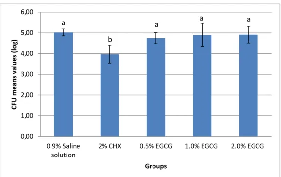
RESULTS
No significant difference between mean values were found for EGCG treatment in different concentrations when compared to saline group (p > 0.05), and there was no statistical difference between EGCG concentrations (p > 0.05). Only CHX group presented statistically different results from the others, as seen in Figure 1 (p < 0.05).
DISCUSSION
21
Many researchers have investigated the effect of catechins on the growth of S. mutans. According to Xu et al. [20], discrepant results of antimicrobial activity of Epigalocatechin-3-gallate can be found, depending on the type of bacterial organization. In planktonic cells, MIC (minimum inhibitory concentration) and MBC (minimum bactericidal concentration) values in a BHI medium were 625 µg/ml and >1,250 µg/ml, respectively. Moreover, epigallocatechin-3-gallate inhibited the formation of S. mutans biofilm in BHI medium with a MBIC50 (the lowest flavonoid concentration that showed at least 50% inhibition of the formation of biofilms compared with control) of 312.5 µg/ml. In the data presented by Mankovskaia et al. [30], the MIC values for epigallocatechin-3-gallate against S. Mutans were within reference ranges of 31.25 µg/ml to 625 µg/ml, depending on the bacterial strain and culture medium. Du et al. [31] concluded that antibacterial activity of the dental adhesives was increased after 200 µg/ml or higher concentration EGCG incorporated. Hirasawa et al. [18] demonstrated that more than 2000 µg/ml of epigallocatechin-3-gallate is require to a mouth-rinsing be effective against S. mutans presents in dental plaque. These researches suggest that epigallocatechin-3-gallate may represent a natural agent against dental biofilm.
The concentrations in which catechins are effective as an antimicrobial agent are wide and one of the causes is the application of different methods of determining bacterial susceptibility. Usually, levels of epigallocatechin-3-gallate to inhibit bacterial culture in planktonic levels are lower than in oral biofilm. This is associated with the fact that in planktonic bacteria cultures find themselves free in suspension, whereas in the biofilms there is an ecosystem structured and dynamic, which acts coordinately. These types of microorganism organization present distinct properties [42,43]. Biofilms are composed of a variety of species mechanisms that establish communication between them, including transfer of genetic material, as virulence traits or bacterial resistance faster than that occurring in culture planktonic [43]. Such characters favors these structures develop resistance to many therapeutic agents [32].
22
surface dentin that remained of the nutrient-rich BHI medium may bind or even precipitate catechins and the binding may jeopardize their antimicrobial activity [18,30]. Therefore, it can be suggested that there were interactions between catechin and specific sites in the molecular structure of collagen. More studies are needed to confirm this hypothesis.
Dentin submitted to cariogenic challenge seems to be more resistant to antimicrobial therapy. In the development of the dental biofilms, salivary proteins and glycoproteins on oral surfaces and other organisms play a decisive role in bacterial adhesion [44]. Dentin is present as a tubular, porous mineralized tissue composed primarily of hydroxyapatite and collagen fibrils [45]. Collagen type I, the main organic component of dentin, is recognized by streptococci that bind to collagen and may facilitate the bacterial adhesion and tissue penetration [46]. Therefore, EGCG concentrations used in this study are shown greater than those used in studies of culture planktonic and biofilm.
This study aimed to evaluate the antimicrobial activity of the flavonoid directly applied in carious dentin, which has not yet investigated in the literature. In order to perform the cariogenic challenge, we employed the microbiological method, in which the dentinal slabs remained immersed for five days in the nutrient medium inoculated with S. mutans. The production of in vitro caries-like lesions may be achieved in different ways, but the microbiological method presents a pattern of collagen degradation morphologically more similar to natural lesions, with an evident infected layer in the dentin caries lesion simulated by that method [33].
The use of chlorhexidine has been employed on caries prevention [34]. This substance has been studied for over twenty years and is considered the most potent therapeutic agent against oral biofilms. This is why the majority of studies investigating the efficacy of new antimicrobial drugs employs chlorhexidine as a positive control [35].
23
catechin remained in contact with dentin for 60 s may preserve resin-dentin bond-strength over time. Therefore, we choose a time of application of 60 s, which it cannot affect the longevity of restorative procedures.
The absence, until this date, of specific answers about the mechanism of EGCG anti-cariogenic points to the need for further research in this direction. There are numerous studies on the anti-cariogenic potential of the flavonoid. However, there is a great variability in the conditions and methods employed, which makes it difficult to attribute this potential to a specific factor or a set of elements.
In conclusion, the data obtained indicate that EGCG should not be used as a cleaning agent cavity in concentrations proposed by this study because it was not efficient in reducing S. mutans levels.
Acknowledgments This project has received financial support (Grant # 131437/2011-9) from CNPq. The authors thank David Queiroz for his help during experimental procedures. The first author received a scholarship during this study from CNPq. This paper was based on a thesis submitted by the first author to the Faculty of Pharmacy, Dentistry and Nursing of the Federal University of Ceará, in partial fulfillment of the requirements for an MS degree in Dentistry.
REFERENCES
1. Tanzer JM, Livingston J, Thompson AM. The microbiology of primary dental caries in humans. J Dent Educ 2001; 65:1028-1037.
2. Eckert R, Sullivan R, Shi W. Targeted Antimicrobial Treatment to Re-establish a Healthy Microbial Flora for Long-term Protection. Advances in Dent Res 2012; 24:94-97.
3. Gambhir RS, Singh S, Singh G, Singh R, Nanda T et al. Vaccine against Dental Caries- An Urgent Need. J Vaccin 2012; 3:136.
24
5. Borges FMC, Melo MAS, Paula DM, Lima JPM, Zanin ICJ, Barros EB, Rodrigues LKA. Antimicrobial effect of chlorhexidine digluconate in dentin: In vitro and in situ study. J Conserv Dent. 2012; 15(1): 22–26.
6. Souza LB, Aquino SG, Souza PPC, Hebling J, Costa CAS. Cytotoxic effects of different concentrations of chlorhexidine. Am J Dent. 2007; 20(6):400-4.
7. Lessa FCR, Aranha AMF, Nogueira I, Giro EMA, Hebling J, Costa CAS. Toxicity of chlorhexidine on odontoblast-like cells. J Appl Oral Sci. 2010; 18(1): 50-8.
8. Serper A, Calt S, Dogan AL, Guc D, Ozielik B, Kuraner T. Comparison of the citotoxic effects and smear layer removing capacity of oxidative potential water, NaOCl and EDTA. J Oral Sci. 2001; 43(4):233-8.
9. McKay DL, Blumberg JB. The Role of Tea in Human Health: an Update. Journal of the American College of Nutrition 2002; 21(1):1–13.
10. Taylor PW, Hamilton-Miller JMT, Stapleton PD. Antimicrobial properties of green tea catechins. Food Sci Technol Bull. 2005 ; 2:71–81.
11. Mitscher LA et al. Chemoprotection: a review of the potential therapeutic antioxidant properties of green tea (Camellia sinensis) and certain of its constituents. Med. Res. Rev.1997; 17:327-365.
12. Hamilton-Miller JM. Antimicrobial properties of tea (Camellia sinensis L.). Antimicrob. Agents Chemother. 1995; 39:2375-2377.
13. Hamilton-Miller JM. Anti-cariogenic effects of tea (Camellia sinensis). Journal of Medical Microbiology 2001; 50:299–302.Wu CD, Wei G. Tea as a functional food for oral health. Nutrition 2002; 18: 443-444.
14. Wu CD, Wei G. Tea as a functional food for oral health. Nutrition 2002; 18: 443-444.
15. Narotzki B, Reznick AZ, Aizenbud D, Levy Y. Green tea: a promising natural product in oral health. Arch of Oral Bio 2012; 57:429-435.
16. Harborne JB, Williams CA. Advances in flavonoid research since 1992. Phytochemistry 2000; 55(6):481-504.
17. Dale GN, Daneel F, Yu-Dong Z. Epigallocatechin-3-gallate (EGCG): Chemical and biomedical perspectives. Phytochemistry 2006; 67(17): 1849–1855. 18. Hirasawa M, Takada K, Otake S. Inhibition of acid production in dental plaque
25
19. Xu X, Zhou XD, Wu CD. The tea catechin epigallocatechin gallate suppresses cariogenic virulence factors of Streptococcus mutans. Antimicrobial agents and chemotherapy 2011; 55-3:1229-1236.
20. Xu X, Zhou XD, Wu CD. Tea catechin epigallocatechin gallate inhibits Streptococcus mutans biofilm formation by suppressing gtf genes. Arch Oral Biol 2012; 57: 678-683.
21. Amaechi B, Higham SM, Edgar W. Efficacy of Sterilisation Methods and Their Effect on Enamel Demineralisation. Caries Res 1998; 32:441-446.
22. Zanin IC, Lobo MM, Rodrigues LKA, Pimenta LA, Hofling JF, Gonçalves RB. Photosensitization of in vitro biofilmes by toluidine blue O combined with a light-emitting diode. Eur J Oral Sci 2006; 114:64-69.
23. Oliveira MPM, Rodrigues LKA, Melo MAS, Nobre dos Santos M. Photodynamic therapy effect in carious bovine dentin – an in vitro study. J Oral Laser Applications 2010; 10(1):29-36.
24. Lima JPM, Melo MAS, Borges FMC, Teixeira AH, Oliveira CS, Nobre dos Santos M et al. Evaluation of the antimicrobial effect of photodynamic antimicrobial therapy in na in situ modelof dentine caries. European Journal of Oral Sciences 2009; 117(5):568-574.
25. Melo MAS, Paula DM, Lima JPM, Borges FMC, Oliveira CS, Nobre dos Santos M et al. In vitro photodynamic antimicrobial chemotherapy in dentine contaminated by cariogenic bacteria. Laser Physics 2010; 20(6):1-10.
26. Newman DJ. Natural products as leads to potential drugs: an old process or the new hope for drug discovery? J Med Chem 2008; 51:2589– 2599.
27. Barbosa CS, Kato MT, Buzalaf MAR. Effect of supplementation of soft drinks with green tea extract on their erosive potential against dentine. Australian Dental Journal 2001; 56:317-321.
28. Kato MT, Magalhães AC, Rios D, Hannas AR, Attin T, Buzalaf MAR. Protective effect of green tea on dentin erosion and abrasion. J Appl Oral Sci 2009; 17(6):560-4.
26
30. Mankovskaia A, Lévesque CM, Prakki A. Catechin-incorporated dental copolymers inhibit growth of Streptococcus mutans. J Appl Oral Sci 2013; 21(2):203-207.
31. Du X, Huang X, Huang C, Wang Y, Zhang y. Epigallocatechin-3-gallate (EGCG) enhances the therapeutic activity of a dental adhesive. Journal of Dentistry 2012; 40:485-492.
32. Costerton JW, Stewart PS, Greenberg EP. Bacterial biofilms: a common cause of persistent infections. Science 1999; 284(5418):1318-22.
33. Marquezan M et al. Artificial methods of dentine caries induction: A hardness and morphological comparative study. Arch Oral Biol 2009; 54:1111-1117. 34. Autio-Gold J. The Role of Chlorhexidine in Caries Prevention. Operative
Dentistry 2008; 33-6:710-716.
35. Kidd EAM. Role of chlorhexidine in the management of dental caries. Int Dent J 1991; 41:279-286.
36. Yun JH, Pang EK, Kim CS, Yoo YJ, Cho KS, Chai JK, et al.Inhibitory effects of green tea polyphenol (_)-epigallocatechin gallate on the expression of matrixmetalloproteinase-9 and on the formation of osteoclasts. Journal of Periodontal Research 2004; 39:300–7.
37. Kato MT, Leite AL, Hannas AR, Buzalaf MAR. Gels containing MMP inhibitors prevent dental erosion in situ. Journal of Dental Research 2010; 89:468–72. 38. Visse R, Nagase H. Matrix metalloproteinases and tissue inhibitors of
metalloproteinases: structure, function, and biochemistry. Circ Res 2003; 2(92):827-39.
39. Pashley DH, Tay FR, Yiu C,Hashimoto M, Breschi L,Carvalho RM, Ito Collagen Degradation by Host-derived Enzymes during Aging. J Dent Res 2004; 83(3):216-221.
40. De Munck J, Van den Steen PE, Mine A, Van Landuyt KL, Poitevin A, Opdenakker G, Van Meerbeek B. Inhibition of Enzymatic Degradation of Adhesive-Dentin Interfaces. J Dent Res 2009; 88:1101-1106.
41. Santiago SL, Osorio R, Neri JR, Carvalho RM, Toledano M. Effect of the Flavonoid Epigallocatechin-3- Gallate on Resin-Dentin Bond Strength. J Adhes Dent 2013; 15: in press.
27
43. Marsh PD. Dental plaque – biological significance of a biofilm and community life-style. J Clin Periodontol 2005; 32 (Suppl 6) 7-15.
44. Gibbons RJ. Microbial ecology: adherent interactions which may affect microbial ecology in the mouth. J Dent Res 198463:378-385.
45. Love RM, Jenkinson HF. Invasion of dentinal tubules by oral bacteria. CritRev Oral Biol Med 2002; 13:171-83.
28
TABLES
TABLE 1: Groups and treatments performed.
GROUP TREATMENT
G I (n= 5) 0.9% Saline solution
G II (n= 5) 2% Chlorhexidine
G III (n= 5) 0.5% EGCG
G IV (n= 5) 1% EGCG
29
FIGURES
0,00
1,00 2,00 3,00 4,00 5,00 6,00
0.9% Saline solution
2% CHX 0.5% EGCG 1.0% EGCG 2.0% EGCG
CFU m
e
an
s
v
al
u
e
s
(l
o
g
)
Groups
a
b
a a a
30
4 CONCLUSÃO GERAL
31
REFERÊNCIAS
COSTA C.A.; EDWARDS C.A.; HANKS C.T. Cytotoxic effects of cleasing solutions recommended for chemical lavage of pulp exposures. Am J Dent, v.14, n.1, p. 25-30, 2001.
DALE G.N.; DANEEL F.; YU-DONG Z. Epigallocatechin-3-gallate (EGCG): Chemical and biomedical perspectives. Phytochemistry, v.67, n.17, p. 1849-1855, 2006.
HIDALGO E.; DOMINGUEZ C. Mechanisms underlying chlorhexidine-induced cytotoxicity. Toxicol In Vitro, v.15, n.4-5, p. 271-6, 2001.
HIRASAWA M.; TAKADA K.; OTAKE S. Inhibition of acid production in dental plaque bacteria by green tea catechins. Caries Res, v.40, n.3, p. 265-270, 2006.
LOESCHE W.J. Role of Streptococcus mutans in Human Dental Decay. Microbiol Rev, v.50, n.4, p. 353-380, 1986.
MEIERS J.C.; KRESIN J.C. Cavity disinfectants and dentin bonding. Oper Dent, v.21, n.4, p.153-9, 1996.
NEWMAN D.J. Natural products as leads to potential drugs: an old process or the new hope for drug discovery? J Med Chem, v.51, n.9, p. 2589-2599, 2008.
SOUZA L.B.; AQUINO S.G.; SOUZA P.P.C.; HEBLING J.; COSTA C.A.S. Cytotoxic effects of different concentrations of chlorhexidine. Am J Dent, v.20, n.6, p. 400-4, 2007.
SOUZA P.P.; ARANHA A.M.; HEBLING J.; GIRO E.M.; COSTA C.A. In vitro
cytotoxicity and in vivo biocompatibility of contemporary resin-modified glass-ionomer cements. Dent Mater, v.22, n.9, p. 838-44, 2006.
TAYLOR P.W.; HAMILTON-MILLER J.M.T.; STAPLENTON P.D. Antimicrobial properties of green tea catechins. Food Sci Technol Bull., v.2, p.71-81, 2005.
TEN CATE J.M. Remineralization of deep enamel dentine caries lesions. Aust Dent J, v.53, n.3, p. 281-285, 2008.
TYAS M.J.; ANUSAVICE K.J.; FRENCKEN J.E.; MOUNT G.J. Minimal intervention dentistry – a review. Int Dent J, v.50,n.1, p.1-12, 2000.
WU C.D.; WEI G. Tea as a functional food for oral health. In: Wilson M, editor. Food constituents and oral health: current status and future prospect. Woodhead
Publishing, Cambridge, United Kingdom, 2009, p.396-417.
XU X.; ZHOU X.; WU C.D. Tea catechin epigallocatechin-gallate inhibits
Streptococcus mutans biofilm formation by suppressing gtf genes. Arch Oral Biol, v. 57, n.6, p.678-683, 2012.
32
ANEXO A - TERMO DE DOAÇÃO DE DENTES
Pelo presente instrumento que atende às exigências legais, o Sr. (a) _________________________________________, após ter tomado
conhecimento do protocolo de pesquisa “ANÁLISE ANTIMICROBIANA DO
FLAVONÓIDE EPIGALOCATEQUINA-3-GALATO COMO AGENTE DE LIMPEZA
CAVITÁRIA EM DENTINA ARTIFICIALMENTE CARIADA”, que tem como objetivo avaliar o efeito do flavonóide epigalocatequina-3-galato como agente de limpeza cavitária em dentina cariada, vem na melhor forma de direito DOAR à CD JORGIANA SILVA DE ASSIS __ dentes, declarando, sob as penas da lei, que os dentes objeto da presente doação foram extraídos por indicação terapêutica, cujos históricos circunstanciados fazem parte dos prontuários dos pacientes de quem se originam.
Data:___/___/___
Assinatura:______________________________________________
33
ANEXO B - ARTIGO 46 DO REGIMENTO INTERNO DO PROGRAMA DE PÓS-GRADUAÇÃO EM ODONTOLOGIA (PPGO) DA UNIVERSIDADE FEDERAL DO
CEARÁ
§3º - O aluno que não obtiver aprovação no Exame Geral de Conhecimentos, terá direito à nova oportunidade, desde que respeitados os artigos 4 e 5 das Normas para os Cursos de Pós-Graduação da UFC.
§4º - O aluno só poderá defender a Dissertação após aprovação no Exame Geral de Conhecimentos de que trata este artigo.
Artigo 46– As dissertações apresentadas ao Programa de Pós-Graduação em Odontologia da Universidade Federal do Ceará poderão ser produzidas em formato alternativo ou tradicional. O formato alternativo estabelece: a critério do orientador e com a aprovação da Coordenação do Programa, que os capítulos e os apêndices poderão conter cópias de artigos de autoria ou co-autoria do candidato, publicados ou submetidos para publicação em revistas científicas, escritos no idioma exigido pelo veículo de divulgação.
§1º - O orientador e o candidato deverão verificar junto às editoras a possibilidade de inclusão dos artigos na dissertação ou tese, em atendimento à legislação que rege o direito autoral, obtendo, se necessária, a competente autorização, deverão assinar declaração de que não estão infringindo o direito autoral transferido à editora.
§2º - A dissertação em formato tradicional ou as sessões gerais do formato alternativo deverão seguir as normas preconizadas pelo Guia para Normalização de Trabalhos Acadêmicos da Biblioteca Universitária disponível no site http://www.biblioteca.ufc.br/servicos.html#apoio. As partes específicas do formato alternativo deverão ser feitas em concordância com o MANUAL DE NORMALIZAÇÃO PARA DEFESA DE DISSERTAÇÃO DE MESTRADO E TESE DE DOUTORADO NO FORMATO ALTERNATIVO do PPGO.
Artigo 47 – Para cada aluno deverá ser constituída uma banca examinadora, que será formada por 03 (três) professores ou especialistas, com o título de Doutor, como membros efetivos e dois suplentes.
34
ANEXO C - NORMAS PARA PUBLICAÇÃO EM PERIÓDICO
Guide for Authors
Editors-in-Chief:
Dr G R Holland, Ann Arbor, MI, USA Professor G B Proctor, London, UK
Archives of Oral Biology is an international journal which aims to publish papers of the highest scientific quality reporting new knowledge from the orofacial region including:
• developmental biology • cell and molecular biology • molecular genetics • immunology • pathogenesis • microbiology
• biology of dental caries and periodontal disease
• forensic dentistry • neuroscience • comparative anatomy • paeleodontology
Archives of Oral Biology will also publish expert reviews and articles concerned with advancement in relevant methodologies. The journal will only consider clinical papers where they make a significant contribution to the understanding of a disease process.
These guidelines generally follow the Uniform Requirements for Manuscripts Submitted to Biomedical Journals.
Online submission of papers
http://ees.elsevier.com/aob
Submission and peer review of all papers is now conducted entirely online. Authors are guided stepwise through the entire process, and can follow the progress of their paper. The system creates a PDF version of the submitted manuscript for peer review, revision and proofing. All correspondence, including the editors' decision and request for revisions, is conducted by e-mail. Authors requesting further information about online submission should follow the tutorial, at http://ees.elsevier.com/aob.
Submission of a paper implies that it has not been published previously, that it is not under consideration for publication elsewhere, and that if accepted it will not be published elsewhere in the same form, in English or in any other language, without the written consent of the publisher. Each manuscript must be accompanied by a statement signed by all authors that the manuscript in its submitted form has been read and approved by them. Authors must supply details of related papers submitted or recently published elsewhere. Submissions lacking this documentation will not be reviewed until it is supplied.
Authors are invited to suggest up to three referees they consider suitable to review their submission. The suggested reviewers should not have collaborated with the authors in the last 5 years. Full postal and email addresses should be included. The editors may or may not, at their discretion, utilize these suggestions.
Authorship
35
Conflict of interest
The potential for conflict of interest exists when an author (or the author's institution), has financial or personal relationships that may influence his or her actions. Authors are specifically asked to reflect on financial conflicts of interest (such as employment, consultancy, stock ownership, honoraria and paid expert testimony) as well as other forms of conflict of interest, including personal, academic and intellectual issues.At the end of the text, under a subheading "Conflict of interest statement" all authors must disclose any financial and personal relationships that could influence their work. Examples of potential conflicts of interest include employment, consultancies, stock ownership, honoraria, paid expert testimony, patent applications/registrations, and grants or other funding. If there are no conflicts of interest a statement confirming such should be included
Ethics
Studies on human beings.
Such studies submitted to Archives of Oral Biology should comply with the principles laid down in the Declaration of Helsinki; Recommendations guiding physicians in biomedical research involving human subjects. The
declaration was adopted by the 18th World Medical Assembly, Helsinki, Finland, June 1964, amended by the 29th World Medical Assembly, Tokyo, Japan, October 1975, the 35th World Medical Assembly, Venice, Italy, October 1983, and the 41st World Medical Assembly, Hong Kong, September 1989 (www.wma.net/e/policy/b3.htm). The manuscript should contain a statement that the work has been approved by the appropriate ethical committees related to the institution(s) in which it was performed and that subjects gave informed consent to the work. A copy of the institutional approval should be included. Submissions lacking these documents will not be reviewed until they are supplied. Patients' and volunteers' names, initials, and hospital numbers should not be used.
Studies on animals.
The experimental procedures and care of animals should be in accordance with the European Convention for the Protection of Vertebrate Animals used for Experimental and Other Scientific Purposes
(http://conventions.coe.int/Treaty/en/Treaties/Html/123.htm). The authors must state that animal care was in accordance with both these and institution guidelines. Signed documents of approval by institutional committees should be included as well as a statement from the authors that the study met the standards described in the European Convention. Submissions lacking these documents will not be reviewed until they are supplied.
Copyright
Accepted papers become the copyright of the Journal and are accepted on the understanding that they have not been published, are not being considered for publication elsewhere and are subject to editorial revision. If papers closely related to the submitted manuscript have been published or submitted for publication elsewhere, the author must state this in their cover letter. Upon acceptance of an article, authors will be asked to sign a 'Journal Publishing Agreement' (for more information on this and copyright see http://www.elsevier.com/authors).
Acceptance of the agreement will ensure the widest possible dissemination of information. An e-mail (or letter) will be sent to the corresponding author confirming receipt of the manuscript together with a 'Journal Publishing Agreement' form.
If excerpts from other copyrighted works are included, the author(s) must obtain written permission from the copyright owners and credit the source(s) in the article. Elsevier has preprinted forms for use by authors in these cases: please consult http://www.elsevier.com/permissions.
Scientific Standards
The aim of Editors and referees is to maintain a high standard of scientific communication. Normally papers are assessed by two referees selected by the Editor, and decisions regarding acceptance are based mainly upon the advice of the referees. Where appropriate, the referees' views are forwarded to the authors for their consideration. Authors may occasionally consider referees' suggestions to be ill-conceived but if their text is misunderstood by referees it is likely to be misunderstood by readers of the journal.
Types of Contribution
Original papers and review articles are welcomed. There will be no differentiation on the basis of length into full or short communications. All submissions will be refereed. Reviews may be submitted in outline prior to full
submission.
Manuscript Preparation
36
findings in the past tense and use the present tense where reference is made to existing knowledge, or where the author is stating what is known or concluded. Original papers should follow the pattern of: Introduction, Materials and Methods, Results or Findings, Discussion.
Authors will gain much assistance by consulting: Council of Biology Editors Style Manual Committee. Scientific Style and Format: The CBE Manual for Authors, Editors, and Publishers, 6th edition. New York: Cambridge University Press, 1994.
We suggest that authors consider using a language editing service to improve the English language usage and quality of a paper. A number of language editing companies will provide their services to our authors at competitive rates. Please follow this link for further
details http://elsevier.com/wps/find/authorsview.authors/languageediting/.
The editors reserve the right to revise the wording of papers in the interest of the Journal's standards of clarity and conciseness.
General
Manuscripts must be word processed (preferably in Word format), double-spaced with wide margins and a font size of 10 or 12 points. The corresponding author should be identified (include a fax number and email address). Full postal addresses must be given for all co-authors. Please check the current style of the journal, particularly the reference style (Vancouver), and avoid excessive layout styling as most formatting codes will be removed or replaced during the processing of your article. In addition, do not use options such as automatic word breaking, justified layout, double columns or automatic paragraph numbering (especially for numbered references). The Editors reserve the right to adjust style to certain standards of uniformity. Authors should retain copies of all versions of their manuscript submitted to the journal. Authors are especially requested to be vigilant over the submission of the correct version of the manuscript at the various stages of the editorial process.
Text
Follow this order when typing manuscripts: Title, Authors, Affiliations, Abstract, Keywords, Main text (Introduction, Materials & Methods, Results, Discussion for an original paper), Acknowledgments, Appendix, References, Figure Captions and then Tables. Do not import the Figures or Tables into your text. The corresponding author should be identified with an asterisk and footnote. All other footnotes (except for table footnotes) should be identified with superscript Arabic numbers.
Title page
As titles frequently stand alone in indexes, bibliographic journals etc., and indexing of papers is, to an increasing extent, becoming computerized from key words in the titles, it is important that titles should be as concise and informative as possible. Thus the animal species to which the observations refer should always be given and it is desirable to indicate the type of method on which the observations are based, e.g. chemical, bacteriological, electron-microscopic, histochemical, etc. A "running title" of not more than 40 letters and spaces must also be supplied. A keyword index must be supplied for each paper.
Structured abstract
The paper should be prefaced by an abstract aimed at giving the entire paper in miniature. Abstracts should be no longer than 250 words and should be structured as per the guidelines published in the Journal of the American Medical Association (JAMA 1995; 273: 27-34). In brief, the abstract should be divided into the following sections: (1) Objective; (2) Design - if clinical, to include setting, selection of patients, details on the intervention, outcome measures, etc.; if laboratory research, to include details on methods; (3) Results; (4) Conclusions.
Received/accepted dates
A received date will be added to all papers when they are received by the Accepting Editor. An accepted date will also be added when the papers are received at the publishing office.
Introduction
This should be a succinct statement of the problem investigated within the context of a brief review of the relevant literature. Literature directly relevant to any inferences or argument presented in the Discussion should in general be reserved for that section. The introduction may conclude with the reason for doing the work but should not state what was done nor the findings.
Materials and Methods
37
and methods were exactly as in a previous paper, it is not necessary to repeat all the details but sufficient
information must be given for the reader to comprehend what was done without having to consult the earlier work.
Authors are requested to make plain that the conditions of animal and human experimentation are as outlined in the "Ethics" and "Studies on Animals" sections above.
Results or Findings
These should be given clearly and concisely. Care should be taken to avoid drawing inferences that belong to the Discussion. Data may be presented in various forms such as histograms or tables but, in view of pressure on space, presentation of the same data in more than one form is unacceptable.
Statistical analysis
Authors should ensure that the presentation and statistical testing of data are appropriate and should seek the advice of a statistician if necessary. A number of common errors should be avoided, e.g.: -
• Use of parametric tests when non-parametric tests are required
• Inconsistencies between summary statistics and statistical tests such as giving means and standard deviations
for data which were analysed with non-parametric tests.
• Multiple comparisons undertaken with multiple t tests or non-parametric equivalents rather than with analysis of variance (ANOVA) or non-parametric equivalents.
• Post hoc tests being used following an ANOVA which has yielded a non-significant result.
• Incomplete names for tests (e.g. stating "Student's t test" without qualifying it by stating "single sample", "paired"
or "independent sample")
• N values being given in a way which obscures how many independent samples there were (e.g. stating simply
n=50 when 10 samples/measurements were obtained from each of 5 animals/human subjects).
• Stating that P=0.000 (a figure which is generated by some computer packages). The correct statement (in this
case) is P<0.0005.
Discussion
This section presents the inferences drawn from the Results: these should be recapitulated only sparingly, sufficient to make the argument clear.
References
All manuscripts should use the 'Vancouver' style for references, which should be numbered consecutively in the order in which they are first cited in the text and listed at the end of the paper.
For journal references, all authors should be included when there are six or fewer (first six followed by 'et al.' when seven or more), followed by the title of article, name of journal abbreviated according to Index Medicus, or left in full, year, volume with part number in brackets, and first and last pages. For example:
1. Walsh NP, Montague JC,Callow N and Rowlands AV. Saliva flow rate, total protein concentrationand osmolality as potential markers of whole body hydration statusduring progressive acute dehydration in humans. Arch Oral Biol2004;49(2):149-154.
For book references, the author(s) should be followed by the chapter title (if appropriate), editor(s) (if applicable), book title, place of publication, publisher, year and page numbers. For example:
Nanci A. Ten Cate's Oral Histology: Development, Structure and Function. 6th ed. St. Louis: Mosby; 2003.
Papers in the course of publication should only be entered in the references if the paper has been accepted by a journal, and then given in the standard manner in the text and list of references but with the words "In press" following the name of the journal.
Units and symbols
In general, Archives of Oral Biology will use the recommended SI (Systeme Internationale) units and symbols.
The use of the litre, usually better written in full, in place of SI dm3 and ml3 in place of SI cm, will continue to be
38
Abbreviations
As Archives of Oral Biology is a journal with a multidisciplinary readership, abbreviations, except those universally understood such as mm, g, min. u.v., w/v and those listed below, should be avoided if possible. Examples of abbreviations which may be used without definition: ADP, AMP, ATP, DEAE-cellulose, DNA, RNA, EDTA, EMG, tris.
Other abbreviations used to improve legibility should be listed as a footnote on the title page. Chemical symbols may be used for elements, groups and simple compounds, but excessive use should be avoided. Abbreviations other than the above should not be used in titles.
Bacterial nomenclature
Organisms should be referred to by their scientific names according to the binomial system. When first mentioned the name should be spelt in full and in italics. Afterwards the genus should be abbreviated to its initial letter, e.g. 'S. aureus' not 'Staph. aureus'. If abbreviation is likely to cause confusion or render the intended meaning unclear, the names of microbes should be spelt in full. Only those names which were included in the Approved List of
Bacterial Names, Int J Syst Bacteriol 1980; 30: 225?420 and those which have been validly published in the Int J
Syst Bacteriol since 1 January 1980 have standing in nomenclature. If there is good reason to use a name that does not have standing in nomenclature, the names should be enclosed in quotation marks and an appropriate
statement concerning the nomenclatural status of the name should be made in the text (for an example see Int J
Syst Bacteriol 1980; 30: 547?556). When the genus alone is used as a noun or adjective, use lower case Roman not italic, e.g.'organisms were staphylococci' and 'streptococcal infection'. If the genus is specifically referred to
use italics e.g. 'organisms of the genus Staphylococcus'. For genus in plural, use lower case roman e.g.
'salmonellae'; plurals may be anglicized e.g.'salmonellas'. For trivial names, use lower case Roman e.g. 'meningococcus'.
Numbers, measurements and statistics.
Numbers one to nine are spelled out unless they are measurements (e.g.5 ml). Numbers greater than nine are spelled out if they begin a sentence, or when clarity requires it. Numbers above and including 10 000 have a space, not a comma. A decimal point is preceded by a number or cypher e.g. '0.5'. Decimal points in columns should be aligned vertically. Dates are usually provided in full: 14 April 1949. Measurements may be expressed in SI or non-metric units. Use 10 ml/h rather than ml.h-1 or ml per h.
Drugs
These should be referred to by their approved and not proprietary names; for guidance, see the British National Formulary. Where it is desirable to indicate a particular brand of preparation, the proprietary name and source should be given in parentheses after the proper name, e.g. testicular hyaluronidase (Testovase, Bovine Enterprises Ltd, London, UK).
Illustrations
In the initial online submission and review stage, authors are required to provide electronic versions of their illustrations. When an article has been accepted, authors must be prepared to provide all illustrations in electronic and camera-ready format, (suitable for reproduction, which may include reduction, without retouching).
The Artwork Quality Control Tool is now available to users of the online submission system. To help authors submit high-quality artwork early in the process, this tool checks the submitted artwork and other file types against the artwork requirements outlined in the Artwork Instructions to Authors on www.elsevier.com/artworkinstructions. The Artwork Quality Control Tool automatically checks all artwork files when they are first uploaded. Each figure/file is checked only once, so further along in the process only new uploaded files will be checked.
General: Information relating to the preferred formats for artwork and illustrations may be found
atwww.elsevier.com/authors. Photographs, charts and diagrams are all to be referred to as "Figure(s)" and should be numbered consecutively in the order to which they are referred. They should accompany the manuscript, but should not be included within the text. All figures are to have a caption. Captions should be supplied on a separate page.
Line drawings: All lettering, graph lines and points on graphs should be sufficiently large and bold to permit reproduction when the diagram has been reduced to a size suitable for inclusion in the journal. Dye-line prints or photocopies are not suitable for reproduction. Do not use any type of shading on computer-generated illustrations.
39
Colour: Certain illustrations will be approved for publication in colour but only if, in the opinion of the Editors, the figures convey information not apparent in monochrome. Please note that figures supplied in colour will appear online in colour at no extra charge, even if the print version is monochrome.
Tables: Tables should be numbered consecutively and given a suitable caption. Begin each table on a separate page. Footnotes to tables should be typed below the table and referred to by superscript lowercase letters. No vertical rules should be used. Tables should not duplicate results presented elsewhere in the manuscript (e.g. in graphs).
Revised manuscripts
Frequently authors are required to submit revised versions of manuscripts in the light of reports from expert reviewers and editorial comments. Revised manuscripts must clearly show revisions and authors must clearly indicate the positions of revisions in a covering letter that addresses the concerns of reviewers/ editors.
Proofs
One set of page proofs in PDF format will be sent by e-mail to the corresponding author which they are requested to correct and return within 48 hours. Only minor corrections are acceptable at this stage. Please use this proof only for checking the typesetting, editing, completeness and correctness of the text, tables and figures. If we do not have an e-mail address then paper proofs will be sent by post. Elsevier now sends PDF proofs that can be annotated; for this you will need to download Adobe Reader version 7 available free
fromhttp://www.adobe.com/products/acrobat/readstep2.html. Instructions on how to annotate PDF files will accompany the proofs. The exact system requirements are given at the Adobe
site:http://www.adobe.com/products/acrobat/acrrsystemreqs.html#70win. If you do not wish to use the PDF annotations function, you may list the corrections (including replies to the Query Form) and return to Elsevier in an e-mail. Please list your corrections quoting the line number. If, for any reason, this is not possible, then mark the corrections and any other comments (including replies to the Query Form) on a printout of your proof and return by fax, or scan the pages and e-mail, or by post.
Please use this proof only for checking the typesetting, editing, completeness and correctness of the text, tables and figures. Significant changes to the article as accepted for publication will only be considered at this stage with permission from the Editor. We will do everything possible to get your article published quickly and accurately. Therefore, it is important to ensure that all of your corrections are sent back to us in one communication: please check carefully before replying, as inclusion of any subsequent corrections cannot be guaranteed. Proofreading is solely your responsibility. Note that Elsevier may proceed with the publication of your article if no response is received.
Offprints
The corresponding author, at no cost, will be provided with a PDF file of the article via e-mail or, alternatively, 25 free paper offprints. The PDF file is a watermarked version of the published article and includes a cover sheet with the journal cover image and a disclaimer outlining the terms and conditions of use. Additional paper offprints can be ordered by the authors. An order form with prices will be sent to the corresponding author.
Funding body agreements and policies
Elsevier has established agreements and developed policies to allow authors who publish in Elsevier journals to comply with potential manuscript archiving requirements as specified as conditions of their grant awards. To learn more about existing agreements and policies please visit http://www.elsevier.com/fundingbodies.
Author enquiries
40
42