ISSN 0001-3765 www.scielo.br/aabc
Detection of
P
element transcripts in embryos
of
Drosophila melanogaster
and
D. willistoni
MONICA L. BLAUTH1,2, RAFAELA V. BRUNO3, ELIANA ABDELHAY4, ELGION L.S. LORETO2,5 and VERA L.S. VALENTE2,5
1Departamento de Ciências Biológicas, Universidade do Estado de Mato Grosso (UNEMAT) Campus Tangará da Serra, Rodovia MT-358, km 07, Bairro Aeroporto
78300-000 Tangará da Serra, MT, Brasil
2Laboratório deDrosophila, Departamento de Genética, Instituto de Biociências Universidade Federal do Rio Grande do Sul (UFRGS), Caixa Postal 15053
91501-970 Porto Alegre, RS, Brasil
3Laboratório de Biologia Molecular de Insetos, Fundação Oswaldo Cruz, Av. Brasil, 4365 Bairro Manguinhos, 21045-900 Rio de Janeiro, RJ, Brasil
4Centro de Transplante de Medula Óssea, Instituto Nacional de Câncer Praça da Cruz Vermelha, 23, 7◦andar, 20230-130 Rio de Janeiro, RJ, Brasil
5Departamento de Biologia, Centro de Ciências Naturais e Exatas, Universidade Federal de Santa Maria, Caixa Postal 5050, Agência Universitária, 97111-970 Santa Maria, RS, Brasil
Manuscript received on August 28, 2008; accepted for publication on March 11, 2009; contributed byVERAL.S. VALENTE*
ABSTRACT
The P element is one of the most thoroughly studied transposable elements (TE). Its mobilization causes the hybrid dysgenesis that was first described inDrosophila melanogaster. While studies of thePelement have mainly been done inD. melanogaster, it is believed thatDrosophila willistoniwas the original host species of this TE and
that P was transposed to the D. melanogastergenome by horizontal transfer. Our study sought to compare the
transcriptional behavior of the P element in embryos ofD. melanogaster, which is a recent host, with embryos of
two strains ofD. willistoni, a species that has contained the P element for a longer time. In both species,
poten-tial transcripts of transposase, the enzyme responsible for the TE mobilization, were detected, as were transcripts of the 66-kDa repressor, truncated and antisense sequences, which can have the ability to prevent TEs mobiliza-tion. The truncated transcripts reveal the truncated P elements present in the genome strains and whose number
seems to be related to the invasion time of the genome by the TE. No qualitative differences in antisense tran-scripts were observed among the strains, even in theD. willistonistrain with the highest frequency of heterochromatic
Pelements.
Key words:Drosophila,D. willistoni, hybrid dysgenesis, RNAi,Pelement, transposable element.
INTRODUCTION
The P element is the most thoroughly studied trans-posable element (TE) in Drosophila. It was first
dis-covered as the causative agent of hybrid dysgenesis in
Drosophila melanogaster(Kidwell 1977). Later, other
*Member Academia Brasileira de Ciências Correspondence to: Vera Lúcia da Silva Valente E-mail: [email protected] / [email protected]
elements, including transposons and retroelements, were identified as also being able to promote a similar syn-drome in D. melanogaster and in other species of the genus (Yannopoulos et al. 1987, Crozatier et al. 1988, Lozovskaya et al. 1990, Petrov et al. 1995, Evgen’ev et al. 1997). The syndrome caused by the P element
be-tween males of strains that contain functionalPelements
in the genome and females that do not (Kidwell and Kidwell 1979, Engels 1989). This syndrome promotes sterility and gonadal atrophy.
The P element is a class II TE that is 2.9 kb long,
including the 31-bp Inverted Terminal Repeats (ITR), and is transposed by the DNA cut and paste mecha-nism (Misra and Rio 1990, Laski et al. 1986). The P
element contains four open reading frames (ORFs) that encode at least two proteins by alternative splicing of the third intron, named IVS3. The transposase, an 87-kDa enzyme necessary for transposition, is expressed exclusively in the germline cells (Siebel and Rio 1990). In somatic cells, the IVS3 sequence is retained in the transcript and a 66-kDa repressor protein is instead produced.
The P element is widely dispersed in Neotropi-cal species of the subgenus Sophophora (Daniels et
al. 1990, Clark and Kidwell 1997, Loreto et al. 1998). This sequence is present in the cosmopolitan species
D. melanogaster, but not in other species ranked under
themelanogastersubgroup, suggesting a recent invasion
of theD. melanogastergenome by horizontal transfer.
This hypothesis is also supported by studies showing that several strains ofD. melanogastercollected before
the 1950s do not have the P element (Bregliano and
Kidwell 1983). The P element sequences ofD. willis-toniandD. melanogasterdiffer by only a single
nucleo-tide substitution (Daniels et al. 1990), suggesting that the donor species belongs to the willistonigroup. The
first contact between the two species is calculated to have occurred around the year 1800, when it is believed thatD. melanogasterarrived in the New World (Engels
1989). Therefore, in a period of about 200 years, the P
element invaded theD. melanogastergenome and
dis-persed across the world.
The invasive capacity of the P element has been
attributed to its ability to regulate its own mobility, in-creasing the chances of host survival in the face of its invasion (Brookfield 1991). Besides the tissue-specific splicing of IVS3, other alternative forms of splicing into this intron, such as those described forD. melanogaster, D. bifasciata and D. helvetica by Chain et al. (1991)
and Haring et al. (1998), as well as deleted copies, are considered as mechanisms that preventPelements from
transposing. The KP protein, encoded by a truncated
P element sequence, is one of the most widely known
repressors of P element mobility. The short KP poly-peptides may interact with thePtransposase and inhibit
its function through the assembly of inactive heteromul-timers (Lee et al. 1996, 1998). Alternatively, short P
polypeptides may interact with thePelement promoter, thus inhibiting transcription.
Simmons et al. (2002) suggested that the nature of the female P cytotype, which refers to the maternal ability to prevent the paternally originated P element
from transposing, is determined by the 66 kDa-repres-sor. A consensus sequence, similar to those found in the maternal genes, was identified in the IVS3 intron and appears to allow only unspliced transcripts to be maternally transmitted by the nurse cells to the oocyte, where they prevent transposase enzyme activity after egg fertilization. On the other hand, the most recent work of Josse et al. (2007) proposes the telomeric Trans-Silencing Effect (TSE), a mechanism by which a transposon inserted in subtelomeric heterochromatin or close to the centromere has the capacity to repress homologous transposons in the genome. The observed sensitivity to mutations in genes that code for the pro-teins AUBERGINE and PIWI suggested that the P cytotype might depend on repeat-associated small in-terfering RNA. The AUBERGINE and PIWI proteins link to small antisense RNAs and trigger the produc-tion of more antisense transcripts, which in turn prevent the expression of homologous sequences by sequence complementarity.
Considering these aforementioned studies, we in-vestigated the presence of transcripts coding for trans-posase and potential repressors of mobilization of the
P element in embryos of two strains of D. willistoni,
which produce offspring affected by hybrid dysgenesis syndrome and whose previous description detected the
P element inserted preferentially in heterochromatic in the Wip strain and in euchromatic in the 17A2 strain. These strains favor the study of the proposed hypothe-sis that correlates heterochromatin sequences with their transcriptions in an antisense way. Apart from this, one strain of D. melanogasterwas included in our studies
MATERIALS AND METHODS
FLYSTOCKS
The strains used were D. melanogaster Harwich (H) from the USA, first collected in the 1960s and normally used as aPelement-positive control in experiments;D. willistoni17A2 from South Brazil (30◦05′S, 51◦39′W),
sampled in the early 1990s, andD. willistoniWip from
Northeast Brazil (12◦54′S, 38◦19′W), sampled in the 1960s. The strains ofD. willistoni were chosen since
they were previously studied forP element
chromoso-mal positioning (Regner et al. 1996). In the 17A2 strain, only 17% of the chromocenters analyzed by the authors have a P element inserted, while the Wip strain have 50% of the chromocenters withP element sites. Some
hybridization signals were detected in the euchromatic arms of the Wip strain, but none had a frequency higher than 15%. The mating of 17A2 males with Wip females generates offspring with a 26% hybrid dysgenesis rate at 29◦C (Regner et al. 1999).
The flies were reared in cornmeal medium (Mar-ques et al. 1966) at constant temperature and humid-ity (17±1◦C; 60% rh).
EXTRACTION OFNUCLEICACIDS
The DNA of 25 adult flies was extracted by the phenol-chloroform method according to Sassi et al. (2005). Total RNA of embryos between 0-18 h of development was extracted by the Trizol method (Invitrogen).
SYNTHESIS OF CDNA
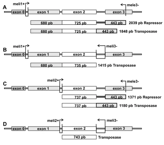
The cDNA synthesis was done according to the proto-col of M-MLV Reverse Transcriptase enzyme method (Invitrogen) using 2 pmol of the specific mele3- primer for the detection of transposase, 66-kDa repressor and truncated transcripts or meli1+ primer for the de-tection of antisense transcripts (Table I lists the primer sequences and Fig. 1 depicts the primers applied and their respective binding sites). The cDNA synthesized from Amd-un2 and Amd-bw primers, specific for the
α-methyldopa gene (Amd) (Tatarenkov et al. 2001),
was used as a DNA contamination control in the assay for antisense transcript detection.
AMPLIFICATION OFDNAAND CDNA
The reaction was carried out in a 15µl volume with
1U Taq DNA polymerase (Invitrogen) in 1X polymerase buffer, 2.5 mM of MgCl2, 10 pmol of each primer and
0.6 mM of dNTP mix. The first PCR round of the cDNA amplification was performed with 2µl of cDNA
sample and the mele3- primer and meli1+ intron-span-ning primers (Fig. 1A). The amplification parameters were a denaturation step at 95◦C for 5 min, followed by 40 cycles of 95◦C for 30 s, 58◦C for 30 s, 72◦C for 2.5 min, and a final extension at 72◦C for 10 min. A 1µl sample of this first PCR amplification was
sub-mitted to a second round of amplification with inter-nal primers combined as follows: meli1+ and meli3-, meli2+ and mele3-, meli2+ and meli3- (Table I; Figs. 1B, 1C, 1D), where meli1+, meli2+ and meli3- are intron-spanning primers. The cycle parameters for the second amplification were a denaturation step at 95◦C for 5 min, followed by 40 cycles at 95◦C for 1 min, 56◦C for 1 min, 72◦C for 1 min, and a final extension at 72◦C for 10 min.
The parameters of amplification of theAmdcDNA
were a denaturation step at 94◦C for 7 min, followed by 40 cycles at 94◦C for 1 min, 56◦C for 30 s, 72◦C for 1 min, and a final extension at 72◦C for 10 min.
Thirty nanograms of genomic DNA were used for PCR amplification with the M-IR primers (Table I). The amplification parameters were a denaturation step at 95◦C for 5 min, followed by 40 cycles at 95◦C for 40 s, 55◦C for 40 s, 72◦C for 1.5 min, and a final exten-sion at 72◦C for 10 min.
SOUTHERNBLOT
The first amplification product, with meli1+ and mele3-primers, was submitted to a Southern blot procedure. The gel preparation was carried out following Sambrook et al. (1989). The hybridization, stringency washes and detection reaction followed the Gene Images Labeling kitr and CPD-Stars Detection Moduler (Amersham
Biosciences). The probe used was the complete canon-icalP element sequence labeled with fluorescein, used
according to the Gene Images Labeling kitr(Amersham
TABLE I
PCR primers used.
Position in canonical Primer Sequence (5’-3’) Pelement ofD. melanogaster
(GenBank accession code X06779) Meli1+ * TACACAAACAGAGTCCTGTT 431–442, 501–508 Meli2+ * GTATATCAGAATCAAAAACCTG 1157–1168, 1222–1231
Meli3- * CATTTCTGTATTCCTGGCTATT 2154–2138, 1947–1943 Mele3- * GTTTATCAACATCGACGTTTC 2581–2561 M-IR ** CATAAGGTGGTCCCGTCG 1–31, 2907–2877
*Haring, Hagemann and Pinsker 1998. **Haring, Hagemann and Pinsker 1995.
Fig. 1 – Map of the canonicalPelement showing exons, introns and primers binding sites used in RT-PCR. (A) meli1+ and mele3- primer binding sites and the probable fragments amplified in RT-PCR amplification. (B) meli1+ and meli3- primer binding sites and the probable fragments amplified in RT-PCR amplification. (C) meli2+ and mele3- primer binding sites and the probable fragments amplified in RT-PCR amplification. (D) meli2+ and meli3- primer binding sites and the fragment and the probable fragments amplified in RT-PCR amplification.
SEQUENCING
The fragments obtained by RT-PCR were sliced off the 0.8% agarose gel and purified with GFX PCR DNA and Band Gel Purification kitr(Amersham Biosciences)
ac-cording to the supplier’s instructions. The purified DNA was sequenced in a MegaBACE 500 automatic sequencer using the DYEnamic ET kitr(Amersham Biosciences)
and the same specific sense and antisense primers used
RESULTS
DETECTION OFPUTATIVETRANSCRIPTS OFTRANSPOSASE AND66-KDAREPRESSOR INEMBRYOS OFD. melanogasterANDD. willistoni
By RT-PCR, we identified a putative transcript of the transposase enzyme and of the 66-kDa repressor in
D. willistoniand D. melanogaster embryos. The
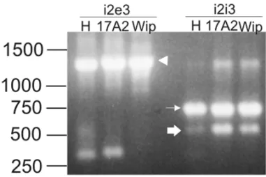
743-bp fragment was amplified with the meli2+ and meli3-primers in both species (Fig. 1D; Fig. 2, indicated by a narrow arrow). This fragment corresponds to tran-scripts that have experienced IVS2 and IVS3 splicing, and would thus code for the functional transposase enzyme (GenBank accession codes DQ486674 and DQ486675).
Two amplification products were expected in a RT-PCR using meli2+ and mele3- primers: 1180 and 1371-bp (Fig. 1C), corresponding to the spliced and unspliced IVS3 transcripts, respectively. In Figure 2, we observe only the unspliced IVS3 fragment (indicated by an ar-rowhead), a result which was confirmed by partial se-quencing of theD. melanogasterfragment. We infer that the transcription of the 66-kDa repressor is higher than the transcription of the transposase, previously identified with the PCR with meli2+ and meli3- primers.
Fig. 2 – Agarose gel electrophoresis with RT-PCR amplification products from embryos ofD. melanogaster (H) and D. willistoni
(17A2 and Wip). The first step of cDNA amplification was carried out with meli1+ and mele3- primers. (i2e3) PCR reamplification with meli2+ and mele3- primers. (i2i3) PCR reamplification with meli2+ and meli3- primers. The narrow arrow indicates the transposase ment; the arrowhead indicates the 66-kDa transposase repressor frag-ment; the thick arrow indicates a fragment with truncated exon 2. A 1-kb DNA ladder is depicted on the left.
ALTERNATIVESPLICING
A 572-bp fragment was amplified by meli2+ and meli3- primers (Fig. 2, indicated by a thick arrow – GenBank accession codes DQ486676, DQ486677 and DQ486678). This sequence lost 198-bp of the 3’ re-gion of exon 2 (1742-1941 nt of canonical P element),
but maintained its splicing donor site (position 1947 nt of the canonical P element) after undergoing the IVS3
splice.
DIFFERENTIALTRANSCRIPTIONBETWEENSPECIES
The transcriptional differences between the species were more evident when PCR primers that amplify longer se-quences were used. Figure 3 displays the amplification products obtained with the meli1+ primer, combined with the mele3- or the meli3- primers (Figs. 1A and B display the expected amplification products).
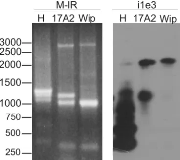
Fig. 3 – Agarose gel electrophoresis with RT-PCR products from embryos ofD. melanogaster(H) andD. willistoni(17A2 and Wip). The first step of cDNA amplification was carried out with meli1+ and mele3- primers. (i1e3) RT-PCR with meli1+ and mele3- primers. (i1i3) PCR reamplification with meli1+ and meli3- primers. The arrow in-dicates the transposase fragment. The fragments indicated by i1e31, i1e32 and i1i34 were gel-purified and sequenced. A 1-kb DNA ladder is depicted on the left.
When meli1+ and meli3- were used in the RT-PCR amplification, the transposase fragment was visi-ble only inD. willistonistrains (Fig. 3, indicated by an
arrow), not inD. melanogaster. In contrast, truncated
transcripts were more evident inD. melanogasterwhen
both combinations of primers were used, as compared toD. willistoni(Fig. 3).
This differential expression between the species appears to result from the larger number of truncated
P element sequences in the D. melanogastergenome
PCR with the M-IR primer (Fig. 4, M-IR). This primer anneals in the ITRs and amplifies the element across the entire length.
The greater number of truncated sequences related to P element in D. melanogasteralso promotes PCR
primer competition, as the complete sequence is not re-alized in the agarose gel (Fig. 4, indicated by an arrow-head).
Fig. 4 – Comparison of the fragments obtained from PCR amplifica-tion of adult genomic DNA and the fragments obtained from RT-PCR of embryos ofD. melanogaster(H) andD. willistoni(17A2 and Wip). (M-IR) Agarose gel electrophoresis with genomic PCR products with M-IR primer. (i1e3) Southern blot, using the canonicalPelement as
probe, of the RT-PCR amplification with meli1+ and mele3- primers. The arrowhead indicate the completePelement sequence and the nar-row arnar-row the repressor of 66-kDa fragment. A 1-kb DNA ladder is shown on the left.
Figure 4 displays a comparison of the amplifica-tion fragments obtained with the M-IR primer and ge-nomic DNA as template and the fragments obtained with the primers meli1+ and mele3- and cDNA as tem-plate, emphasizing the presence and transcription of truncated P element sequences in theD. melanogaster
genome. The Southern blot procedure (Fig. 4, i1e3) also permitted the visualization of fragments not apparent in Figure 3 (i1e3), including the expected 2039-bp fragment (Fig. 1A; Fig. 4, indicated by an arrow), which encodes the 66-kDa repressor. This discrepancy may de-rive from PCR primer competition among the high num-ber of transcribed sequences related to the Pelement.
Since truncated sequences of the P element are
potential repressors of transposition (Pinsker et al. 2001), three of the highly expressed fragments in D. melanogaster were sequenced. They are indicated as
i1e31, i1e32 and i1i34 in Figure 3 and 377, 611 and 284 nt of their sequence were determined, respectively (GenBank accession codes DQ486679, DQ486680 and DQ486681). The sequences align with 3’ exon 1 re-gion, which is also present in the KP repressor sequence (Black et al. 1987), but they show little nucleotide con-servation. The i1e32 sequence has a duplication of the 5’ exon 1 region, and the disparity of the fragment size estimated in the agarose gel and the sequence determi-nants can be the result of a secondary structure acquired by the sequence.
PRESENCE OFANTISENSEPELEMENTTRANSCRIPTS
DURINGEMBRYOGENESIS
The PCR procedure using total embryo cDNA, synthe-sized starting from a sense primer (meli1+), suggested the presence of antisense transcripts (Fig. 5) in embryos of both species.
Fig. 5 – Agarose gel electrophoresis of sense and antisensePelement
fragments obtained by RT-PCR of embryos ofD. melanogaster(H)
andD. willistoni(17A2 and Wip). The sense cDNA was synthesized starting from the meli1+ sense primer and the antisense cDNA was synthesized starting from mele3- antisense primer. The first cDNA amplification was done with meli1+ and mele3- primers and the re-amplification reaction with meli2+ and mele3- primers. (AS) Anti-sense fragments. (S) Sense fragments. A 1-kb DNA ladder is shown on the left.
Unlike the P element, no transcript amplification
was observed in the total embryo cDNA synthesized starting from the sense primer Amd-un2 of theAmdgene (data not shown), suggesting the absence of antisense transcripts of theAmdgene and of DNA contamination
DISCUSSION
The present work aimed to characterize the transcrip-tion of P element sequences during embryogenesis of
one recently invaded species,D. melanogaster, as well
as in D. willistoni, a long-standing host of this
trans-posable element, and obtain data about the antisense transcription of P element comparing specifically the two strains of D. willistoni. The work of Haring et al. (1998) proposes the utilization of a combination of four primers to detect the transposase and the 66-kDa repressor. Three of these primers anneal only in spliced sequences, assuring that only cDNA sequences are used as template and also makes it possible to distinguish the transcripts of the transposase and the 66-kDa repressor by the alternative splicing of the IVS3 intron. Using this procedure, we detected transcription of the trans-posase gene and the 66-kDa repressor in embryos of the studied strains. The absence of transposase transcripts in the amplification using meli2+ and mele3- primers, which was designed to reveal transcripts of both the transposase and the 66-kDa repressor, suggests that there is a greater number of transcripts of the 66-kDa repressor available, as this transcript was more readily used as the template in the RT-PCR reaction. However, transcription of the transposase is specific to germline cells, at least in adults, and these cells are less well rep-resented in the sample than somatic cells.
It is believed that the 66-kDa repressor can bind toPelement cleavage sites and block subsequent
bind-ing of the transposase enzyme, thus avoidbind-ing excision of the TE (Gloor et al. 1993, Laski et al. 1986). Addi-tionally, antisense transcripts and polypeptides encoded by truncated copies offer alternative hypotheses to ex-plain howPelement mobilization can be silenced (Ras-musson et al. 1993, Simmons et al. 1996). These au-thors tested a group of antisense P element constructs and showed that these arebona fide repressors of pu-pal lethality, a condition established by the existence of P element sequences in which the IVS3 intron is
deleted. These mutated sequences allow transcription of the transposase only, not the 66-kDa repressor, even in somatic cells (Engels et al. 1987). In these studies, the antisense P element sequences were inserted in
embryos by a transformation procedure while, in the
present work, we suggested the synthesis of antisense
Pelement in un-manipulated organisms.
Antisense transcriptional silencing or RNA inter-ference (RNAi) has been described for other TEs (Kleckner et al. 1996, Joanin et al. 1997, Jensen et al. 1999a). InD. hydei(Lankenau et al. 1994) and species of therepletagroup (Almeida and Carareto 2004), two antisense transcripts of TE micropia, starting from an internal promoter, can be expressed in a testis-specific fashion. These antisense transcripts are not expressed in D. melanogaster, suggesting that D. melanogaster
has developed a different mechanism of regulation thanD. hydei. In our work, two distantly related species,
one of the melanogaster group and the other of the willistoni group, shared an antisense P element
tran-script of a similar size, suggesting the conservation of the transcription of the antisense sequence and its im-plied importance for the organism.
In further support of the idea that antisense tran-scripts regulate transposon mobilization inDrosophila
embryos, Blumenstiel and Hartl (2005) proposed that siRNAs (small interfering RNA, composed of dsRNA of 21-25 nt) homologous to thePenelopeTE are
mater-nally loaded into embryos ofD. virilisand act as a
silenc-ing machinery againstPenelopeand other unrelated TEs.
Moreover, P element regulation relies on a
chro-mosomal component. Ronsseray et al. (1991, 1996) sug-gested that the insertion of the P element in
telomeric-associated sequence on the X chromosome ofD. mela-nogaster strongly promotes the induction of the
cyto-type inhibitor. The authors also showed that this capac-ity is sensitive to mutant alleles of the geneSu(var)205,
which encodes the Heterochromatin Protein 1, involved in the heterochromatin formation, and of the gene
aubergine, which encodes the AUBERGINE protein, a protein that favors P element silencing in a hetero-chromatin loci-dependent manner, through a mechan-ism triggered by RNAi (Aravin et al. 2001, Reiss et al. 2004). Also, Josse et al. (2007) determined the sensitiv-ity of the P cytotype to mutations in the geneSu(var)205
and in the geneaubergine,piwi,armitageandhomeless,
The TEsgypsy,ZamandIdefixof Drosophilaare
also regulated by heterochromatic loci: theflamenco
lo-cus ofgypsy(Sarot et al. 2004), and theCOMlocus of Zam and Idefix (Desset et al. 2003). PIWI is one of
the proteins involved in the RNAi-mediated mobiliza-tion silencing of these TEs (Desset et al. 2003, Sarot et al. 2004). Through immunopurification, various kinds of TEs were observed as being linked to PIWI, as were sense and antisense sequences of the TE roo(Saito et
al. 2006). It is interesting that this protein is strongly ex-pressed in ovaries and early embryos, including the pole cells where mobilization of the Pelement promotes the
gonadal atrophy that characterizes hybrid dysgenesis. When evaluating the two strains of D. willistoni
with regard to their heterochromatic sites and the silenc-ing mechanisms described previously, we expected that the Wip strain would have a more efficient RNAi silenc-ing mechanism than the 17A2 strain, in which P
ele-ments are preferentially arranged in euchromatic sites. However, we did not observe significant qualitative ex-pression differences between the two strains of D. wil-listoni by RT-PCR. Considering the low percentage of hybrid dysgenesis (26%) observed in their offspring (Regner et al. 1999), we can suppose that the 17A2 strain has already developed a mobilization control for the P element and, as described by Josse et al. (2007),
it is possible to register variegation in ovaries when si-lencing is incomplete. Yet, as cited by Johnson (2008) in a recent commentary about transposon silencing, one or two insertions of the P element at the telomeric
silencing site can efficiently suppress as many as 80 homologues copies elsewhere in the genome. Quanti-tative evaluation of the transposase and antisense tran-scripts is necessary for a better comprehension of the mechanisms and to shed more light on the possibility that it really acts in the repression of mobilization.
In the present work, truncated transcripts related to the P element and amplified by RT-PCR were
se-quenced. These truncated copies could function as a sta-ble source of repressive proteins (Pinsker et al. 2001), by creating heteromultimers (Rio 1999), or by triggering a mechanism of homology-dependent silencing (Jensen et al. 1999a, b). A greater number of truncated copies were amplified in D. melanogaster than in D. willis-toni. This difference seems to reflect the sequences
avail-able in the genome of each species, rather than differen-tial expression. Apart from this, the larger number of truncated copies exhibited byD. melanogaster
demon-strates the process of stabilization of the P element in
the genome of this species (Kidwell and Lisch 2001). It is possible that truncated copies that play an important role in the regulation of TE mobility would be main-tained in the genome, while others would be lost due to successive mutations. When the two strains ofD. willis-toniare observed more carefully, it is possible to infer
the same: the 17A2 strain, where we can visualize one extra truncated sequence as compared to the Wip strain (Fig. 4, M-IR), is under a process of silencing that is more recent than that of the Wip strain.
Three highly expressed truncated P element
tran-scripts fromD. melanogasterwere sequenced. The most
conserved region among them is an exon 1 sequence, also present in the KP repressor. However, we do not know whether these transcripts maintain the same DNA binding and dimerization motifs of the KP repressor (Lee et al. 1996, 1998), two important sites for the in-hibition of transcription, because the ORF0, which en-codes these motifs, was not amplified and sequenced. Independent of the sequence conservation, the activity of the truncated transcripts as repressors of mobility should be tested, as the UP1 and NP2 sequences, with 212 and 159 amino acids similar to the KP, do not si-lence P element mobilization (Simmons et al. 1996).
Investigation of these sequences could also be valu-able due to their high expression in embryos of D. melanogaster and because non-translatable sequences
can act via a homology-dependent silencing mechan-ism, related to the copy number and homology of the sequence, as suggested by theIretrotransposon (Jensen et al. 1999a, b).
In addition, we observed a truncated sequence shared by D. willistoni and D. melanogaster species
that could be the result of another horizontal transfer between these species, similar to that suggested for the complete P element (Daniels et al. 1990). It is also
possible to conjecture that we detected a deletion hot-spot or a splicing variant site in the sequence of P
ele-ment. Variant splicing sites in P element were already
sites, however, lie within the IVS3 intron sequence, while our data suggest an alternative splicing site into exon 2, at 1741 nt of the canonicalPsequence (GenBank
accession code X06779). The transcripts described by Haring et al. (1998), originated by variant splicing, are 120 and 105-bp longer than the transposase transcript, and were not observed in embryos of either species ex-amined in our study, in spite of their nucleotide conser-vation. The ability of these splicing variant sequences to transpose and their capacity to function as a repressor are still unknown.
The presence of a putative transposase transcript during embryogenesis of a non- hypermutable strain indicates the existence of a post-transcriptional control mechanism, possibly including the 66-kDa repressor, the truncated copies, mainly in D. melanogaster, and
the antisense transcripts. The last option is the most re-cently proposed mechanism ofP element silencing and
the role of these transcripts in the repressor cytotype, which should be further investigated. The inclusion of
D. willistoniin studies of P element regulation could improve the understanding of silencing mechanisms, as well as how they are broken in the offspring of some strains, resulting in hybrid dysgenesis.
ACKNOWLEDGMENTS
We are grateful to Dr. Marco Silva Gottschalk for his revision of the manuscript and for his help with the fig-ures. This project was supported by Conselho Nacional de Desenvolvimento Científico e Tecnológico (CNPq), Coordenação de Aperfeiçoamento de Pessoal de Nível Superior (CAPES) and Fundação de Amparo à Pesquisa do Estado do Rio Grande do Sul (FAPERGS).
RESUMO
O elemento P é um dos elementos transponíveis (TE) mais amplamente estudado. Sua mobilização causa a disgenesia do híbrido que foi primeiramente descrita emD. melanogaster.
Apesar dos estudos sobre o elemento P terem sido
realiza-dos principalmente comD. melanogaster, acredita-se queD. willistoni foi a espécie hospedeira original deste TE e que
ele se transpôs para o genoma deD. melanogasterpor
trans-ferência horizontal. Nosso estudo visou a comparação do com-portamento transcripcional do elementoPem embriões deD.
melanogaster, que é a hospedeira recente, com o de embriões de duas linhagens deD. willistoni, uma espécie que é, a longo tempo, hospedeira do elemento P. Em ambas as espécies fo-ram detectados transcritos potenciais da transposase, enzima responsável pela mobilização do TE, bem como transcritos do repressor de 66-kDa e de seqüências truncadas e antisenso, os quais podem ter a habilidade de prevenir a mobilização de TEs. Os transcritos truncados refletem os elementos P
trun-cados presentes no genoma das linhagens e cujo número parece relacionado com o tempo de invasão do genoma pelo TE. Nenhuma diferença qualitativa de transcritos antisenso foi ob-servada entre as espécies, mesmo na linhagem deD. willistoni
com alta freqüência de elementoPheterocromático.
Palavras-chave: Drosophila,D. willistoni, disgenesia do hí-brido, RNAi, elementoP, elemento transponível.
REFERENCES
ALMEIDA LMANDCARARETOCM. 2004. Identification of two subfamilies ofmicropiatransposable element in
species of therepletagroup ofDrosophila. Genetica 121:
155–164.
ARAVIN AA, NAUMOVA NM, TULIN AV, VAGIN VV, ROZOVSKY YM AND GVOZDEV VA. 2001.
Double-stranded RNA-mediated silencing of genomic tandem re-peats and transposable elements in the D. melanogaster
germline. Curr Biol 11: 1017–1027.
BLACK DM, JACKSON MS, KIDWELL MG ANDDOVER
GA. 1987. KP elements repress P-induced hybrid dysgen-esis inDrosophila melanogaster. EMBO J 6: 4125–4135.
BLUMENSTIEL JP AND HARTL BL. 2005. Evidence for
maternally transmitted small interfering RNA in the re-pression of transposition inDrosophila virilis. Proc Natl Acad Sci USA 102: 15965–15970.
BREGLIANO JC AND KIDWELL MG. 1983. Hybrid
dys-genesis determinants. In: SHAPIRO JA (Ed), Mobile Genetic Elements. Academic Press, New York, p. 363– 410.
BROOKFIELD JF. 1991. Models of repression of
transpo-sition in P-M hybrid dysgenesis by P cytotype and by zygotically encoded repressor proteins. Genetics 128: 471–486.
CHAIN AC, ZOLLMAN S, TSENG JC AND LASKI FA. 1991. Identification of a cis-acting sequence required for germ line-specific splicing of the P element
CLARKJBANDKIDWELLMG. 1997. A phylogenetic per-spective onP transposable element evolution in Droso-phila. Proc Natl Acad Sci USA 94: 11428–11433.
CORPETF. 1988. Multiple sequence alignment with
hierar-chical clustering. Nucleic Acids Res 16: 10881–10890. Technical tips online
(http://prodes.toulouse.inra.fr/multalin/multalin.html).
CROZATIER M, VAURY C, BUSSEAU I, PELISSON A
AND BUCHETON A. 1988. Structure and genomic or-ganization of I elements involved in I-R hybrid
dysge-nesis in Drosophila melanogaster. Nucleic Acids Res 16: 9199–9213.
DANIELSSB, PETERSON KR, STRAUSBAUGH LD, KID -WELLMGANDCHOVNICKA. 1990. Evidence for hor-izontal transfer of the P transposable element between
Drosophilaspecies. Genetics 24: 339–355.
DESSET S, CONTE C, DIMITRI P, CALCO V, DASTUGUE
BANDVAURYC. 2003. COM, a heterochromatic locus governing the control of independent endogenous retro-viruses from Drosophila melanogaster. Genetics 164:
501–509.
ENGELSWR. 1989. Pelements inDrosophila melanogaster.
In: BERGDE AND HOWE MM (Eds), Mobilie DNA.
American Society for Microbiology Publication, Wash-ington DC, p. 437–484.
ENGELS WR, BENZ WK, PRESTON CR, GRAHAM PL,
PHILLISRWANDROBERTSONHM. 1987. Somatic ef-fects ofP element activity inDrosophila melanogaster: pupal lethality. Genetics 117: 745–757.
EVGEN’EVMB, ZELENTSOVAH, SHOSTAKN, KOZITSINA
M, BARSKYI V, LANKENAU DH AND CORCES VG. 1997. Penelope, a new family of transposable elements
and its possible role in hybrid dysgenesis inDrosophila virilis. Proc Natl Acad Sci USA 94: 196–201.
GLOORGB, PRESTONCR, JOHNSON-SCHLITZDM, NAS -SIF NA, PHILLIS RW, BENZ WK, ROBERTSON HM ANDENGELSWR. 1993. Type I repressors ofPelement mobility. Genetics 135: 81–95.
HARING E, HAGEMANN S ANDPINSKERW. 1995.
Dif-ferent evolutionary behaviour of P element subfamilies: M-type and O-type elements inDrosophila bifasciataand
D. imaii. Gene 163: 197–202.
HARINGE, HAGEMANNS ANDPINSKE RW. 1998. Tran-scription and splicing patterns of M- and O-type P ele-ments inDrosophila bifasciata, D. helvetica, and Scap-tomyza pallida. J Mol Evol 46: 542–551.
JENSENS, GASSAMAMPANDHEIDMANNT. 1999a. Co-suppression of I Transposon Activity inDrosophilaby
I-Containing Sense and Antisense Transgenes. Genetics
153: 1767–1774.
JENSENS, GASSAMAMPANDHEIDMANNT. 1999b.
Tam-ing of transposable elements by homology-dependent gene silencing. Nat Genet 21: 209–212.
JOANINP, HERSHBERGERRJ, BENITOMI ANDWALBOT
V. 1997. Sense and antisense transcripts of the maize MuDR regulatory transposon localized byin situ hybrid-ization. Plant Mol Biol 33: 23–36.
JOHNSONL. 2008. The extraordinary epigenetics of a trans-poson trap. Heredity 100: 5.
JOSSET, TEYSSETL, TODESCHINIAL, SIDORC, ANXO
-LABÉHÈRE DANDRONSSERAYS. 2007. PLoS Genet
(doi:10.1371/journal.pgen.0030158.eor).
KIDWELLJFANDKIDWELLMG. 1979. Dynamics of natural selection on a lethal fourth chromosome ofDrosophila. Twelve-generation study of experimental populations of
D. melanogaster. J Hered 70: 123–126.
KIDWELLMG. 1977. Reciprocal differences in female recom-bination associated with hybrid dysgenesis inDrosophila melanogaster. Genet Res 30: 77–88.
KIDWELLMGANDLISCH DR. 2001. Perspective:
Trans-posable elements, parasitic DNA, and genome evolution. Evolution 55: 1–24.
KLECKNERN, CHALMERSRM, KWON D, SAKAIJAND
BOLLAND S. 1996. Tn10 and IS10 transposition and chromosome rearrangements: mechanism and regulation
in vivoandin vitro. Curr Top Microbiol Immunol 204: 49–82.
LANKENAU S, CORCES VG ANDLANKENAU DH. 1994. The Drosophila micropia retrotransposon encodes a
testis-specific antisense RNA complementary to reverse transcriptase. Mol Cell Biol 14: 1764–1775.
LASKIFA, RIODC ANDRUBIN GM. 1986. Tissue speci-ficity ofDrosophila Pelement transposition is regulated
at the level of mRNA splicing. Cell 44: 7–19.
LEECC, MULYM ANDRIO DC. 1996. TheDrosophila P-element KP repressor protein dimerizes and interacts with multiple sites onP-element DNA. Mol Cell Biol 16: 5616–5622.
LEECC, BEALLELANDRIODC. 1998. DNA binding by
the KP repressor protein inhibits P-element transposase activityin vitro. EMBO J 17: 4166–4174.
LOZOVSKAYAER, SCHEINKER VSANDEVGEN’EVMB. 1990. A hybrid dysgenesis syndrome in Drosophila virilis. Genetics 126: 619–623.
MARQUESEK, NAPPM, WINGEHANDCORDEIRO AR.
1966. A corn meal, sorbean flour, wheat germ medium forDrosophila. Dros Inf Serv 41: 187.
MISRA SANDRIODC. 1990. Cytotype control of Droso-phila P element transposition: the 66-kDa protein is a
repressor of transposase activity. Cell 62: 269–284. PETROVDA, SCHUTZMANJL, HARTLDLANDLOZOVS
-KAYAER. 1995. Diverse transposable elements are
mo-bilized in hybrid dysgenesis inDrosophila virilis. Proc Natl Acad Sci USA 92: 8050–8054.
PINSKERW, HARINGE, HAGEMANNSANDMILLERWJ. 2001. The evolutionary life history of P transposons:
from horizontal invaders to domesticated neogenes. Chro-mosoma 110: 148–158.
RASMUSSONKE, RAYMONDJDANDSIMMONSMJ. 1993.
Repression of hybrid dysgenesis inDrosophila melano-gasterby individual naturally occurringPelements. Ge-netics 133: 605–622.
REGNER LP, PEREIRA MS, ALONSO CE, ABDELHAY E AND VALENTE VL. 1996. Genomic distribution of P
elements inDrosophila willistoniand a search for their relationship with chromosomal inversions. J Hered 87: 191–198.
REGNERLP, ABDELHAYE, RODHEC, RODRIGUESJSAND
VALENTE VL. 1999. Temperature-dependent gonadal hybrid dysgenesis inDrosophila willistoni. Genet Mol
Biol 22: 205–211.
REISSD, JOSSET, ANXOLABEHEREDANDRONSSERAYS. 2004. Aubergine mutations inDrosophila melanogaster
impairPcytotype determination by telomericPelements inserted in heterochromatin. Mol Genet Genomics 272: 336–343.
RIO DC. 1999. Molecular mechanisms regulating Droso-phila P element transposition. Annu Rev Genet 24: 543–578.
RONSSERAY S, LEHMANN M AND ANXOLABÉHÈRE D.
1991. The maternally inherited regulation of Pelements
in Drosophila melanogaster can be elicited by two P
copies at cytological site 1A on the X chromosome. Ge-netics 129: 501–512.
RONSSERAY S, LEHMANN M, NOUAUD D AND ANXO
-LABEHERE D. 1996. The regulatory properties of au-tonomous subtelomeric P elements are sensitive to a
Suppressor of variegation in Drosophila melanogaster. Genetics 143: 1663–1674.
SAITOK, NISHIDA KM, MORIT, KAWAMURAY, MIYO
-SHI K, NAGAMI T, SIOMI H AND SIOMI MC. 2006.
Specific association of Piwi with rasiRNAs derived from retrotrasnposon and heterochromatic regions in the Dro-sophilagenome. Genes and Dev 20: 2214–2222.
SAMBROOKJ, FRITSHEFANDMANIATIST. 1989.
Molec-ular cloning: a laboratory manual. Cold Spring Harbor, New York.
SAROT E, PAYEN-GROSCHÊNE G, BUCHETON A AND
PÉLISSONA. 2004. Evidence for a piwi-dependent RNA silencing of the gypsy endogenous retrovirus by the
Drosophila melanogaster flamencogene. Genetics 166:
1313–1321.
SASSIAK, HERÉDIAF, LORETOELS, VALENTEVLSAND
RODHEC. 2005. Transposable elements Pandgypsyin
natural populations ofDrosophila willistoni. Genet Mol
Biol 28: 734–739.
SIEBELCWANDRIODC. 1990. Regulated splicing of the
Drosophila P transposable element third intronin vitro:
somatic repression. Science 248: 1200–1208.
SIMMONSMJ, RAYMONDJD, GRIMES CD, BELINCOC, HAAKE BC, JORDAN M, LUND C, OJALA TA AND
PAPERMASTERD. 1996. Repression of hybrid
dysgen-esis inDrosophila melanogasterby heat-shock-inducible sense and antisenseP-element constructs. Genetics 144: 1529–1544.
SIMMONSMJ, HALEYKJANDTHOMPSONSJ. 2002.
Ma-ternal transmission of P element transposase activity in
Drosophila melanogasterdepends on the last P intron.
Proc Natl Acad Sci USA 99: 9306–9309.
STADEN R. 1996. The Staden sequence analysis package. Mol Biotechnol 5: 233–241.
TATARENKOV A, ZUROVCOVÁ M AND AYALAFJ. 2001.
Ddc and Amd sequences resolve phylogenetic
relation-ships ofDrosophila. Mol Phylogenet 20: 321–325. YANNOPOULOS G, STAMATIS N, MONASTIRIOTI M,
HATZOPOULOSPANDLOUISC. 1987. hobois