Endocrine cells and nerve ganglia of the small intestine of the Opossum
Didelphis aurita
Wied-Neuwied, 1826 (Mammalia: Didelphidae)
GLÁUCIA M. FREITAS-RIBEIRO1*, CLÁUDIO C. FONSECA2, SIRLENE S.R. SARTORI4, ALAN LOURES-RIBEIRO3 and CLÓVIS A. NEVES5
1
Departamento de Biologia Molecular, CCEN, Cidade Universitária, Castelo Branco, Universidade Federal da Paraíba, 58051-900 João Pessoa, PB, Brasil
2Departamento de Veterinária, Campus Universitário, Universidade Federal de Viçosa, Avenida Peter Henry Rolfs, s/n, 36570-000 Viçosa, MG, Brasil
3
Departamento de Sistemática e Ecologia, CCEN, Cidade Universitária, Castelo Branco, Universidade Federal da Paraíba, 58051-900 João Pessoa, PB, Brasil
4Departamento de Biologia Animal, Campus Universitário, Universidade Federal de Viçosa, Avenida Peter Henry Rolfs, s/n, 36570-000 Viçosa, MG, Brasil
5
Departamento de Biologia Geral, Campus Universitário, Universidade Federal de Viçosa, Avenida Peter Henry Rolfs, s/n, 36570-000 Viçosa, MG, Brasil
Manuscript received on June 7, 2010; accepted for publication on March 22, 2011
ABSTRACT
The nervous and endocrine systems jointly control intestinal movements, secretions of their glands and also participate of the processes of nutrient digestion and absorption. Therefore, the central objective of this study was to verify the existence of a possible relationship between the number of nervous cells and ganglia of the submucosal and myenteric plexuses and the number of endocrine cells in the small intestine of adult D. aurita. The utilized staining techniques were Grimelius, modified Masson-Fontana, direct immunoperoxidase and H-E. Argyrophillic, argentaffin and insulin immunoreactive endocrine cells do not numerically vary between the initial, mid and final regions of the duodenum, jejunum and ileum (P>0.05), except for argyrophillic cells in the jejunum (P>0.05). No numerical relationship has yet been verified between the number of nerve ganglia and endocrine cells, and also between nervous and endocrine cells. We recommended the use of new immunohistochemical techniques to confirm the numerical correlation between the nervous and endocrine systems in the small intestine. The morphology and distribution of endocrine cells and the nerve ganglia studied were similar to those encountered in eutherian mammals.
Key words: argyrophillic cells, argentaffin cells, endocrine cells, ganglia, insulin.
Correspondence to: Gláucia M. Freitas-Ribeiro Email: glauciamfr@yahoo.com.br
*Financial support CNPq
INTRODUCTION
Marsupials of the genus
Didelphis
have already
been utilized as a model in studies of
enteroen-docrine cells, aiding in aspects of their identification,
distribution, quantification (Krause et al. 1985,
Barbosa et al. 1987, Takagi et al. 1990) and
ontogenesis (krause et al. 1989, Fonseca et al. 2002).
Enteroendocrine cells can be classified as
argyrophillic or argentaffin by their capacity to
retain and reduce silver salts in both open and closed
cells, in accordance with the apical communication
(Annals of the Brazilian Academy of Sciences)
with the lumen and in hormone producing cells
(insulin, secretin, somatostatin and others),
princi-pally based on the characteristics and content of their
secreting granules (Grimelius and Wilander 1980,
Polak et al. 1993, Santos and zucoloto 1996).
Insulin producing endocrine cells have been
immunolocalized in extrapancreatic regions, as in
the prostate, nephron, central nervous system, retina
and intestine (Coutinho et al. 1985, Stahler et al.
1988, Bendayan and Park 1991, Devaskar et al. 2002,
Meimaridis et al. 2003). It is speculated that in the
intestine, insulin may precipitate glucose not absorbed
from the digestive residue, reducing peristalsis to
provide sufficient time for a more efficient absorption
as well as control secretion of other peptides of the
same cell or neighboring cells, mortality and intestinal
absorption (kendzierski et al. 2000).
In addition to the endocrine control of the
gastrointestinal functions performed by the
entero-endocrine cells, there is also the neural control
performed by nerve cells present on the wall of
the digestive tube, pancreas and biliary system,
constituting the enteric nervous system (Furness
2000). This system contains two ganglion plexuses
in the intestine, the myenteric and submucosal
plexuses, where the majority of intrinsic nerve cells
reside. Neurons in the enteric nervous system of the
small intestine have been identified in terms of their
morphologies, projections, primary neurotransmitters
and physiological identifications. In this region there
are 14 functionally defined neuron types, each with
a characteristic combination of morphological,
neurochemical and biophysical properties. Most
neurons are characterized as sensorials (afferent),
motors (efferent) and interneurons (Furness 2000).
The nervous and endocrine systems jointly control
intestinal movements, secretion of its glands and
participate indirectly in the processes of nutrient
digestion and absorption (Rodrigues et al. 2005).
The enteric and endocrine nervous systems
are involved in many physiological processes and
even in pathological processes of the digestive tract,
which are essential to normal life (De Giorgio et al.
2000). Therefore, it is necessary to better understand
the distribution of ganglions and endocrine cells
dispersed along the small intestine of mammals.
Based on these factors, the objective of
the present work was to quantify argyrophillic,
argentaffin and insulin immunoreactive endocrine
cells in the initial, mid and final regions of the
duodenum, jejunum and ileum of opossums
(
Didelphis aurita
) weighing more than 400
g (adults), and to quantify the ganglia of the
submucosal and myenteric plexuses and nervous
cells of ganglia in segments of the small intestine.
Since regulation of events linked to digestion is
performed by the integrated action of the nervous
and endocrine systems, we verified that endocrine
and nervous cells are somehow related to ganglia.
Finally, we performed a descriptive analysis of
several morphometric parameters of the mucous,
submucous and muscle layers of the small intestine.
METHODS
Ten adult opossums, both male and female,
from the species
D. aurita
were used in this
experiment. The animals were captured between
January and June 2007 at Viçosa, Minas Gerais
State, Brazil. Hook traps were placed to capture
the opossums using bates composed of pineapple
and cotton impregnated with cod liver oil. The
animals weighing more than 400 g (adults) were
euthanized with a general anesthetic (sodium
pentabarbital), followed by the administration of
potassium chloride. The capture of these animals
was authorized by IBAMA (license n. 10168-1)
and the experiment was evaluated by the Ethics
Commission of the Veterinary Department of the
Federal University of Viçosa (process n. 56/2007).
Two fragments (1 cm
2) were collected from
the initial, mid and final regions of each intestinal
segment per animal. They were fixed for 24h
technique (Grimelius 1968), the direct
immuno-peroxidase technique (Sternberger 1979) and the
H-E techniques (Bancroft and Stevens 1996),
and in 10% buffered formalin for the modified
Masson-Fontana technique (Barbosa et al. 1984).
The fragments were then dehydrated, diaphanized,
embedded in paraffin and sectioned 5 µm width with
the assistance of a manual rotating microtome (model
Leica, RM2155). Each slide had four sections of
the same fragment, each one being placed 30 µm
far from each other during microtomy. It produced
a total of six slides for each region. Therefore, a
total of 24 sections were analyzed. After removing
the paraffin, the histological sections were
hydrated and stained. The staining techniques
aimed to identify and quantify argyrophillic cells
(Grimelius), argentaffin cells (modified
Masson-Fontana), insulin immunoreactive (IR) cells (direct
immunoperoxidase), submucosal and myenteric
ganglia and nervous cells (H-E techniques).
In the argyrophillic reaction (Grimelius
technique), the silver salts in ammoniacal, aqueous
or alcoholic solution bond to the cytoplasmic
granules, being then reduced to silver metal by the
exposure to an exogenous reducing substance. In
the argentaffin reaction (modified Masson-Fontana
technique), a reduction in the ammoniacal silver
nitrate is a result of the reducing capacity of its
cellular components (Rodrigues et al. 2005).
The antibodies used in the
immunohisto-chemical technique were produced by the Bethyl
laboratory, lot n. A90-117P-4, and the opossum
pancreas was used as a positive control. Processing
of the material was performed at the Laboratory
of Structural Biology of the General Biology
Department, Federal University of Viçosa.
Quantification of the argentaffin, argyrophillic
and insulin immunoreactive cells was performed in
six random fields of the mucosa sections, which were
defined by the extension of the ocular micrometric
scale coupled to a 10X ocular, equivalent to 300 μm
in extension and with an objective of 40X. Likewise
we counted the nervous cells in the submucous and
myenteric ganglia. The area of the mucosa was
obtained from the average thickness multiplied by
the extension of the micrometric scale. The average
number of endocrine cells was registered for each
mm
2of the mucosa layer, and likewise the average
number of nervous cells in muscular and submucous
layers. The mucous, submucous and muscle areas
were obtained from its average thickness multiplied
by the length of the micrometric scale.
For the quantification of submucosal and
myenteric ganglia, a 10X objective was used, where
six fields of submucous and muscular layers were
studied, respectively. These fields were delimited
by the extension of the ocular micrometer scale
equivalent to 1000 μm in extension. An average
number of endocrine cells per mm
2of the mucous
layer area was registered, as well as the number of
submucosal and myenteric ganglia for each mm
2of submucous and muscular layers, respectively.
The mucous, submucous and muscular areas were
obtained by multiplying the average width by
micrometric scale.
To verify differences in terms of the number of
endocrine cells in the initial, mid and final regions of
each segments, and the number of submucosal and
myenteric ganglia between the intestinal segments
(duodenum, jejunum and ileum), the kruskal-Wallis
test (
H
) was employed. The possible relationship
between the number of endocrine cells and
submucosal and myenteric ganglia, and the number
of endocrine and nervous cells was tested by a
regression analysis (
P
< 0.05) where the
rs
described
in the tables represent the Spearman Coefficient.
RESULTS
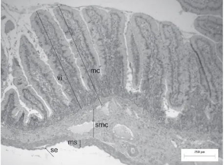
The mucous layer of the small intestine of
D. aurita
heads were found in all segments. The submucous
layer with a connective tissue of mucous showed a lot
of neurons, forming the Meissner or submucous plexus
(Figure 2). The muscular layer also showed neurons
of myenteric plexus (Auerbach plexus) (Figure 3).
This layer was constituted by two layers of smooth
muscles, an inner circular layer and other external
longitudinal layer. The neurons of the myenteric
plexus were located between these two layers.
Regarding the morphometry of the intestinal
tube (Figure 1), the mucous layer had an average
thickness of 508.93 ± 177.69 µm; the average
Figure 2 - Submucous nervous plexus in the small intestine of D. aurita. mci-inner circular
muscular layer (H-E).
Figure 1 - Tube of the small intestine of D. aurita. vi-villus; mc-mucous layer; smc-submucous
height of villi was 395.40 ± 170.59 µm; inner and
external muscular layers had 146.47 ± 78.05 µm;
and the submucous layer 102.03 ± 74.20µm.
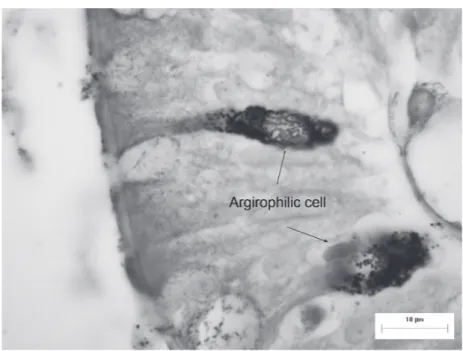
The endocrine cells (Figures 4 and 5) distributed
along the digestive tube of
D. aurita
were located
in the epithelium lining of the mucous and in the
glandular epithelium interspersed with exocrine
cells. They had a varied morphology despite most
them seeming to be oval in shape. Two cell types were
observed, one in which the cells reached the luminal
surface (the open type), and the other presenting no
continuity with the lumen (the closed type).
Figure 3 - Myenteric nervous plexus in the small intestine of D. aurita. mci-inner circular
muscular layer; mle-external longitudinal muscular layer; se-serous (H-E).
Figure 4 - Argirophilic endocrine cells in the mucous layer of the ileum of D. aurita
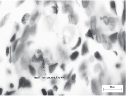
Figure 5 - Immunoreactive endocrine cells to insulin in the mucous layer of the ileum of D. aurita (Imunoperoxidase technique).
1a argyrophillic argentaffin Insulin I R 2b Submucosal ganglia
Myenteric
ganglia Nervous Cells Di 61.11±13.38a 17.63±7.14a 0.86±0.51a
Dm 65.82±16.93a 15.36±7.13a 0.56±0.42a D 5.49±2.60a 5.79±1.42 a 29.36±10.06ab Df 68.02±11.40a 14.81±6.06a 0.72±0.68a
Ji 48.41±7.58ab 10.18±4.26a 0.71±0.65a
Jm 54.71±11.56a 10.78±3.68a 0.49±0.36a J 5.21±2.19a 8.03±3.13 a 25.40±10.54a Jf 40.20±13.40b 7.19±4.43a 0.62±0.67a
Ii 54.11±15.56a 6.92±2.54a 1.46±1.45a
Im 45.56±11.72a 7.61±4.08a 1.34±1.27a I 6.59±3.80a 9.95±5.54 a 35.09±15.54b If 51.36±18.57a 5.98±4.43a 0.96±1.04a
Endocrine and nervous cells per mm2 (average ± standard deviation) and the submucosal and myenteric ganglia (average ± standard deviation) per mm2 of the submucous and muscular layers. (1a: regions) D, J and I represent the duodenum, jejunum and ileum segments, respectively. (2b: segments) Lower case letters i, m and f represent, in this order, the initial, mid and final regions of each segment. The average followed by
the same letter, in the same column, does not differ among themselves by the Kruskal-Wallis test at the significance level of 5% (n = 10).
TABLE I
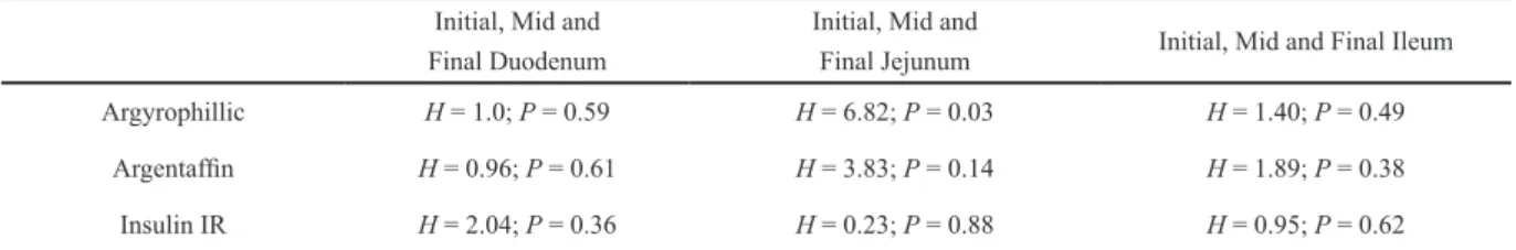
No variation was observed between different
endocrine cells in the initial, mid and final regions
of the segments, except for argyrophillic cells in the
jejunum (Tables I and II). Most of them presented an
oval shape. Open and closed types of endocrine cells
were observed interspersed with exocrine cells.
Nerve cells were observed in groups and also
isolated, both in the submucous layer and between the
muscular layers. The number of submucosal (
H
=
0.54; df = 2;
P
= 0.76) and myenteric (
H
= 5.44; df =
2;
P
= 0.06) ganglia did not vary between all small
intestine segments of
D. aurita
(Table I). The number
of nervous cells varied only between jejunum and
ileum (
H
= 8.70 ; df = 2;
P
= 0.01) (Table I). In average,
17.29 submucosal ganglia/mm
2were encountered in
the submucous, and 23.77 myenteric ganglia/mm
2in the muscular region. The myenteric plexus was
apparently greater than that of the submucous.
There is no relationship between the number
of ganglia and nervous cells, and endocrine cells
in different segments of the small intestine of
D.
aurita
(
P
> 0.05) (Table III).
DISCUSSION
The difference among duodenum, jejunum and
ileum of
D. aurita
was similar to that described for
rodents (Altmann and Leblond 1970, Allogninouwa
et al. 1996), pigs (Mitjans and Ferrer 2004) and
capybaras (Velásquez et al. 2003, Rodrigues et al.
2006). The duodenum was highlighted by the presence
of Brünner glands in the submucous layer. These
glands were not observed in the submucous layer of
the jejunum and ileum. The ileum was identified by
the presence of associated lymph nodes (Peyer dishes)
for all mucous layer, as well by submucous layer.
In descending order, the argyrophillic cells
were more numerous than argentaffin cells, and
argentaffin than insulin immunoreactive cells.
Such results can be explained by the employed
technique since argentaffin reactions appears only
in enterochromaffin cells, while the argyrophillic
reaction occurs in nearly all enteroendocrine
cells, except in cholecystokinin, somatostatin and
insulin-producing cells (Polak et al. 1993). The
Initial, Mid andFinal Duodenum
Initial, Mid and
Final Jejunum Initial, Mid and Final Ileum
Argyrophillic H = 1.0; P = 0.59 H = 6.82; P = 0.03 H = 1.40; P = 0.49
Argentaffin H = 0.96; P = 0.61 H = 3.83; P = 0.14 H = 1.89; P = 0.38
Insulin IR H = 2.04; P = 0.36 H = 0.23; P = 0.88 H = 0.95; P = 0.62
kruskal-Wallis test. gl = 2. The adopted significance level was 5% (n=10).
Argyrophillic Cells Argentaffin Cells Insulin IR Cells
Submucosal ganglia rs = 0.12; P = 0.51 rs = 0.20; P = 0.27 rs = 0.19; P = 0.29
Myenteric ganglia rs = -0.34; P = 0.06 rs = -0.16; P = 0.37 rs = -0.19; P = 0.30
Nervous cells rs = 0.03; P = 0.79 rs = 0.07; P = 0.53 rs = 0.20; P = 0.05
Regression Analysis. The adopted significance level was 5% (n=10).
TABLE III
Relationship between the number of enteroendocrine cells and submucous and myenteric ganglia, and endocrine and nervous cells in the duodenum, jejunum and ileum of adult opossums.
TABLE II
immunohistochemical technique used to stain the
insulin immunoreactive cells is more specific than
those used for other cells. This is due to the fact
that the employment of monoclonal antibodies
increases the sensibility of this method.
In our work, insulin immunoreactive cells
were found insolated in intestinal crypts and villi.
kendzierski et al. (2000) encountered intracellular
immunoreactivity to insulin in glandular cells of
the stomach and the colon of rats, but not in the
small intestine. Contrarily, Coutinho et al. (1984)
observed a positive insulin staining in the brush
border and in some cells isolated in segments near
the small intestine of adult opossums (
Didelphis
albiventris
), although no evidence was observed in
the mid and distal segments. Likewise, Bendayan
and Park (1991) located extrapancreatic islets
between the duodenum crypts and the muscle layer
of the mucosa in rats. These islets were restricted
to the duodenum region and crossed by the final
portion of the bile duct before its opening in the
intestinal lumen. The islets were surrounded by
conjunctive tissue and exhibited no direct contact
with the epithelial cells of the ducts and crypts. Ito
et al. (1988) also encountered a small number of
insulin immunoreactive cells in the pyloric antrum
and duodenum of pigs between 32 and 41 days old.
Pancreatic insulin reduces glucose in blood,
increases the deposit of glycogen in the muscles
and the metabolic use of the glucose, while enteral
insulin controls the intestinal motility (kendzierski
et al. 2000).
Studies with endocrine cells distributed along
the digestive tube of mammals generally refer only
to those located in one of the segment regions,
therefore disconsidering the initial, mid and final
regions of each segment (krause et al. 1989, Takagi
et al. 1990, Fonseca et al. 2002). It was believed
that differences in the number of endocrine cells
can be encountered between regions of the small
intestine segments of
D. aurita
. However, this fact
was not statistically proven in this work.
Ganglia of the submucosal plexus were
concentrated near the muscularis mucosa and
myenteric ganglia located between the circular
internal layer and the longitudinal external layers
together with nerve clusters as related to eutherian
mammals (Young et al. 1993, Brehmer et al. 1994,
Liberti et al. 1994, 1998). The plexuses were
prominent, being the myenteric one apparently
greater than that of submucous, that is, with a
greater population of neuron bodies, similar to those
encountered in the small intestine of the ginea-pig
(Liberti et al. 1994). According to Bressan et al.
(2004), the greater size of the myenteric ganglia
reflects its importance in the intestinal motility
control. The propulsion and mixing movements of
the small intestine include an assembly of neurons
in the plexus. In the small intestine of pigs, the
contraction of a single 10 mm unit includes roughly
6,500 intrinsic primary afferent neurons in the
myenteric ganglion, 1,200 ascending interneurons,
3,000 descending interneurons, 4,000 inhibitory
motor neurons of the circular musculature and 3,000
excitatory motor neurons for the circular muscle
(Clerc et al. 1998, kunze and Furness 1999).
The clusters formed by the neuron bodies
had varying sizes and forms in the two plexuses.
Nerve cells were observed isolated, not grouping
together in both plexuses. Leaming and Cauna
(1961) also encountered isolated nerve cells and
grouped nerve cells in the duodenum and jejunum
of cats, being that the myenteric plexus of the
jejunum contains more nerve cells forming groups
than the myenteric plexus of the duodenum, with
an average of 37 cells/mm
2in the jejunum and
12.17 cells/mm2 in the duodenum.
myenteric neurons than the one of pigs, and
approximately 80 times more than that of mice
(Gabella 1987).
Our data indicate a greater quantity of myenteric
ganglia (23.77 ganglia/mm
2) when compared to
those of submucous (17.29 ganglia/mm
2) in the
small intestine of
D. aurita
. Rodrigues (unpublished
data) also obtained this relation, in which in the small
intestine of capybaras, 2.29 myenteric ganglia were
found in the muscular layer and 0.69 submucosal
ganglia in the submucous layer, both with 1.2 mm
length. There was no difference between the quantity
of myenteric and submucosal ganglia in the segments
in the small intestine of capybaras. However, it
seems that a greater quantity of ganglia was found in
the ileum. According to this author, since the number
of ganglia is more frequent in portions with greater
motility, it is possible that there exists no significant
difference among the duodenum, jejunum and ileum
of capybaras, as well as the capacity of muscle
peristalsis. The myenteric ganglia control peristaltic
movements of the intestine and the submucous, as
well as participate in the innervation of the muscle
layer, regulate the gastrointestinal secretion and
blood flow (Hens et al. 2002).
Neuro-histological studies have been carried
out on the myenteric plexus of the duodenum and
jejunum of adult cats using silver method (Leaming
and Cauna 1961). In this study, these authors found
122 and 30.7 cells/mm
2in the duodenum and
jejunum, respectively. In our work, we found 29.36
and 25.40 cells/mm
2in the duodenum and jejunum,
respectively. These differences probably occurred
in the number of plexuses analyzed. We investigated
two plexuses (submucosal and myenteric), while
Leaming and Cauna (1961) have analyzed only the
myenteric plexus.
Polak et al. (1993) reported some similarities
between endocrine cells and enteric nerve cells,
such as the presence of morphologically similar
secretion granules, the presence of a sensorial
and a transmitter part that liberates granules to the
synaptosomal area and vascular pole. However,
the peptides liberated by the nerve cells do not
integrally meet the criteria for conceptual harmony,
therefore employing the peptide-regulator term. A
large portion of these peptides present a paracrine
signaling effect locally stimulating neighboring
cells, although they can interact with receptors and
transform intracellular signals in other ones similar
to those produced by circulating hormones (Santos
and zucoloto 1996). In the intestine, regulatory
peptides can be encountered in the ganglia cells,
nerve fibers and endocrine cells.
Immunohistochemical and molecular biology
studies have revealed functional interactions
between endocrine and neuronal cells (Delellis and
Dayal 1992). However, the present study did not
demonstrate this relationship in the morphometric
field. We did not identify any relationship between
the number of endocrine cells and submucous
nervous and myenteric ganglia, and between
endocrine and nervous cells of the opossum’s small
intestine. Bressan et al. (2004) who did not find any
relationship between the number of endocrine cells
and submucous and myenteric nervous ganglia of
the caecum of capybara. Various techniques can be
used in the identification of endocrine and ganglion
cells, including histochemical (Masson-Fontana,
Grimelius, Servier-Munger, Hellerstrom-Hellman)
and immunocytochemical utilizing humoral
and non-humoral markers (membrane proteins,
enzymes and acid glycoproteins). Therefore,
other techniques should probably be employed to
confirm the quantitative interrelationship between
the endocrine and nerve cells studied in this work.
ACKNOWLEDGMENTS
The authors would like to thank to many
colleagues who gave important contributions to the
manuscript. The current research was funded by
Conselho Nacional de Desenvolvimento Científico
RESUMO
Os sistemas nervoso e endócrino controlam integra-damente os movimentos intestinais, a secreção de suas glândulas e também participam dos processos de digestão e absorção de nutrientes. Portanto, o objetivo central deste estudo foi verificar a existência de uma possível relação entre o número de células nervosas e gânglios dos plexos submucosos e mioentéricos e o número de células endócrinas no intestino delgado de adultos de D. aurita. As técnicas de coloração utilizadas foram Grimelius, Masson-Fontana modificada, imunoperoxidase direta e H-E. As células endócrinas argirófilas, argentafins e imunorreativas à insulina não variaram numericamente entre as regiões inicial, média e final do duodeno, jejuno e íleo (P>0,05), exceto as células argirófilas no jejuno (P<0,05). Nenhuma relação numérica foi verificada entre o número de gânglios nervosos e células endócrinas, e também entre células nervosas e endócrinas. Nós recomendamos o emprego de novas técnicas imunohistoquímicas para confirmar a correlação numérica entre os sistemas nervoso e endócrino no intestino delgado. A morfologia e a distribuição das células endócrinas e dos gânglios nervosos estudados foram similares àqueles encontrados em mamíferos eutérios.
Palavras-chave: células argirófilas, células argentafins, células endócrinas, gânglios, insulina.
REFERENCES
ALLOGNINOUWA T,AGBAkC,AGOSSOU E AND kPODEkON M.1996. Anatomical, histological and functional speci-ficities of the digestive tract in the male Grasscutter (Thryonomys swinderianus, Temminck 1827). Anat Histol Embryol 25: 15-21.
ALTMANN GG AND LEBLOND CP.1970. Factors influencing villus size in the small intestine of adult rats as revealed by transposition of intestinal segments. Am J Anat 127: 15-36. BANCROFT JD AND STEVENS A.1996. Theory and practice
of histological techniques. New York: Churchill Livingstone. USA, 766 p.
BARBOSA AJA, CASTRO LPF AND NOGUEIRA AMF. 1984. A simple and economical modification of the Masson-Fontana method of staining melanin granules and enterochromaffin cells. Stain Technol 59: 193-196.
BARBOSA AJA, NOGUEIRA JC, PENNA FJ AND POLAk JM. 1987. Distribution of enteroglucagon- and polypeptide YY-immunoreactive cells in the gastrointestinal tract of the white-belly opossum (Didelphis albiventris). J Histoch 88: 37-40.
BENDAYAN M AND PARk I-S.1991. Presence of extrapan-creatic islets of Langerhans in the duodenal wall of the rat. Diabetologia 34: 604-606.
BREHMER A,STACH W AND ADDICkS k.1994. Fine Structural Distinction between Ganglia of the Outer and Inner Submucosal Plexus in Porcine Small Intestine. Acta Anat 151: 188-193.
BRESSAN MS, FONSECA CC, MENIN E AND PAULA TAR. 2004. Identificação e quantificação de gânglios nervosos, células argentafins, argirófilas e imunorreativas à serotonina no ceco de capivara (Hydrochoerus hydro-chaeris). Rev Ceres 51: 29-739.
CHRISTENSEN J.1988. The forms of argyrophilic ganglion cells in the myenteric plexus throughout the gastrointestinal tract of the opossum. J Auton Nerv Syst 24: 251-260. CLERC N,FURNESS JB,LI zS,BORNSTEIN JC AND kUNzE
WAA. 1998. Morphological and immunohistochemical identification of neurons and their targets in the guinea-pig duodenum. Neuroscience 86: 679-694.
COUTINHO HB, SEWEL HF AND COUTINHO VB. 1985. Immunocytochemical demonstration of insulin in the mesonephros and metanephros of the brazilian opossum
Didelphis albiventris. Anat Anz Jena 159: 97-103. COUTINHO HB, SEWEL HF, SMITH DI, COUTINHO VB AND
PINHEIRO PBN.1984. Demonstration of insulin in the pancreas of the Didelphis albiventris (Opossum) by immunocytochemical techniques. Anat Anz Jena 157: 167-175.
DE GIORGIO R,STANGHELLINI V,BARBARA G, CORINALDESI R, DE PONTI F, TONINI M, BASSOTI G AND STERNINI C. 2000. Primary enteric neuropathies underlying gastrointestinal motor dysfunction. Scand J Gastroenterol 35: 114-122.
DELELLIS RA AND DAYAL Y.1992. Neuroendocrine system. In: STERNBERG SS (Ed), Histology for pathologists. New York: Raven Press, USA, p. 347-362.
DEVASkAR SU,SINGH BS,CARNAGHI LR,RAJAkUMAR PA AND GIDDINGS SJ.2002. Insulin II gene expression in rat central nervous system. Regul Pept 48: 55-63.
FONSECA CC, NOGUEIRA JC AND BARBOSA AJA. 2002. Argyrophilic and Glucagon-Immunoreactive cells in the ileum and colon of the developing opossum Didelphis albiventris (Marsupialia). Cells Tissues Organs 170: 20-33. FURNESS JB.2000. Types of neurons in the enteric nervous
system. J Auton Nerv Syst 81: 87-96.
GABELLA G. 1987. The number of neurons in the small intestine of mice, guinea-pigs and sheep. Neuroscience 22: 737-752.
GRIMELIUS L.1968. A silver nitrate for α2 cells in human pancreatic islets. Acta Soc Med Upsal 73: 243-270. GRIMELIUS L AND WILANDER E.1980. Silver stains in the study
HENS J,GAJDA M,SCHEUERMANN DW AND ADRIAENSEN D. 2002. The longitudinal smooth muscle layer of the pig small intestine is innervated by both myenteric and submucous neurons. Histochem Cell Biol 117: 481-492. ITO H,HASHIMOTO Y,kITAGAWA H, kON Y AND kUDO N.
1988. Ontogeny of gastroenteropancreatic (GEP) endocrine cells in mouse and porcine embryos. Jap J Vet Sci 50: 99-110.
kENDzIERSkI kS,PANSkY B, BUDD GC AND SAFFRAN M. 2000. Evidence for biosynthesis of preproinsulin in gut of rat. Endocrine 13: 353-359.
kRAUSE WJ, YAMADA J AND CUTTS H. 1985. Quantitative distribution of enteroendocrine cells in the gastrointestinal tract of adult opossum, Didelphis virginiana. J Anat 140: 591-605.
kRAUSE WJ,YAMADA J AND CUTTS H.1989. Enteroendocrine cells in the developing opossum small intestine and colon. J Anat 162: 83-96.
kUNzE WAA AND FURNESS JB.1999. The enteric nervous system and regulation of intestinal motility. Annu Rev Physiol 61: 117-142.
LEAMING DB AND CAUNA N. 1961. A qualitative and quantitative study of the myenteric plexus of the small intestine of the cat. J Anat 95: 160-168.
LIBERTI EA,GASPAR LP,CARVALHO CAF, FUJIMURA I AND SOUzA RR. 1998. A morpho-quantitative study of the myenteric ganglia throughout the human digestive tract. Rev Hosp Clin Fac Med Univ 53: 55-60.
LIBERTI EA, QUEIROz LM, POPEU E, PERITO MAM, MINARELLI AM,MORAES JOR AND SOUzA RR.1994. A quantitative and comparative study of the ganglionic neurons in the myenteric and submucous plexuses of the small intestine, and in the intramural plexus of the gall bladder of the ginea-pig. Rev Bras Cienc Morfol 11: 106-114.
MEIMARIDIS DG,MORSE DE,PANSkY B AND BUDD GC.2003. Insulin immunoreactivity in the fetal and neonatal rat retina. Neurosci Lett 118: 116-119.
MITJANS M AND FERRER R.2004. Morphometric study of the guinea pig small intestine during development. Microsc Res Techn 63: 206-214.
POLAk JM,BISHOP AE,BARBOSA AJA AND BLOOM SR.1993. Hormônios gastrointestinais. In: DANI R AND CASTRO LP (Eds), Gastroenterologia Clínica. Rio de Janeiro: Guanabara-koogan, Brasil, p. 1446-1465.
RODRIGUES SS,FONSECA CC AND NEVES MTD.2005.Células endócrinas do sistema gastropancreático: conceitos, distribuição, secreções, ação e controle. Arq Cienc Vet zool 8: 171-180.
RODRIGUES SS,FONSECA CC,PAULA TAR AND PEIXOTO JV. 2006. Aspectos biométricos corporais e do intestino delgado da capivara Hydrochoerus hydrochaeris
Linnaeus, 1766 (Mammalia, Rodentia, Hydrochaeridae). Biotemas 19: 79-86.
SANTOS GC AND zUCOLOTO S. 1996. Células endócrinas gastrointestinais: breve histórico e principais métodos de identificação à microscopia óptica. Arq Gastroenterol 33: 36-44.
STAHLER MS, PANSkY B AND BUDD GC. 1988. Immuno cytochemical demonstration of insulin-like immuno-reactivity in the rat prostate gland. Prostate 13: 189-198. STERNBERGER LA.1979. Immunocytochemistry. New York:
J Wiley & Sons, USA, 104 p.
TAFURI WL AND BRENNER z.1967. Lesões dos plexos de Meissner e de Auerbach do intestine de camundongo albino na fase crônica da tripanossomíase cruzi experimental. Rev Inst Med Trop 9: 149-154.
TAkAGI C, YAMADA J, kRAUSE WJ, kITAMURA N AND YAMASHITA T. 1990. An immunohistochemical study of endocrine cells in the proximal duodenum of eight marsupial species. J Anat 168: 49-56.
VELÁSQUEz JC,FONSECA CC, MENIN E AND PAULA TAR. 2003. Estudo histológico do intestino delgado de capivaras adultas (Hydrochoerus hydrochaeris). Arq Ciência Vet zool 6: 21-25.