www.scielo.br/aabc
Differentially displayed expressed sequence tags in
Melipona scutellaris
(Hymenoptera, Apidae, Meliponini) development
FLÁVIA A. SANTANA1, FRANCIS M.F. NUNES2, CARLOS U. VIEIRA1,
MARIA ALICE M.S. MACHADO1, WARWICK E. KERR1, WILSON A. SILVA Jr2 and ANA MARIA BONETTI1
1Universidade Federal de Uberlândia, Instituto de Genética e Bioquímica, Laboratório de Genética, Campus Umuarama, Bloco 2E sala 33, rua Acre s/n, 38400-902 Uberlândia, MG, Brasil 2Departamento de Genética, Faculdade de Medicina de Ribeirão Preto, Universidade de São Paulo,
Av. Bandeirantes 3900, 14040-900 Ribeirão Preto SP, Brasil
Manuscript received on January 31, 2005; accepted for publication on August 31, 2005; contributed byWARWICKE. KERR*
ABSTRACT
We have compared gene expression, using the Differential Display Reverse Transcriptase – Polymerase Chain Reaction (DDRT-PCR) technique, by means of mRNA profile inMelipona scutellarisduring ontogenetic postembryonic development, in adult worker, and in both Natural and Juvenile Hormone III-induced adult queen. Six, out of the nine ESTs described here, presented differentially expressed in the phases L1 or L2, or even in both of them, suggesting that key mechanisms to the development ofMelipona scutellarisare regulated in these stages. The combination HT11G-AP05 revealed in L1 and L2 a product which matches to thioredoxin reductase protein domain in theClostridium sporogenes, an important protein during cellular oxidoreduction processes. This study represents the first molecular evidence of differential gene expression profiles toward a description of the genetic developmental traits in the genusMelipona.
Key words:Melipona scutellaris, gene expression, DDRT-PCR.
INTRODUCTION
Except forApis mellifera, the knowledge about
bio-logical, cellular, and molecular traits in Hymeno-ptera is still meager, although they are an excel-lent model for studying social characteristics, be-havior, neurobiology, development, and gene ex-pression regulation. ForApis mellifera, it is known
that differences in food quality during the larval period determine the caste phenotypes. There are differentially expressed genes, both in queens and workers, which are involved in that mechanism. On
*Member, Academia Basileira de Ciências Correspondence to: Flávia Assumpção Santana E-mail: [email protected]
the other hand, in Melipona, the caste
determina-tion is decisively influenced by a genetic predispo-sition. Diploid larvae will become queens or work-ers; brood cells are operculated soon after the egg is laid, eliminating in this way any possibility of differential feeding (Kerr 1950). Some studies were reported based on phenotypic differences associated with Juvenile Hormone (JH) topical administration. When applied to larvae from genusMelipona, this
hormone promotes the appearance of complete fe-male (queen) features (Campos 1978, Bonetti 1983, 1984, Bonetti et al. 1995). Genes up- or down-regulated in response to JH during the Melipona
The identification of differentially expressed genes has been used to understand the function of genes and the basic molecular mechanisms of dis-tinct biological processes (Vedoy et al. 1999). Dif-ferential display reverse transcriptase – polymerase chain reaction (DDRT-PCR) has innovated the gene expression field (Liang and Pardee 1992). This is an easy, widely employed and reliable PCR-based method used to detect differentially expressed genes in cells, tissues, and organisms (Warthoe et al. 1995, Liu and Raghothamna 1996, Poirier et al. 1997). Using this approach, Hepperle and Hartfelder (2001) identified, in honeybee queens and workers, an Ftz-F1 homolog and a Cut-like transcript, two strong candidates to participate in processes like the meta-morphic ecdysone response cascade regulation and ovariolos degradation, respectively.
In this work, the DDRT-PCR technique was used to study differential gene expression profiles during post-embryonic development of Melipona scutellaris. The differentially expressed cDNA
fragments detected were cloned, sequenced and an-alyzed by comparison using public databases. The results reported here are the first pieces of evidence of differential gene expression profiles inMelipona scutellarisduring its ontogenetic development.
MATERIALS AND METHODS
BIOLOGICALMATERIAL
Stingless bees Melipona scutellaris come from
Bahia, Brazil, and were kept in Meliponário Uberlândia, Uberlândia – MG, Brazil (S 180◦55′ /
W 480◦17′). Larvae of different stages were used:
larva 1 (L1), larva 2 (L2), larva 3 (L3), predefecat-ing (LPD) and defecatpredefecat-ing (LD); pupae from workers (WP) and queens (QP); and adults: newborn work-ers (AW), natural virgin queens (AQ) and JH III (Sigma) -induced virgin queen (AIQ). The samples were collected, put immediately in liquid nitrogen and preserved at−80◦C. Just before RNA extraction
they were allowed to thaw in ice.
RNAEXTRACTION
Total RNA of whole body was extracted using TRI-zol reagent (Invitrogen) following the recommended instructions and was treated with DNase I (Invitro-gen). RNA quality was verified in 1% agarose gel electrophoresis
SCREENING FOR DIFFERENTIAL EXPRESSION BY
DDRT-PCR
Reverse transcription reactions were performed in triplicate using 200 ng of total RNA, 8 pmols of one of the oligo-DT primers containing an anchored base (Table I), 12U RNAsin (Amersham Biosciences), 1x RT buffer, 10 mM DTT, 200 mM dNTPs and 200U RT Superscript II (Invitrogen), brought to a final volume of 20µl. Reverse transcriptase was not
added to negative control, in order to verify possible DNA contamination. PCR amplification was car-ried out according to the following protocol: 1µL
of the RT reaction, 50µM of each dNTP, 1U Taq
DNA polymerase (Invitrogen), 1x buffer, 2.5 mM MgCl2, 8pmol of one of the arbitrary primers (Ta-ble I) in a final volume of 20µL. cDNA profiles were
loaded in 6% denaturing polyacrylamide gels con-taining 8M urea, run at 150V for 13 hours. After the electrophoresis, gels were stained with AgNO3 ac-cording to Bassan et al. (1991). Bands representing differentially expressed genes were cut out of the gels, the cDNA eluted, and precipitated according to Sambrook et al. (1989). A total of 15 purified fragments with the respective primer combinations, under the same PCR conditions were obtained. Puri-fied fragments were cloned into pUC18 (SureClone Ligation kit, Amersham Biosciences) and compe-tentE. coliDH10B-cells were transformed by heat
PCR products on 1% agarose gel electrophoresis. Sequencing reactions were performed in both directions, using 3.2 pmol M13 primer (forward or reverse), 2µL Big Dye and 1µL of PCR product, to
a final volume of 5µL and submitted to ABI
PRISM 377 DNA automatic sequencer (Applied Biosystems).
TABLE I
Sequences of oligo-dT (HT11) and arbitrary (AP, OPA and OPF) primers employed for
DDRT-PCR experiments.
Primers Sequence 5′ →3′
HT11A AAGCTTTTTTTTTTTA HT11C AAGCTTTTTTTTTTTC HT11G AAGCTTTTTTTTTTTG
AP04 AAGCTTCTAACG AP05 AAGCTTAGTAGGC OPA18 AGGTGACCGT OPA17 GACCGCTTGT OPA16 AGCCAGCGAA OPF12 ACGGTACCAG
SEQUENCE ANALYSES
Eletropherograms were analyzed and assembled with Sequencher 3.1 program. Sequences of at least 180 bp after both vector and quality trimming were considered high-quality ESTs (nine of 15 clones). Nine 3′ Expressed Sequence Tags (ESTs) were
de-posited in dbEST database and were compared to GenBank data using NCBI-Blast algorithms (stan-dard parameters) and to Apis mellifera Genome
Project
(http://www.hgsc.bcm.tmc.edu/projects/honeybee/)
version 2.0 (Blast N algorithms). In some cases, the following NCBI analyses tools were used: 1) CDD, to search conserved domains evidences and 2) Entrez Gene, looking for Gene Ontology terms to annotate functional assignments.
RESULTS
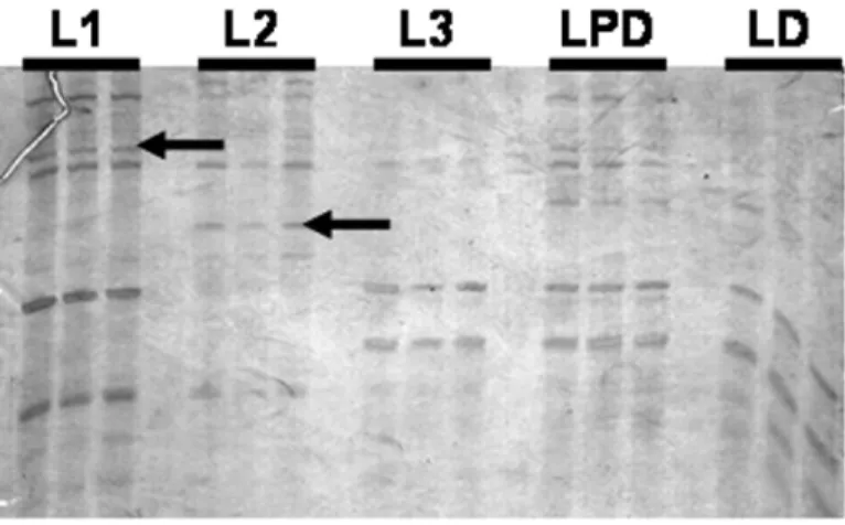
We tested 18 primer combinations, an example of
M. scutellarisexpression profile obtained in a
poly-acrylamide gel is shown in Figure 1. We could verify differences of intensity in fragments during larval stages and also, bands exclusively found only in one of the samples.
Of nine analyzed ESTs (Table II), only 2 se-quences, D01 e B03, had significant results for Blast N-EST of humans. By its turn, results from Blast N-nr for five of our ESTs (D01, B03, D08, C07 and B08) presented high similarity with human se-quences, mainly chromosomal but, only for D01, B03 and B08, the best Blast X match was corre-spondent to the same species.
The possibility of contamination with human material during sample manipulation ofM. scutel-lariswas discarded because the technique was
rig-orously followed and, beyond this, of the only two
M. scutellarisESTs, B03 and B04, that had
similar-ity with the GroupUn.6210 and Group1.45, respec-tively, in theApisGenome Assembly version 2.0, the
B03 (which matched human) showed highly con-served among bees, with e-value = 1e-22. Melipona
sequences that didn’t have conclusive matches with the ones deposited in databases were obtained in this study and they could represent genes not yet described.
Six of the nine ESTs described here were differentially expressed in the phases L1 or L2, or even in both of them, suggesting that key mecha-nisms to the development of M. scutellariscould be
regulated in these stages.
The primer combinations HT11G-AP05 re-vealed differentially displayed product, EST B01, that matched with a thioredoxin-disulphide reduc-tase from Clostridium sporogenes (Blast X best
match result) with e-value equals to 5e-15.
func-Fig. 1 – Profiles of DDRT-PCR in larval stages ofMelipona scutellarisgenerated using HT11A-AP04 primer combination. 6% denaturing polyacrylamide gel, stained with AgNO3. Arrows indicate differentially expressed fragments (performed in
trip-licate) in L1 and L2. The in L1 is respected to EST D01 and in L2 to EST B09. Some bands, not stage-specific, with differences of intensity can also be observed during development. Larvae of different stages: larva 1 (L1), larva 2 (L2), larva 3 (L3), predefecating (LPD) and defecating (LD).
TABLE II
Summary of information about the nineMelipona scutellarisESTs
differen-tially expressed. ESTs B03 and B04 show significant similarity withApis mel-liferagenome version 2.0, using Blast N algorithms. Developmental stages:
L1 (larva 1), L2 (larva 2), LD (defecating larva), AW (newborn workers), AQ (natural virgin queens) and AIQ (JH III-induced virgin queen).
EST GenBank accession Length Developmental identification number (bp) expressed stage
D01 CK722151 256 L1
D08 CK722150 212 L1 and L2
C07 CK722149 690 LD
B03 CK722148 448 L1 and L2
B01 CK722147 204 L1 and L2
B09 CK722146 197 L2
B08 CK722145 469 L1 and L2
B07 CK722144 248 AQ
B04 CK722143 232 AQ, AIQ and AW
tional classification, this sequence had domain iden-tity with the human domain KOG2424 (e-value = 8e-12), which interacts with RNA polymerase II, involved in transcription star site selection.
activity. The EST is similar to CAAX prenyl pro-tease 1, described inDrosophila as CG7573 gene
product, putatively involved in cell surface receptor linked to signal transduction. The EST B08 pre-sented weak similarity to Signaling Lymphocytic Activation Molecule, described in mammals T-cells surface antigen CD2 proteins, involved in cell-cell adhesion and immune response modulation.
DISCUSSION
Relatively slight changes inM. scutellarisgene
ex-pression could be monitored by DDRT-PCR. The data presented here suggest that we detected ESTs that are up- or down-regulated, depending on the de-velopmental stage of this bee. Some ESTs seem to be specifically expressed in one or more stages, sug-gesting that they can correspond to genes involved in stage or caste determination.
InA. mellifera, the differences already reported
(Evans and Wheeler 2001) could reflect, in part, a common impact of regulatory hormones on gene ex-pression in young larvae and in larvae destined to become workers.
We also detected some ESTs which play house-keeping roles, with similar or variant band inten-sity. Evans and Wheeler (1999) found differen-tially expressed genes inA. melliferaworkers and
queens, related to metabolic, nutritional, and sig-naling diverse processes underlying development, potentially involved differentiation processes of fe-male castes and, suggested that distinct morpholog-ical phenotypes are based on differential gene ex-pression rather than genetic polymorphism.
Our data lead us to three arguments. The first one may indicate new sequences that for the first time entered public databases. The second premise indicates that such sequences are specific to those species, regulating biological processes restrict to the development of that bee. The third one can pos-sibly indicate that we are not really dealing with po-tentially coding sequences, because these EST were generated by 3’ strategy using oligodT primer, fa-voring access to non-translated 3’ regions (3’UTRs).
Human genome is, by far, the best character-ized, with the greater number of sequences deposited in GenBank. The hugely available data for this species may account for a bias in the analyses of little known organisms, like M. scutellaris. Even
with genomes of other available insects, such infor-mation seemed not enough for us to do, for example, phylogenetic inferences.
Our data reflect the identification of ESTs that may be lacking in the Apis genome, as well as Anopheles andDrosophila. Previous work withA. melliferashowed a high level of sequence with weak
or no match to the Drosophila genome, predicted
proteins or ESTs sequence, but plausible orthologs were found for proteins from human, mouse, and other non-Arthropoda (Whitfield et al. 2002, Kucharski and Maleszka 2002). In the same way, Nunes et al. (2004) also found a set of sequences more similar to mammals (especially humans) than to other vertebrates or invertebrates, indicating that some genes diverged less between social bees and this group.
Lobo et al. (2003) sequenced 81kb genomic region from honey bee and, all 13 putative genes described have lacked similarity to known inverte-brate or verteinverte-brate data, differences that may show evolution of traits such as social behavior and hap-lodiploidy. In this context, the similarities found betweenMeliponaESTs and non-insect sequences
cannot be considered artifacts; contrarily, they re-flect new biological interpretations and also the need for additional efforts to characterize other genomes. Considering thatApisgenome is not yet
finished, there are still possibilities that such se-quences will soon become more closely shared.
of a system that protects against cytotoxic reactive oxygen species (for review, see Williams Jr 2000). InDrosophila, TrxR acts as an intracellular
antioxi-dant, and mutants for this enzyme have their capac-ity of cell protection against cytotoxic damage re-duced, which results in larvae death. Such mutants have this enzyme reduced; affected pupal eclosion and a diminished life span (Missirlis et al. 2001). A similar enzyme inM. scutellaris may be important
to the early embryonic development when the food is offered only once (massal feeding) to the new-ecloded larvae, implicating in an intensive energetic metabolism.
Drosophila Mef-2 is a direct regulator of
Actin57B transcription in cardiac, skeletal and vis-ceral muscle lineages and also, based on GO terms; it is involved in cell proliferation, transcrip-tion activities and ovarian follicle cell development. Specifically, this last function is in accordance with our findings, because genes that regulate oogene-sis during embryogeneoogene-sis can also perform impor-tant roles related to vitellogenesis and physiological processes ascribed to adult females, associated to reproduction and social behavior of the colonies.
The differentiation that occurs in an animal is driven by “TURN ON” and “TURN OFF” genes and, depending on the active genes in a cell, the difference between one cellular type and the other appears. Developing programs depend on signal-ing and answersignal-ing cascades, which have been kept highly conserved in the animal kingdom.
In order to disclose these developmental func-tions and understand differences and similar-ities among bees, other insects and taxa, we are designing studies using RNA blots, gene-by-gene approach,in situhybridization, real time PCR and
RNAi methods. The analysis proposed in this work shows an overview of genome activity during the development, mainly the larval one. Further ap-proaches are now needed in order to delineate: com-plete Melipona transcriptome, a novel set of
dif-ferentially expressed genes, the relationship among novel genes described here and the genetic hi-erarchy that leads to life cycle regulation.
ACKNOWLEDGMENTS
Maria Cristina Ramos Costa, Caetano Costa, Karine Sá Ferreira for critical reading of the manuscript. This research was supported by a grant from Co-ordenação de Aperfeiçoamento de Pessoal de Nível Superior (CAPES), Conselho Nacional de Desen-volvimento Científico e Tecnológico (CNPq), Fun-dação de Amparo à Pesquisa do Estado de São Paulo (FAPESP) and Fundação Hemocentro de Ribeirão Preto.
RESUMO
Nesse estudo nós usamos a técnica de Differential Dis-play Reverse Transcriptase – Polymerase Chain Reaction (DDRT-PCR) para comparamos o perfil de mRNA em Melipona scutellaris durante o desenvolvimento onto-genético pós-embrionário e em operárias adultas, rainha natural e induzida pelo Hormônio Juvenil III. Fragmen-tos diferencialmente expressos foram detectados usando as seguintes combinações de primers: HT11G-AP05; HT11C-AP05; HT11G-OPF12; HT11G-OPA16. Dos 9 ESTs descrito nesse trabalho, 6 tiveram expressão dife-rencial nas fases de larva L1 e L2, sugerindo serem meca-nismos chave no regulação do desenvolvimento larval em Melipona. A combinação HT11G-AP05 revelou em L1 e L2 um produto com similaridade à proteína tioredoxina redutase deClostridium sporogenes, uma proteína impor-tante durante os processos de oxidoredução. Esse estudo representa as primeiras evidências moleculares do perfil de expressão durante o desenvolvimento ontogenético em abelhas do gêneroMelipona.
Palavras-chave: Melipona scutellaris, expressão gêni-ca, DDRT-PCR.
REFERENCES
BASSANBJ, CAETANO-ANOLESGANDGRESSHOFF
PM. 1991. Fast and sensitive silver staining of DNA in polyacrylamide gels. Anal Biochem 196: 80–83. BONETTI AM. 1983. Action of Juvenile hormone on
gene expression inMelipona(Hymenoptera, Apidae, Meliponinae). Rev Bras Genet 6: 583–585. BONETTIAM. 1984. Efeitos do Hormônio Juvenil no
BONETTI AM, KERRWEANDMATUSITASH. 1995. Effects of Juvenile Hormones I, II and III, in sigle and fractionated dosage in Melipona bees. Rev Bras Biol 55: 113–120.
CAMPOSLAO. 1978. Sex determination in bees. VI.
Effects of juvenile hormone analog in males and females of Melipona quadrifasciata (Apidae). J Kansas Entomol Soc 51: 228–234.
EVANSJDANDWHEELERDE. 1999. Differential gene
expression between developing queens and workers in the honey bee, Apis mellifera. Proc Natl Acad Sci USA 96: 5575–5580.
EVANSJDANDWHEELERDE. 2001. Expression pro-files during honeybee caste determination. Genome Biol 2: RESEARCH0001.
HEPPERLE C AND HARTFELDER K. 2001.
Differen-tially expressed regulatory genes in honey bee caste development. Naturwissenschaften 88: 113–116. KERRWE. 1950. Genetic determination of castes in the
genus Melipona. Genetics 35: 143–152.
KUCHARSKIR ANDMALESZKAR. 2002. Evaluation of differential gene expression during behavioral de-velopment in the honeybee using microarrays and northern blots. Genome Biol 3: RESEARCH0007. LIANG PANDPARDEE AB. 1992. Differntial display
of eukaryotic Messenger RNA by means of the poly-merase chain reaction. Science 257: 967–971. LIU C AND RAGHOTHAMNA KG. 1996. Practical
method for cloning cDNAs generated in mRNA differential display. Biotechniques 20: 576–580. LOBONF, TONLQ, HILLCA, EMOREC, ROMERO
-SEVERSON J, HUNTGJANDCOLLINSFH. 2003.
Genomic analysis in the sting-2 quantitative trait lo-cus for defensive behavior in the honey bee, Apis mellifera. Genome Res 13: 2588–2593.
MISSIRLISF, PHILLIPSJPANDJACKLEH. 2001. Co-operative action of antioxidant defense systems in Drosophila. Cirr Bio 11: 1272–1277.
NUNESFMFET AL. 2004. The use of Open Reading
frame ESTs (ORESTES) for analysis of the honey bee transcriptome. BMC Genomics 5(84): 1–12. POIRIERGM, PYATIJ, WANJSANDERLANDERMG.
1997. Screening differentially expressed cDNA clones obtained by differential display using ampli-fied RNA. Nucleic Acids Res 25: 913–914. SAMBROOKJ, FRITSCHEF ANDMANIATIS T. 1989.
Molecular cloning: a laboratory manual, 583 p. VEDOY CG, BENGTSON MH AND SOGAYAR MC.
1999. Hunting for differentially expressed genes. Braz J Med Biol Res 32: 877–884.
WARTHOE P, BAUER D, ROHDE MAND STRAU SS.
1995. Detection and identification of expressed genes by differential display. In: PCR Primer: A laboratorial manual. Dieffenbach CH, Dveskler GS. Cold Spring Harbor Laboratory Press, New York, USA.
WHITFIELDCW, BANDMR, BONALDOMF, KUMAR
CG, LIU L, PARDINAS JR, ROBERTSON HM,
SOARES MB AND ROBINSON GE. 2002.
Anno-tated expressed sequence tags and cDNA microar-rays for studies of brain and behavior in the honey bee. Genome Res 12: 555–566.