Hunting fo r diffe re ntially
e xpre sse d ge ne s
Instituto de Q uímica, Universidade de São Paulo, São Paulo, SP, Brasil
C.G. Vedoy, M.H. Bengtson and M.C. Sogayar
Abstract
Differentially expressed genes are usually identified by comparing steady-state mRNA concentrations. Several methods have been used for this purpose, including differential hybridization, cDNA subtrac-tion, differential display and, more recently, DNA chips. Subtractive hybridization has significantly improved after the polymerase chain reaction was incorporated into the original method and many new protocols have been established. Recently, the availability of the well-known coding sequences for some organisms has greatly facilitated gene expression analysis using high-density microarrays. Here, we describe some of these modifications and discuss the benefits and drawbacks of the various methods corresponding to the main ad-vances in this field.
Co rre spo nde nce
M.C. Sogayar
Instituto de Q uímica, USP Caixa Postal 26077 05599-970 São Paulo, SP Brasil
Fax: + 55-11-818-3820 E-mail: mcsoga@ quim.iq.usp.br
Presented at the I International Symposium on “Signal Transduction and Gene Expression in Cell Proliferation and Differentiation”, São Paulo, SP, Brasil,
August 31-September 2, 1998.
Research supported by FAPESP, CNPq, PADCT III SBIO -CAPES, ICGEB, FBB and PRP-USP. C.G. Vedoy and M.H. Bengtson are recipients of FAPESP pre-doctoral fellowships.
Received November 26, 1998 Accepted January 15, 1999
Ke y wo rds
·Differential gene expression ·Differential hybridization ·Differential display (DDRT) ·Subtraction hybridization ·Suppressive PCR ·DNA microarrays (chips)
Intro ductio n
The identification of differentially ex-pressed genes has been used as an experi-mental approach to understand not only gene function but also the molecular mechanisms underlying several biological processes. This approach has been used in a wide range of studies including cell cycle control in mam-malian cells (1-4), signal transduction in Dro-sophila (5), and circadian rhythms (6). To fully describe the differential gene expres-sion of a given biological system, it is impor-tant to ensure that most (or all) differentially expressed mRNAs are represented in the cDNA library, i.e., both abundant and rare mRNA transcripts.
Several methods have been used to ana-lyze differential gene expression, namely,
differential hybridization, electronic subtrac-tion (including serial analysis of gene ex-pression; SAGE), differential display reverse transcriptional-polymerase chain reaction (DDRT-PCR), cDNA subtraction and, more recently, DNA chips.
D iffe re ntial hybridizatio n
The general scheme for differential colony hybridization is based on generation of a cDNA library containing the gene sequences of interest. These cDNA clones are trans-ferred to bacterial plates, in an orderly array, and replica plated onto duplicate membrane filters (7). Each filter is then hybridized to two different 32P-labelled cDNA probes,
made from polyA+
pre-pared from any two cell populations that are expected to display differences in gene ex-pression. This strategy was successfully uti-lized to isolate and characterize the first platelet-derived growth factor (PDGF)-regu-lated genes (1), genes expressed during the G0-G1 transition in mouse cells (2), the early genetic response to growth factors in mouse fibroblasts (3) and glucocorticoid-regulated genes in C6/ST1 rat glioma pheno-typic reversion (8). Although the isolation of differentially expressed genes of unknown sequences in several systems was first achieved by differential screening, this pro-cedure is very laborious and time consum-ing. DDRT-PCR and subtractive hybridiza-tion coupled to PCR appeared as less labori-ous, more rational and promising approaches.
D iffe re ntial display
DDRT-PCR relies on randomly primed amplification of a sub-fraction of total mRNA from two cell populations, with the amplicons run side by side on sequencing gels, and with the isolation of cDNA fragments which are expressed at different levels under both con-ditions. Since the introduction of DDRT-PCR in 1992 (9) over 100 reports regarding improvements and/or successful applications have been published. Although DDRT-PCR seems to be technically simple, the road from band on the gel to a positive clone can be treacherous. The primary criticisms are: 1) a high false-positive rate, 2) questioned ability of DDRT-PCR to identify both abun-dant and rare mRNAs, 3) coding regions of mRNAs are usually not cloned, and 4) the verification process is time consuming and usually requires a fair amount of RNA. Meth-odological modifications have since been introduced to streamline the techniques. Major efforts have centered on how to elimi-nate false positives as approached from a variety of angles, ranging from RNA sample preparation, Northern blot confirmation and primer length variation. A detailed review
can be found in Ref. 10.
Subtractive hybridizatio n
There are numerous protocols for sub-tractive hybridization, but the principle re-mains the same. The most common methods have employed subtraction based on synthe-sized cDNAs instead of mRNA. This proce-dure improves the final efficiency because it minimizes RNA degradation that may occur during the hybridization procedure. In gen-eral, cDNAs from the target cells/tissues are hybridized using a vast molar excess of driver cDNA (control cells/tissue) followed by sepa-ration of the double-stranded nucleic acid hybrids from the single-stranded cDNAs (cor-responding to differentially expressed mRNAs) by hydroxyapatite or streptavidin-biotin interaction and, more recently, by sup-pression PCR. The resulting subtracted cDNA is then used either as a labelled probe to screen libraries or for the construction of a subtracted cDNA library.
Separation of single-stranded cDNA by hydroxyapatite has considerable disadvan-tages. In addition to requiring large amounts of mRNA, the unhybridized mRNA, recov-ered after chromatography, is very diluted. Moreover, the chromatographic separation procedure requires a high temperature (60oC)
which presumably increases the probability of mRNA degradation.
A profound modification of cDNA sub-traction was obtained by coupling it to am-plification by PCR to increase the starting material to be subtracted or to select the resulting subtracted products. This method was first applied by Duguid and Dinauer (11) to identify differentially expressed genes in scrapie infection. An interesting modifi-cation of this method (12) utilizes oligo-(dT)30-latex particles and PCR. The fine
oligo-(dT)30-latex within a short reaction period
and cDNA synthesis is carried out using the annealed mRNA as a template. This allows subtractive hybridization to be carried out in an Eppendorf tube and the unhybridized mRNA to be separated by brief centrifuga-tion at low temperature. The resulting mRNA can be enriched by successive hybridization reactions of unhybridized mRNA to the cDNA-oligo-(dT)30-latex in a relatively short
period of time and, subsequently, amplified by PCR after conversion to cDNA. This method has been successfully employed in the isolation of cDNA clones that are specif-ic for undifferentiated human embryonal carcinoma cells (12).
A similar approach utilizing the biotin/ streptavidin affinity to separate subtracted cDNAs requires no RNA isolation and has been applied to cells removed from cryostat tissue sections of different cell populations (13). This was possible because the reverse transcription-PCR (RT-PCR) technique al-lows the use of a very small amount of RNA that is reverse-transcribed to cDNA and am-plified. The procedure involves homopoly-meric A tailing of cDNA synthesized from released RNA using an anchored oligo-dT primer. PCR amplification is then carried out using a biotinylated (X)nT16
primer-adap-tor in the presence of biotin-dATP. This biotinylated driver cDNA is twice hybrid-ized, in 50-fold excess, to heterologous tar-get cDNA made with a non-biotinylated primer. Common driver and excess driver cDNA are magnetically removed following the addition of streptavidin-coated magneto-spheres which bind to biotinylated strands, leaving behind the enriched target popula-tion sequences.
Another important feature added to the subtraction technology arose with the ability to rapidly reduce the number of candidate genes to a few which could be easily charac-terized. Two techniques with this potential have been described, namely DDRT-PCR and RDA (representational difference
anal-ysis), both of which employ PCR to amplify messages to detectable levels, but their mode of operation is fundamentally different.
sequence representation (15,16).
Normalization not only increases the dis-criminating power of differential cloning strategies but also provides access to the functionally important class of poorly ex-pressed sequences. Sequences are generally
normalized by submitting thermally dena-tured cDNAs to a self-reassociation reaction and separating the abundant, re-annealed se-quences from the rare single stranded spe-cies. Additional methods to normalize cDNA libraries have been described (16). Since it is
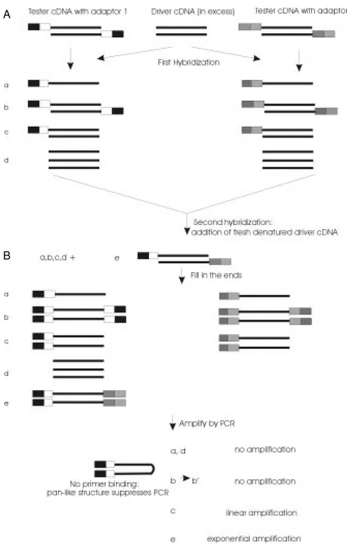
Figure 1 - Schematic diagram of the cDNA subtraction procedure using the PCR suppression ef-fect (18). Boxes represent the outer and inner portions of adap-tors 1 and 2. Solid lines repre-sent the RsaI-digested tester or driver cDNA.
A
well known that physical methods for the separation of single- and double-stranded DNA are both cumbersome and unreliable, novel approaches which use molecular se-lection by magnetic beads have been used to eliminate redundant sequences in the malization procedure (17). However, nor-malization prior to subtraction is only ac-ceptable when target molecules are entirely absent from the driver cDNA population.
Apparently, the best approach is to apply normalization during the subtraction proce-dure, as proposed by Gurskaya et al. (18). This method, illustrated in Figure 1, utilizes the suppression PCR effect (19), allowing the development of a high-efficiency sub-traction procedure that avoids laborious and ineffective physical separation methods (18). This technology uses an adaptor primer which is shorter in length than the adaptor and is capable of hybridizing to the outer primer-binding site. If any PCR products are gener-ated containing the double-stranded adaptor sequences at the both ends, the individual DNA strands will form pan-like structures following every denaturation step due to the presence of inverted terminal repeats. These structures are more stable than the primer-template hybrid and, therefore, will suppress exponential amplification. The use of this technology has allowed the isolation of tran-scripts activated upon induction of Jurkat cells by phytohemagglutinin and phorbol 12-myristate 13-acetate (18) and of glucocorti-coid-regulated genes in C6/ST1 rat gliomas transformed to normal phenotypic reversion (Vedoy CG and Sogayar MC, unpublished results).
D NA micro arrays (D NA chips)
Currently, DNA chips constitute the most promising and revolutionary technique ever developed to study differential gene expres-sion. The basic idea is remarkably simple and elegant consisting of the arrangement of different DNA sequences (ESTs or
deoxyo-ligonucleotides) in an organized array on a small glass surface.
The two mRNA populations that are to be compared are first converted to cDNA, tagged with different fluorochromes (green and red, for example), denatured and then simultaneously hybridized to the immobi-lized DNA samples. Upon hybridization, the so-called DNA chip (glass plate) is scanned at the appropriate wavelengths following excitation of the fluorochromes. Compari-son of the images generated by the two wave-lengths allows the identification of the dif-ferentially expressed sequences.
Due to the very small area occupied by the array, the volume of the hybridization reaction can be reduced, with consequent probe concentration and high sensitivity. According to some authors (20), one in 1.5-3.0 x 10⁵ molecules can be detected.
More-over, the use of glass instead of porous mem-branes significantly reduces the background. Basically, there are 2 kinds of arrays, according to the nature of the DNA: a) cDNA fragments (ESTs) and b) in situ synthesized deoxy-oligonucleotides.
de-creasing its affinity for the DNA to be hy-bridized in solution. Chemically synthesized oligonucleotides can also be robotically spot-ted, as an alternative to cDNA fragments.
The second category comprises 2 differ-ent methods, i.e., photolithography and pi-ezoelectric printing. In the former, photolabile protecting groups are used in oligonucle-otide 3OH terminals. In the first step, the glass slide containing the OH-bearing spacer group is illuminated through a lithographic mask, which allows de-protection of pre-determined regions on the glass slide. Upon losing the photolabile group, the compounds hydroxyl group is free to react with the first type of protected deoxynucleotide. Thus, by successively varying the photolithographic masks, and subsequent reaction of free OH groups with different nucleotides, it is pos-sible to synthesize thousands of different nucleotides of up to 30 mer at known
loca-tions on the glass slide (24). In these arrays, each oligonucleotide has an almost perfect copy physically adjacent to it, differing in only one base. This method, developed by Affymetrics (http://www.affymetrix.com), has the advantage of eliminating the need to deal with thousands of PCR products that have to be purified, quantitated and properly stored. In addition, synthesis can be directed from data bases, and therefore it is possible to direct it so as to differentiate members of the same gene family. This allows the high-est density of oligonucleotides, but the pho-tolithographic masks are very expensive and difficult to generate. In view of the costly process involved, synthesis of these arrays is limited to the industry.
Figure 2 illustrates some approaches used to generate microarrays.
Another method of in situ synthesis is the piezoelectric printing technique utilized by
ink jet printers, in which the printing head moves along a glass surface, spotting drop-lets of one type of deoxynucleotide triphos-phate. Upon reaction, washing and depro-tection, droplets of another type of nucle-otide triphosphate are added, until up to 50-mer oligonucleotides are synthesized (25). Currently, this technique is not as potent as photolithography or microarrays but it is certainly very promising.
DNA chips have been widely used. Ex-pression of cytokines induced by phorbol ester in murine 2D6 helper T cell has been studied by photolithography (26). Signifi-cant induction of gamma interferon and al-terations in IL-3, IL10, granulocyte macro-phage-colony-stimulating factor (GM-CSF) and tumor necrosis factor (TNF)-alpha were found. However, as expected, no alterations were found in the expression levels of house-hold genes like beta actin and GAPDH. Cali-bration experiments pointed to a dynamic range of 1:300,000 to 1:300.
Spotted saccharomyces DNA chips have been used to reveal the genes related to glucose depletion in the anaerobic to aerobic transition (21). At least two-fold induction was found for 710 genes and approximately 2-fold repression was detected for 1,030 genes. In addition, 183 genes were induced 4-fold and 203 genes were repressed 4-fold. Half of the differentially expressed genes had no known function and more than 400 had no apparent homology with known genes. A correlation of 0.87 was found when 2 different microarrays were used and differ-ences between duplicates were lower than a factor of 2 for 95% of the genes (21).
Genes differentially expressed in S. cerevisae growing in minimum versus rich medium were sought using DNA chips gen-erated by photolithography (20). In rich me-dium, 36 RNAs were found to be more abun-dant, 16 of them by a factor of 10. In mini-mum medium, more than 140 RNAs were found to be more abundant, by at least 5-fold. Fifty-seven of the 140 were at least 10
times more abundant. The detection speci-ficity was estimated to be 1:150,000 to 1:300,000. Hybridization of the same RNA with 2 different microarrays resulted in more than a 2-fold difference in 14 of a total of 6,200. In 2 independent experiments, 74 RNAs showed differences greater than 2-fold and 6 (less than 0.1% of the total) showed differences of at least 3-fold.
Some companies are concentrating on achieving DNA chips containing ESTs cor-responding to all genes expressed by a given organism. This would allow identification of genes that are expressed in different cell types, physiological conditions and/or treat-ment conditions, in this organism. DNA chips with up to 40,000 ESTs are already avail-able.
Co ncluding re marks
In the few instances in which the genome coding sequences are well known, the search for differentially expressed genes is greatly facilitated. However, in spite of the efforts put into several genome projects, the ge-nomes of most organisms have yet to be elucidated.
One major approach to gaining insight into the differentially expressed sequences is to construct cDNA libraries using differ-ential hybridization or cDNA subtraction. The quality of these libraries has significant-ly improved with the introduction of cDNA fractionation and normalization by Soares and colleagues (15,16). These authors have generated a set of normalized cDNA librar-ies with improved representation of larger (full length) cDNAs that have been widely distributed for sequencing and mapping, con-stituting the integrated molecular analysis of genomes and their expression (IMAGE) con-sortium (27).
the inefficient process of examining all cDNAs/mRNAs expressed in order to find those that change each time a new compari-son is desired. At any rate, both DNA chips and other methods (differential hybridiza-tion, cDNA subrachybridiza-tion, DDRT-PCR) involve
confirmation and functional characterization of isolated sequences. Although still some-what conceptual, DNA chips should provide a more versatile tool to understand the alter-ations of gene expression and the molecular basis of several diseases.
Re fe re nce s
1. Cochran BH, Reffel AC & Stiles CD (1983). M olecular cloning of gene sequences regulated by platelet-derived grow th fac-tor. Cell, 33: 939-947.
2. Lau LF & Nathans D (1985). Identification of a set of genes expressed during G0/G1 transition of cultured mouse cells. EM BO Journal, 4: 3145-3151.
3. Almendral JM , Somer D, M acDonald-Bravo H, Burckhardt J, Perera J & acDonald-Bravo R (1988). Complexity of the early genetic response to grow th factors in mouse fi-broblasts. M olecular and Cellular Biology, 8: 2140-2148.
4. el-Deiry WS (1993). WAF1, a potential me-diator of p53 tumor suppressor. Cell, 75: 817-825.
5. Smith DP, Shieh BH & Zuker CS (1990). Isolation and structure of an arrestin gene from Drosophila. Proceedings of the Na-tional Academy of Sciences, USA, 87: 1003-1007.
6. Lorus JJ, Denome SA & Dunlap JC (1989). M olecular cloning of genes under control of circadian clock in Neurospora. Science, 243: 385-388.
7. Cochran BH, Zumstein P, Zullo J, Rollins B, M ercola M & Stiles CD (1987). Differ-ential colony hybridization: M olecular clon-ing from a zero data base. M ethods in Enzymology, 147: 64-85.
8. Valentini SR & Armelin M CS (1996). Clon-ing of glucocorticoid-regulated genes in C6/ST1 rat glioma phenotypic reversion. Journal of Endocrinology, 147: 11-17. 9. Liang P & Pardee AB (1992). Differential
display of eukaryotic messenger RNA by means of the polymerase chain reaction. Science, 257: 967-971.
10. Liang P & Pardee AB (1995). Recent ad-vances in differential display. Current Opinion in Immunology, 7: 274-280. 11. Duguid JR & Dinauer M C (1990). Library
subtraction of in vitro cDNA libraries to identify differentially expressed genes in
scrapie infection. Nucleic Acids Research, 18: 2789-2792.
12. Hara E, Kato T, Nakada S, Sekiya S & Oda K (1991). Subtractive cDNA cloning using oligo(dT)30-latex and PCR: isolation of cDNA clones specific to undifferentiated human embryonal carcinoma cells. Nucle-ic Acids Research,19: 7097-7104. 13. Luqmani YA & Lymboura M (1994).
Sub-traction hybridization cloning of RNA am-plified from different cell populations microdissected from cryostat tissue sec-tions. Analytical Biochemistry, 222: 102-109.
14. Hubank M & Schatz DG (1994). Identifica-tion differences in mRNA expression by representational difference analysis of cDNA. Nucleic Acids Research, 22: 5640-5648.
15. Soares M B, Bonaldo M F, Jelene P, Su L, Law ton L & Efstratiadis A (1994). Con-struction and characterization of a normal-ized cDNA library. Proceedings of the Na-tional Academy of Sciences, USA, 91: 9228-9232.
16. Bonaldo M F, Lennon G & Soares M B (1996). Normalization and subtraction: tw o approaches to facilitate gene discov-ery. Genome Research, 6: 791-806. 17. Coche T & Dew ez M (1994). Reducing
bias in cDNA sequence representation by molecular selection. Nucleic Acids Re-search, 22: 4545-4546.
18. Gurskaya NG, Diatchenko L, Chenchik A, Siebert PD, Khaspekov GL, Lukyanov KA, Vagner LL, Ermolaeva OD, Lukyanov SA & Sverdlov E (1996). Equalizing cDNA sub-traction based on selective suppression of polymerase chain reaction: cloning of Jurkat cell transcripts induced by phyto-hemagglutinin and phorbol 12-myristate 13-acetate. Analytical Biochemistry, 240: 90-97.
19. Siebert PD, Chenchik A, Kellogg DE, Lukyanov KA & Lukyanov SA (1995). An
improved PCR method for w alking in uncloned genomic DNA. Nucleic Acids Research, 23: 1087-1088.
20. Wodicka L, Dong H, M ittmann M , Ho M & Lockhart DJ (1997). Genome-w ide expres-sion monitoring in Saccharomyces cerevi-siae. Nature Biotechnology, 15: 1359-1367.
21. DeRisi JL, Iyer VR & Brow n PO (1997). Exploring the metabolic and genetic con-trol of gene expression on a genomic scale. Science, 278: 680-686.
22. Schena M , Shalon D, Davis RW & Brow n PO (1995). Quantitative monitoring of gene expression patterns w ith a comple-mentary DNA microarray. Science, 270: 467-470.
23. Shalon D, Smith SJ & Brow n PO (1996). A DNA microarray system for analyzing complex DNA samples using tw o-color fluorescent probe hybridization. Genome Research, 6: 639-645.
24. Pease AC, Solas D, Sullivan EJ, Cronin M T, Holmes CP & Fodor SPA (1994). Light-generated oligonucleotide arrays for rapid DNA sequence analysis. Proceed-ings of the National Academy of Sciences, USA, 91: 5022-5026.
25. M arshal A & Hodgson J (1998). DNA chips: an array of possibilities. Nature Bio-technology, 16: 27-31.
26. Lockhart DJ, Dong H, Byrne M C, Follettie M T, Gallo M V, Chee M S, M ittmann M , Wang C, Kobayashi M , Horton H & Brow n EL (1996). Expression monitoring by hy-bridization to high-density oligonucleotide arrays. Nature Biotechnology, 14: 1675-1680.