Assessment of
Duguetia furfuracea
genotoxic and
cytotoxic activity in bacteria and mice
CAROLINA R. SILVA1, PABLINE M. VIEIRA1, SUZANA C. SANTOS2 and LEE CHEN-CHEN1 1Departamento de Biologia Geral, Instituto de Ciências Biológicas, Universidade Federal de Goiás,
Caixa Postal 131, Campus-II, 74001-970 Goiânia, GO, Brasil
2Instituto de Química, Universidade Federal de Goiás, Caixa Postal 131, Campus-II, 74001-970 Goiânia, GO, Brasil
Manuscript received on January 21, 2011; accepted for publication on June 6, 2011
ABSTRACT
Duguetia furfuracea (St. Hil.) Benth & Hook f. (1862), popularly known as “sofre-do-rim-quem-quer” and “araticum-seco”, is a shrub of the Annonaceae family that occurs in several regions of Brazil. In folk medicine the infusion of its leaves and twigs is used to treat rheumatism and renal colic, whereas the seed powder is mixed with water to treat pediculosis. Previous studies have described biological activities of this plant with cytotoxic, antitumoral, trypanocidal, leishmanicidal, antiplasmodial and antiprotozoal effects. In the present work, genotoxicity and cytotoxicity of Duguetia furfuracea lyophilized leaf extract were evaluated using the prophage λ induction test (SOS-Inductest) and mouse bone marrow micronucleus test. Our results showed that Duguetia furfuracea lyophilized leaf extract did not present an increase either in the induction of prophage λ (P>0.05) using the SOS-inductest or in the micronucleated polychromatic erythrocytes (P>0.05) using the micronucleus test, suggesting absence of genotoxicity in both tests. On the other hand, a significant decrease in the number of bacteria, (P<0.05), as well as a significant decrease in the polychromatic erythrocytes and normochromatic erythrocytes ratio, (P<0.05), were observed, showing the cytotoxic action of Duguetia furfuracea lyophilized leaf extract. Thus, Duguetia furfuracea did not present the genotoxic action, but showed a cytotoxic effect in both assays utilized in the present work.
Key words: cytotoxicity, Duguetia furfuracea, genotoxicity, micronucleus test, SOS-Inductest.
Correspondence to: Lee Chen-Chen E-mail: [email protected]
natural antioxidants, such as tocopherols, vitamin C,
carotenoids, and phenolic compounds, which are
responsible for human health maintenance, reduction
of oxidative damage, and protection against coronary
heart diseases and cancer (Kilani et al. 2008).
Despite the increasing research on flora, only a
small percentage of about 250,000 species of higher
plants has been chemically and pharmacologically
investigated (Ruffa et al. 2002), and data on the
mutagenic properties of plants commonly used
INTRODUCTION
In the last few decades, many health claims have
been made for compounds derived from vegetables,
fruits and plants (Veiga Junior et al. 2005). The
active substances of the natural products are used as
remedies for the treatment of many diseases (Pimenta
and Nepomuceno 2005). A large number of plants
have been screened as viable sources of a variety of
in folk medicine are limited (Ruiz et al. 1996).
Green plants in general contain mutagenic and
carcinogenic substances (Pimenta and Nepomuceno
2005, Sandermann Junior 1988, Velemi’nsky’
and Gichner 1988, Kanaya et al. 1992, Plewa and
Wagner 1993), but there is little information on
the biological effects of the compounds present
in these plant species (Pimenta and Nepomuceno
2005, Basaran et al. 1996). Therefore it is of vital
importance to evaluate the genotoxic and cytotoxic
effects of phytotherapeutic substances since their
usage has major impacts on the health of a large
number of people (Marques et al. 2003).
Duguetia furfuracea (D. furfuracea) (St. Hil.)
Benth & Hook f. (1862), popularly known as
“sofre-do-rim-quem-quer” and “araticum-seco”, is a shrub of
the Annonaceae family that occurs in several regions in
Brazil (Carollo et al. 2006a, Rodrigues and Carvalho
2001). In folk medicine, the infusion of leaves
and twigs of this species is used in the treatment of
rheumatism and mainly in the treatment of renal colic
(Rodrigues and Carvalho 2001). D. furfuracea seed
powder is also used to treat pediculosis
(Silberbauer-Gottsberger 1981/82). Alkaloids and flavonoids
have already been isolated from the aerial parts of
D. furfuracea
and identified (Carollo et al. 2006a).
Furthermore, many biological activities of this plant
species were reported, such as toxicity in mice embryo
(Toledo et al. 2006), trypanocidal, antiplasmodial, and
antiprotozoal properties (Carollo et al. 2006a, Fischer
et al. 2004, Mesquita et al. 2007).
Due to many biological activities presented by
this plant, as well as to its widespread use in folk
medicine by Brazilian people, the aim of this work
was to assess the genotoxic and cytotoxic activities
of
D. furfuracea lyophilized leaf extract (DFE)
using the lysogenic induction test (SOS-Inductest)
(Moreau et al. 1976) and the mouse bone marrow
micronucleus assay (Heddle 1973).
A combination of tests is generally applied to
investigate the effect on the main types of DNA
damage. The SOS-Inductest is a short term assay
performed with Escherichia coli lysogenic strains
allowing the quantitative evaluation of one SOS
function. When bacteria are exposed to damaging DNA
agents leading to the inhibition of DNA replication,
this causes prophage to enter the lytic cycle as an
expression of SOS induction (Moreau 1981).
The
in vivo mouse bone marrow micronucleus
test is a mutagenicity test system for the detection
of agents that induce chromosome fragments
(clastogenic effect) and/or aneuploidy (aneugenic
effect) (Kirsch-Volders et al. 1997). Therefore, data
obtained with both assays (one prokaryotic and other
eukaryotic) in the present study represent a step further
to the appropriate evaluation on the genotoxicity and
cytotoxicity of D. furfuracea extract.
MATERIALS AND METHODS
PLANT MATERIAL
Samples of D. furfuracea leaves were collected in
the district of Itanhangá, municipality of Goiânia, in
the state of Goiás, Brazil. The plant was identified by
Prof. Heleno Dias Ferreira, and a voucher specimen
(no. 29975) was deposited in the Central Herbarium
of the Universidade Federal de Goiás, in Goiânia,
GO, Brazil. The leaves of this plant were dried at
40◦C in a forced ventilation stove and ground in a
fraction mill to a dry powder that was submitted to
the hot aqueous extraction process (85 g/1000 mL)
and later to lyophilization. The lyophilized extract
was stored at –18°C until further use. Tests were
done with the total lyophilized extract dissolved in
water just before use.
INDUCTEST
Strains
The SOS-Inductest tester strains WP2s(λ) (lysogenic
the Instituto de Biofísica Carlos Chagas Filho,
Universidade Federal do Rio de Janeiro, Rio de
Janeiro, RJ, Brazil.
SOS-Inductest – prophage λ induction
The experiments were performed according to
Moreau et al. (1976). The lysogenic strain
Escherichia coli
WP2s(λ), which contains a
mutation in the gene uvrA (Fonseca et al. 1994),
was cultured on LB medium (1% bacto tryptone, lot
no. 9117660, Difco, Sparks, USA; 0.5% bacto yeast
extract, lot no. 1551325, Biobras, Montes Claros,
Brazil; 1% NaCl, lot no. 87642, Vetec Química Fina
Ltda, Duque de Caxias, Rio de Janeiro, Brazil) up
to the exponential phase of growth. At this point,
15 mL of the culture were centrifuged at 3000× g for
15 min and resuspended on an equal volume of M9
buffer (0.6% Na
2HPO4
, lot no. 19117, Dinâmica
Química Contemporânea, São Paulo, Brazil; 0.3%
KH
2PO4, lot no. 1644, Cinética Química Ltda.,
São Paulo, Brazil; 0.5% NaCl, lot no. 87642,
Vetec Química Fina Ltda, Duque de Caxias, Rio de
Janeiro, Brazil; 1 mL 1M MgSO4
, lot no. 871243,
Vetec Química Fina Ltda, Duque de Caxias, Rio de
Janeiro, Brazil). Following this, 1 mL aliquots of
the bacterial culture were incubated with different
doses of DFE (1, 2, 5 and 10 mg/ 0.1 mL –
stock-solution 100 mg/mL), the negative control (100
μ
L
sterile distilled water), and the positive control
[0.5
μ
g mitomycin C (C
15H
18N
4O5), MMC, lot
no. 237AEL, Bristol, Mayers Squibb, São Paulo,
Brazil], for 25 min at 37°C, after what they were
diluted in M9 buffer to undergo the assays. In order
to assess the cytotoxicity of this plant, 0.1 mL of
the dilutions in M9 buffer was inoculated into
LB plates and incubated for 24 h at 37°C. After
this period, the total number of colonies was
counted. To evaluate D. furfuracea genotoxicity,
0.1 mL lysogenic strain E. coli
WP2s(λ) diluted
in M9 buffer was added to 0.3 mL RJF013 culture
(indicator strain) and 2.5 mL top agar (0.6
% agar,
lot no. 5294419, Difco, Sparks, USA; 0.5% NaCl,
lot no. 87642, Vetec, Duque de Caxias, Brazil). This
mixture was poured into LB
(1/2)(malt/amp)plates and
incubated for 24
h at 37°C, after what the number of
plaques was counted.
MOUSE BONE MARROW MICRONUCLEUS TEST
Animals
This study was approved by the Human and Animal
Research Ethics Committee of the Universidade
Federal de Goiás (CEPMHA/HC/UFG no. 044/09).
Healthy, young male adult (8-12 weeks) outbred
mice (Mus musculus, Swiss Webster), weighing
25-30
g and obtained from the Central Animal House of
the Universidade Federal de Goiás were used in the
study. All animals were brought to the laboratory
five days before the experiments and housed in
plastic cages (40 cm × 30 cm × 16 cm) at 24 ± 2°C
and 55 ± 10% of humidity, with a light-dark natural
cycle of 12 h. Food (appropriate commercial rodent
diet Labina, Ecibra Ltda., Santo Amaro, Brazil) and
water were given ad libitum.
Experimental procedure
The experiments were performed according to
Von Ledebur and Schmid (1973). Doses of DFE
(100, 200, and 300 mg/kg body weight) were
orally administered to groups of five animals for
each treatment. A positive control group (4 mg/
kg i.p. MMC, lot no. 237AEL, Bristol, Mayers
Squibb, São Paulo, Brazil) and a negative control
group (sterile distilled water) were also included.
The animals were euthanized 24 or 48 h after the
administration of DFE by cervical dislocation, and
their bone marrow cells were flushed from both
and fixed with absolute methanol (CH
4O, lot no.55026, Synth, Diadema, Brazil) for 5 min at room
temperature. The smears were stained with Giemsa
(lot no. 1081, Doles, Goiânia, Brazil), dibasic sodium
phosphate (Na
2HPO4
12H
2O, lot no. 982162, Vetec,Duque de Caxias, Brazil) and monobasic sodium
phosphate (NaH
2PO4H
2O, lot no. 983831, Vetec,Duque de Caxias, Brazil) to detect micronucleated
polychromatic erythrocytes (MNPCE). For each
animal, three slides were prepared and a minimum
of 2,000 polychromatic erythrocytes (PCE) were
counted to determine the frequency of MNPCE. In
order to assess DFE cytotoxic activity, we determined
the polychromatic erythrocytes (PCE) and
normochromatic erythrocytes (NCE) ratio (PCE/
NCE). The slides were analyzed by microscopy
(Olympus BH-2 10 × 100, Tokyo, Japan).
STATISTICAL ANALYSIS
The results of the survival and genotoxicity
assays by SOS inductest were generated by four
independent experiments carried out in duplicates.
All the data obtained from the test plates and the
negative control plates (cytotoxicity assay), as well
as the number of plaques obtained in the test plates
and the number of plaques recorded in the negative
control plates (genotoxicity assay) were expressed
as mean (m) ± standard deviation (sd). After this,
they were evaluated using ANOVA tests and
posthoc Tukey to compare the differences among
the means (Vieira 2004), considering the results
significant when P<0.05.
In order to analyze the genotoxic activity of
DFE using the mouse bone marrow micronucleus
test, the frequencies of MNPCE of the treated
groups were compared with the results obtained
for the negative control groups using one-way
ANOVA. P values lower than 0.05 (P<0.05) were
considered indicative of significance. To assess
its cytotoxicity, the PCE/NCE ratio obtained at
different concentrations of DFE was compared with
the negative controls by the qui-square test (χ
2). P
values lower than 0.05 (P<0.05) were considered
indicative of significance.
RESULTS
SOS-INDUCTEST
The results of DFE cytotoxicity and genotoxicity
are presented in Table I.
Assessing the cytotoxicity of DFE, it was
possible to observe a small increase in the number
of survivors at the dose of 1 mg compared with the
negative control group, although not presenting
a significant difference (P>0.05). At doses 2, 5,
and 10 mg of DFE, we detected a decrease in the
number of survivors compared with the negative
control, which showed a significant difference
(P<0.05) only at the doses of 5 and 10 mg.
In the assessment of DFE genotoxicity, we
noted that the dose of 1 mg increased the induction
of prophage λ compared with the negative control
group, but not presenting significant difference
(P>0.05). Applying 2, 5 and 10 mg of DFE, we
observed a small decrease in the induction of
prophage λ compared with the negative control
group, with no significant difference (P>0.05). The
small induction observed at the 1 mg dose of DFE
may be related to the increased number of survivors
at this dose (Table I).
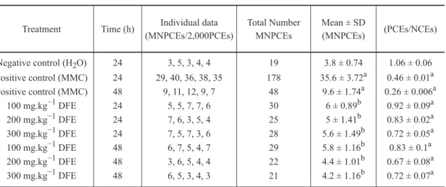
MOUSE BONE MARROW MICRONUCLEUS TEST
MNPCE frequencies and PCE/NCE ratio at 24 and
48 h at different doses of DFE are shown in Table II.
The results for all tested doses (100, 200,
and 300 mg/kg) do not indicate any significant
The PCE/NCE ratio can be an indicator of
cytotoxicity. In this work, all tested doses (100,
200, and 300 mg/kg) at both times of treatment (24
and 48 h) caused a significant reduction of PCE/
NCE ratio compared with the negative control
group at 24 and 48 h (P<0.05). We observed that,
24 h after the treatment with 100 mg of DFE, the
reduction of PCE/NCE ratio was small, although
statistically significant (P<0.05), demonstrating
moderate cytotoxicity of this plant extract.
DISCUSSION
The aim of this study was to evaluate the cytotoxic
and genotoxic potential of DFE using the
SOS-Inductest and mouse bone marrow micronucleus
test. The SOS-Inductest identifies agents capable of
producing DNA lesions, which block its replication
and they detects the induction of the lytic cycle of
prophage λ, which is one of the manifestations of
SOS functions (Moreau et al. 1976). On the other
hand, the bacterial survival assay is useful and
necessary to define basal cytotoxicity, allowing the
observation of the intrinsic ability of a compound
to cause cellular death as a consequence of damage
to basic cellular functions (Einsenbrand et al. 2002).
The mouse bone marrow micronucleus test is a
short-term assay widely employed to detect the genotoxic
(clastogenic and/or aneugenic) and cytotoxic effects
of extracts of other plants (Vilar et al. 2008).
Non-significant increases in λ prophage
induction (P>0.05) in the SOS-Inductest or in
MNPCE (P>0.05) in the micronucleus test were
seen to occur, therefore suggesting that DFE lack
genotoxic effect under the present testing conditions.
In contrast, we observed a significant decrease
in the number of survivors at the doses of 5 and
10 mg extract (P<0.05) by the SOS-Inductest, as
well as a decrease in the PCE/NCE ratio using
the mouse bone marrow micronucleus test, which
shows the cytotoxic action of DFE. This finding is
in accordance with the results already demonstrated
for extracts made from this plant species in mouse
embryos (Toledo et al. 2006).
It has been already known that cytotoxic agents
can induce a great variety of events in the cell: DNA
or protein damage, inhibition of growth, apoptosis,
etc. The nature of the interaction between a drug
and its target is of critical importance in determining
the cell fate. Many cytotoxic drugs employed in
chemotherapy, such as MMC and cyclophosphamide
(CP), have their mechanism of action based on this
genotoxic/mutagenic potential activity (Hickman
et al. 1992, Estream and Vanleeuwen 2000, Kraut
and Drnovsek-Olup 1996). However, the cytotoxic
action presented in this work suggests that this
activity did not result from genotoxic mechanisms.
Possibly the toxic effects caused by exposition
to DFE override any genotoxic and mutagenic
effects. This fact suggests that DFE is not a
pro-carcinogenic agent. Thus, this compound may
cause degenerative pathways. Similar results were
observed in the evaluation of the mutagenic and
cytotoxic activities of ethanolic extracts of araticum
(Annona crassiflora), which also had used the mice
bone marrow micronucleus test (Vilar et al. 2008).
The phytochemical analysis of the aerial parts of
D. furfuracea showed the presence of sesquiterpenes
(Carollo et al. 2005), flavonoids, and several
alkaloids (Carollo et al. 2006a, b). Moreover, recent
studies demonstrated that the alkaloid extract and five
TABLE I
Survival fraction (SF) and standard deviation (SD) of number of colonies of
E. coli WP2s(λ), infective centers (IC) and standard deviation (SD) in RJF013
E. coli cultures formed after treatment with different doses of D. furfuracea
TABLE II
MNPCE frequencies and PCE/NCE ratio after treatment of the animals using different doses
of Duguetia furfuracea lyophilized leaf extract and time.
Treatment
Negative control (H2O) Positive control (MMC) Positive control (MMC)
100 mg.kg−1 DFE
100 mg.kg−1 DFE 200 mg.kg−1 DFE
200 mg.kg−1 DFE 300 mg.kg−1 DFE
300 mg.kg−1 DFE
All results were compared to their respective negative control. aSignificant difference compared
to the negative control (P<0.05). bNo significant difference compared to the negative control
(P>0.05). DFE, D. furfuracea lyophilized leaf extract; MMC, mitomycin C.
Time (h)
24
24 48
48 48 48 24
24 24
19
30 48
29 22 21 178
25 28
3.8 ± 0.74
6 ± 0.89b 9.6 ± 1.74a
5.8 ± 1.16b 4.4 ± 1.01b 4.2 ± 1.16b 35.6 ± 3.72a
5 ± 1.41b 5.6 ± 1.49b
1.06 ± 0.06
0.92 ± 0.09a 0.26 ± 0.006a
0.83 ± 0.1a 0.67 ± 0.08a 0.72 ± 0.07a 0.46 ± 0.01a
0.83 ± 0.02a 0.72 ± 0.05a 3, 5, 3, 4, 4
5, 5, 7, 7, 6 9, 11, 12, 9, 7
6, 7, 5, 4, 7 3, 6, 5, 4, 4 6, 5, 3, 4, 3 29, 40, 36, 38, 35
7, 6, 3, 5, 4 7, 5, 7, 3, 6 Individual data (MNPCEs/2,000PCEs)
Total Number MNPCEs
Mean ± SD
(MNPCEs) (PCEs/NCEs) Treatment SF ± SD(survival) IC ± SD(induction)
Negative control (H2O) 1 mg DFE 2 mg DFE 5 mg DFE 10 mg DFE Positive control (MMC)
1.94 × 10⁸ ± 3.6 × 10⁷ 2.1 × 10⁸ ± 2.5 × 10⁷b 1.6 × 10⁸ ± 6 × 10⁷b 1.02 × 10⁸ ± 1.2 × 10⁷a
8.1 × 10⁷ ± 1.9 × 10⁷a 2.18 × 10⁶ ± 1.67 × 10⁶a
4.13 × 10⁷ ± 1.47 × 10⁷ 9.7 × 10⁷ ± 4.3 × 10⁷b 3.48 × 10⁷ ± 1.39 × 10⁷b 4.47 × 10⁷ ± 2.76 × 10⁷b 2.09 × 10⁷ ± 5.8 × 10⁶b
9.7 × 10⁸ ± 4 × 10⁷a
All results were compared to their respective negative control group. aSignificant difference compared to the negative control group
(P<0.05). bNo significant difference compared to the negative control group (P>0.05). MNPCE, micronucleated polychromatic erythrocyte;
In summary, D. furfuracea lyophilized leaf
extract presented cytotoxic activity, but did not
present genotoxic activity under the experimental
conditions followed in this study.
ACKNOWLEDGMENTS
We are thankful to all the sponsors of this research
project: Fundação de Amparo à Pesquisa do
Estado de Goiás (FAPEG), Fundação de Apoio
à Pesquisa (FUNAPE), Conselho Nacional de
Desenvolvimento Científico e Tecnológico (CNPq),
and Universidade Federal de Goiás (UFG).
RESUMO
Duguetia furfuracea (St. Hil.) Benth & Hook f.
(1862), popularmente conhecida como
“sofre-do-rim-quem-quer” e “araticum-seco”, é um arbusto
da família Annonaceae que ocorre em várias
regiões do Brasil. Na medicina popular a infusão
de suas folhas e galhos é usada no tratamento de
reumatismo e cólica renal, enquanto que o pó da
semente é misturado com água para o tratamento
de pediculose. Estudos anteriores têm descrito
atividades biológicas desta planta com ação
citotóxica, antitumoral, tripanomicida, leishmanicida,
antiplasmódica e antiprotozoárica. No presente
trabalho, a genotoxicidade e citotoxicidade do
extrato liofilizado de folhas de
Duguetia furfuracea
foram avaliadas pelo teste de indução profago λ
(Induteste-SOS) e pelo teste do micronúcleo em
medula óssea de camundongos. Nossos resultados
mostraram que o extrato liofilizado de folhas
Duguetia furfuracea não apresentou um aumento na
indução do profago λ (P>0,05) no Induteste-SOS e
também não foi observado aumento do número de
eritrócitos policromáticos micronucleados (P>0,05),
no Teste do Micronúcleo, sugerindo ausência de
genotoxicidade em ambos os testes. Por outro lado,
uma diminuição significativa no número de bactérias,
(P<0,05), bem como uma diminuição significativa na
relação entre eritrócitos policromáticos e eritrócitos
normocromáticos, (P<0,05), foram observadas,
mostrando ação citotóxica do extrato liofilizado de
folhas de Duguetia furfuracea. Assim, Duguetia
furfuracea não apresentou ação genotóxica, mas
mostrou um efeito citotóxico em ambos os testes
utilizados no presente trabalho.
Palavras-chave: citotoxicidade, Duguetia
furfuracea, genotoxicidade, teste do micronúcleo,
Induteste-SOS.
REFERENCES
BASARAN AA, YU TW, PLEWA MJ AND ANDERSON D. 1996. An investigation of some Turkish herbal medicines in Salmonella typhimurium and in the Comet assay in human lymphocytes. Teratog Carcinog Mutagen
16: 125–138.
CAROLLO CA, HELLMANN AR AND IQUEIRA JM. 2005. Sesquiterpenoids from the essential oil from leaves of Duguetia furfuraea (Annonaceae). Biochem Syst Ecol 33: 647–649.
CAROLLO CA, HELLMANN-CAROLLO AR, SIQUEIRA JM AND ALBUQUERQUE S. 2006a. Alkaloids and a flavonoid from aerial parts (leaves and twigs) of Duguetia furfuracea – Annonaceae. J Chil Chem Soc 51: 837–841.
CAROLLO CA, SIQUEIRA JM, GARCEZ WS, DINIZ R AND FERNANDES NG. 2006b. N-nitrosoanonaine and N-nitrosoxylopine, aporphine alkaloids from Duguetia furfuracea. J Nat Prod 69: 1222–1224.
EINSENBRAND G ET AL. 2002. Methods of in vitro toxicology. Food Chem Toxicol 40: 193–236.
ESTREAM SA AND VANLEEUWEN RN. 2000. Use of mitomycin-C for maintaining myringotomy patency. Otolaryngol Head Neck Surg 122: 8–10.
FECHINE IM, NAVARRO VR, CUNHA EVL, SILVA MS, MAIA JGS AND BARBOSA-FILHO JM. 2002. Alkaloids and volatile constituents from Duguetia flagellaris. Biochem Syst Ecol 30: 267–269.
FISCHER DCH ET AL. 2004. In vitro screening for antiplasmodial activity of isoquinoline alkaloids from Brazilian plant species. Acta Trop 92: 261–266.
FONSECA CAS, LEAL J, COSTA SS AND LEITÃO AC. 1994. Genotoxic and mutagenic effects of guarana (Paullinia cupana) in prokaryotic organisms. Mut Res 321: 165–173. HEDDLE JA. 1973. A rapid in vivo test for chromosomal
damage. Mutat Res 18: 187–190.
KANAYA N, TAKEHISA S, NICOLOFF H, NIKOLOVA T AND DAMIANOVA V. 1992. Plant extracts induce chromosome aberrations and sister-chromatid exchanges in Chinese hamster ovary cells and human lymphocytes. Mutat Res 281: 47–54.
KILANI S ET AL. 2008. In vitro evaluation of antibacterial, antioxidant, cytotoxic and apoptotic activities of the tubers infusion and extracts of Cyperus rotundus. Bioresour Technol 99: 9004–9008.
KIRSCH-VOLDERS M, ELHAJOUJI A, CUNDARI E AND VAN HUMMELEN P. 1997. The in vitro micronucleus test: a multi-endpoint assay to detect simultaneously mitotic delay, apoptosis, chromosome breakage, chromosome loss and non-disjunction. Mutat Res 392: 19–30.
KRAUT A AND DRNOVSEK-OLUP B. 1996. Instillation of mytomicin c after recurrent pterygium surgery. Eur J Ophthalmol 6: 264–267.
LEBOEUF M, CAVÉ A, BHAUMIK PK, MUKHERJEE B AND MUKHERJEE R. 1980. The phytochemistry of the Annonaceae. Phytochemistry 21: 2783–2813.
MAIA JGS, ANDRADE EHA, CARREIRA LMM AND OLIVEIRA J. 2006. Essential oil composition from Duguetia species (Annonaceae). J Essent Oil Res 18: 60–63.
MARQUES RCP, MEDEIROS SRB, DIAS CS, BARBOSAFILHO JM AND AGNEZ-LIMA LF. 2003. Evaluation of the mutagenic potential of yangambin and of the hydroalcoholic extract of Ocotea duckei by the Ames test. Mutat Res 536: 117–120. MESQUITA ML, GRELLIER P, MAMBU L, PAULA JE AND
ESPINDOLA LS. 2007. In vitro antiplasmodial activity of Brazilian Cerrado plants used as traditional remedies. J Ethnopharmacol 110: 165–170.
MOREAU PL. 1981. Mécanismes de la mutagénèse et de l’induction lysogénique. Principe des tests bactériens pour la détection des cancérogènes et antitumoraux potentiels. Sci Techn Animaux Lab 6: 267–277.
MOREAU PL, BAILONE A AND DEVORET R. 1976. Prophage lambda induction in Escherichia coli K12 envA, uvrB: a highly sensitive test for potential carcinogens. Proc Natl Acad Sci USA 73: 3700–3704.
MUHAMMAD I, DUNBAR DC, TAKAMATSU S, WALKER LA AND CLARK AM. 2001. Antimalarial, cytotoxic, and antifungal alkaloids from Duguetia hadrantha. J Nat Prod 64: 559–562.
PEREIRA NFG, CAROLLO CA, GARCEZ WS AND SIQUEIRA JM. 2003. Novel santalane sesquiterpenoids from the stem bark of Duguetia glabriuscula – Annonaceae. Quim Nova 26: 512–516.
PIMENTA VMSD AND NEPOMUCENO JC. 2005. Genotoxicity Testing of Plantago major Extracts in Somatic Cells of Drosophila melanogaster. Environ Mol Mutagen 45: 56–61.
PLEWA MJ AND WAGNER ED. 1993. Activation of promutagens by green plants. Annu Rev Genet 27: 93–113.
RODRIGUES VEG AND CARVALHO DA. 2001. Levantamento etnobotânico de plantas medicinais no domínio do cerrado na região do Alto Rio Grande – Minas Gerais. Cienc agrotec 25: 102–123.
RUFFA MJ, FERRARO G, WAGNER ML, CALCAGNO ML, CAMPOS RH AND CAVALLARO L. 2002. Cytotoxic effect of Argentine medicinal plant extracts on human hepatocellular carcinoma cell line. J Ethnopharmacol 79: 335–339.
RUIZ AR, DE LA TORRE RA, ALONSO N, VILLAESCUSA A, BETANCOURT J AND VIZOSO A. 1996. Screening of medicinal plants for induction of somatic segregation activity in Aspergillus nidulans. J Ethnopharmacol 52: 123–127. SANDERMANN JUNIOR H. 1988. Mutagenic activation of
xenobiotics by plant enzymes. Mutat Res 197: 183–194. SILBERBAUER-GOTTSBERGER I. 1981/82. O cerrado como
potencial de plantas medicinais e tóxicas. Oréades 8: 15–30. SILVA DB, TULLI ECO, MILITÃO GCG, COSTA-LOTUFO LV, PESSOA C, MORAES MO, ALBUQUERQUE S AND SIQUEIRA JM. 2009. The antitumoral, trypanocidal and antileishmanial activities of extract and alkaloids isolated from Duguetia furfuracea. Phytomedicine 16: 1059–1063. TEMPONE AG, BORBOREMA SET, DE ANDRADE JÚNIOR HF, GUALDA NCA, YOGI A, CARVALHO CS, BACHIEGA D, LUPO FN, BONOTTO SV AND FISCHER DCH. 2005. Antiprotozoal activity of Brazilian plant extracts from isoquinoline alkaloid-producing families. Phytomedicine 12: 382–390.
TOLEDO MRS ET AL. 2006. Fitotoxicidade do extrato aquoso de Duguetia furfuraceae (St. Hill) B et H em ratas (Rattus norvegicus). RBPM 8: 218–222.
VEIGA JUNIOR VF, PINTO AC AND MACIEL MAM. 2005. Plantas Medicinais: cura segura? Quim Nova 28: 519–528. VELEMI’NSKY’ J AND GICHNER T. 1988. Mutagenic activity of promutagens in plants: indirect evidence of their activation. Mutat Res 197: 221–242.
VIEIRA SM. 2004. Bioestatística: tópicos avançados, 2nd ed., Rio de Janeiro: Campus, 216 p.
VILAR JB, FERREIRA FL, FERRI PH, GUILLO LA AND CHEN CHEN L. 2008. Assessment of the mutagenic, antimutagenic and cytotoxic activities of ethanolic extract of araticum (Annona crassiflora Mart. 1841) by micronucleus test in mice. Braz J Biol 68: 141–147.
VON LEDEBUR M AND SCHMID W. 1973. The micronucleus test: methodological aspects. Mutat Res 19: 109–117. WOO SH, SUN NJ, CASSADY JM AND SNAPKA RM. 1999.