Vol.48, Special : pp. 127-135, October 2005
ISSN 1516-8913 Printed in Brazil
BRAZILIAN ARCHIVES OF
BIOLOGY AND TECHNOLOGY
A N I N T E R N A T I O N A L J O U R N A L
Metastasic Bone Pain Management with Radioactive
Isotopes
Juan Coya Viña
*Departamento de Medicina Nuclear; Hospital Universitario La Paz; Paseo de la Castellana, 261; 28046; jcoya@telefonica.net; Madrid - Spain
ABSTRACT
Pain is the commonest clinical manifestation of bone metastases. Its treatment is palliative in nature, and consists of chemotherapy, radiotherapy, hormonotherapy, diphosphonates, and drug therapy (i. e., opiates). Radioactive isotopes represent an appealing alternative to conventional treatment modalities. Among the different types of isotopes, wide clinical experience with 153Sm has been obtained in this laboratory. In the present study, 94 patients (mean age = 65 years), who had been diagnosed of having breast, prostate and other malignant tumors, were evaluated. These patients were treated with 37 MBq/Kg 153Sm-EDTMP. All of them complained of bone pain and had scintigraphic evidence of metastatic bone dissemination. Treatment efficacy was evaluated both objectively and subjectively. Eighty-five per cent (85%) of the patients reported pain relief, and analgesia was reduced by 55%. Twenty-two per cent (22%) had a complete response. Bone marrow toxicity was not a concern, with mild transient hematologic derangements in 30% of the patients. It was concluded that 153Sm-EDTMP results in relief or cessation of metastatic bone pain in a majority of patients.
Key words: Metastasic bone, 153Sm-EDTMP
*
Author for correspondence
INTRODUCTION
Bone metastases are the commonest contributor to morbidity in cancer patients, especially in those with breast and prostate cancer1,2. Metastatic bone dissemination give rises to pain, with pain being the most frequent clinical manifestation from bone metastases3. It is estimated that about 75% of patients with advanced cancer report pain at some time point along course of their disease4.
Metastatic bone pain results from the increment in size of bone metastases and destruction of bone tissue by osteoclasts, which, in turn, result in increased osteolytic activity. This gives rise to the occurrence of a certain number of substances such as prostaglandins, bradykinin, citokines, tumor
growth factors, tumor necrosis factor, platelet growth factor, potasium ions, and osteoclast triggering factors5,6,7,8. All these substances are associated with nerve end sensitization at the periostium in response to chemical and thermic stimulii, whereby the neuron membrane threshold decreases and peripheral nociceptive receptors are stimulated. Since these receptors are usually silent, the above substances cause primary hiperalgesia9,10,11,12.
Metastatic bone pain is somatic in nature13.
note, the psychological and emotional component of metastatic bone pain should also be borne in mind14.
Metastatic bone pain is associated with a major reduction in patients’ life quality. Its treatment is palliative in nature, the major aim not being complete eradication, but rather relief. A multidisciplinary team (i. e., Departments of Medical Oncology, Urology, Radiation Therapy, Surgery and Nuclear Medicine and Palliative Care Unit) should be involved in metastatic bone pain management.
Classically, metastatic bone pain has been successfully treated by chemotherapy, hormonotherapy, radiotherapy, diphosphonates, and drug therapy (i.e., opiates). Currently, radioactive isotopes are also favored as a treatment modality for metastatic bone pain.
Chemotherapy, with the use of tamoxifen, can reduce tumor size, thereby relieving bone pain in most cancer patients. Its major backdraw is bone marrow toxicity15.
Hormotherapy is effective only for metastatic bone pain patients with breast or prostate cancer. However, the pain may become refractory to this kind of treatment15.
Radiotherapy is usually the first-line treatment modality for focal lesions. The radiotherapist may achieve complete pain control in 50% of the patients, and pain control duration is approximately 3 months16. Radiation therapy results in decreased inflammation and lesion size. However, radiotherapy efficacy is often limited by the inability to deliver the scheduled dose because of the attendant toxicity to adjacent organs.
Diphosphonates are especially efficient for breast cancer-associated metastatic bone pain, and alliviate the pain through their osteoclast inhibitory action1. First-line drug therapy modalities for metastatic bone pain include opiates, which initially control pain. They are associated with important adverse effects, and may cause tolerance18. Radiopharmaceuticals are currently an appealing alternative for standard metastatic bone pain treatment modalities.
TREATMENT OF METASTATIC BONE PAIN WITH RADIOACTIVE ISOTOPES
In patients with advanced cancer in whom both radiotherapy and hormonoterapy fail (as far as radiotherapy is concerned, this failure results either because the patient has already been treated
or it is not indicated because the patient presents múltiple metastatic bone dissemination), drug therapy is not sufficiently efficient for palliative care because patients often develop tolerance or adverse effects that are unacceptable. In these cases, systemic coadjuvant treatment with radioactive isotopes may be an option.
Before initiating treatment with radioactive isotope, the patient should be evaluated to determined that the pain is occurs in the bone and cannot be explained by other causes. Radioactive isotopes have been used for metastatic bone pain palliation for more than 50 years. The isotope first used was phosphorus (32P) in the form of sodium phosphate19. Its use in clinical practice is now discouraged because it is associated with severe hematologic severity.
In the recent 15 years, an upsurge of the interest in radioactive isotopes as an alternative treatment modality for metastatic bone pain palliation has been witnessed because, currently, many isotopes are known that bind to bone structures and exert their therapeutic action locally. The characteristics of the most relevant radioactive isotopes used for metastatic bone pain treatment20,21 are presented in Table 1.
The ideal isotope to be used for metastatic bone pain is the one that is a β-emissor and able to be taken up by metastases, so that high doses will be reached locally with minimal involvement of adjacent tissues. It should also selectively exert a cytotoxic effect on metastases by acting at peripheral nerve ends, where inflammatory cells, tumor cells and cells with inmmunitary activity and chemical substances modulating the pain accumulate22. It is necessary to achieve a rapid soft tissue depuration and a peak energy exceeding 0.8 MeV and lower than 2 MeV23.
Renium (186Re)24 and tin (117mSn)25 are not included in the present review on radioactive isotopes for metastatic bone pain management since they have not been commercialized as yet in Spain. Therefore we lack clinical experience with these isotopes.
STRONTIUM-89
view, it is similar to calcium; thereby it fixes on bone areas in which the maximum calcium absorption occurs. Bone uptake is proportional to the bone regenerative activity, so that uptake peaks in the most ostogenic sites. Peak and mean energy are 1.46 MeV and 0.58 MeV, respectively, with a soft tissue penetration range of 2.4 mm. Between 30 and 40% of the administered dose is excreted in the urine within 48 hours, with the remaining dose being taken up by the bone. Biological half-life in metastases is somewhat longer than 50 days. Biological half-life in the healthy bone is 14 days30. The use of this isotope was traditionally limited to metastatic bone pain from prostate cancer. Our clinical experience consists of only 15 patients. All these patients were given each a single dose of 148 MBq. One patient had a complete response. In 7 patients, response was partial, with different degrees of pain relief. Six patients did not have any pain palliation, and one could not be evaluated. In patients who positively responded to 89Sr treatment, improvement started between 20 and 40 days, with a mean duration of about 4 months.
SAMARIUM-153
Samarium-153 (153Sm) is the isotope with which the widest clinical experience in the field of metastatic bone pain has been obtained in this laboratory. In Spain, it was commercialized in the year 2000. 153Sm has a number of advantages over
89
Sr. It is belief of the authors that 153Sm is the most suitable isotope currently available for metastatic bone pain management.
It is a sodium salt composed of a radioactive complex consisting of samarium bound to lexidronam
(ethylenediaminetetramethylenephosphonate [EDTMP]). It is commercialized as Quadramet31. The physical half-life is 46.3 hours. It emits β particles, with peak energies of 810 (20%), 710 (50%), and 640 KeV (30%). Mean β emission is of 233 KeV, with a peak soft tissue penetration range of 3.1 mm, and a therapeutic bone penetration range of 1.7 mm32,33. In addtion, it emits γ photons, with a energy of 103 KeV (29%)31,33.
Lexidronam shows a high affinity for bone tissue, this being similar to that of technetiated agents (e. g., diphosphonates) used for conventional bone scintigraphy. These pharmacokinetic features make 153Sm bound to EDTMP useful for pain from
bone metastases34. Thus, Lexidronam concentrates in sites with bone metabolic activity in both osteoblastic and osteolytic lesions, that is, in sites wherein there exists a high bone turnover, with a relationship of healthy bone to lesion equal to that of 99mTc-diphosphonates. Therefore, the agent can be specifically taken up by bone metastases, with a concentration of the metastases to that within the healthy bone of 5/135,36.
OUR CLINICAL EXPERIENCE
153
Sm is undoubtably indicated for palliative metastatic bone pain treatment in patients with osteoblastic bone metastases, like those seen in prostate cancer, and in those presenting osteolytic metastases with a osteoblastic component, as is the case in many breast cancer patients. However,
153
Sm treatment should not be started if metastasis uptake of technetiated agents (i.e., those routinely used for bone scintigraphy) is not objectively visualized.
Herein, the clinical experience with Quadramet at the Department of Nuclear Medicine of La Paz University Hospital (Madrid, Spain) from November, 2000 through May, 2005 is reported. Within this period, 113 doses of 153 Sm-Lexidronam to a total of 103 patients were administered.
MATERIALS AND METHODS
greater than 3,200; serum bilirubine level not exceedingly 2 mg/dl; and serum creatinine level lower than 2 mg/dl. Also, patients were required not to have received chemotherapy, radiotherapy or hormonotherapy within six weeks before initiation of treatment. In the case of child-bearing-age women, a negative pregnancy test was required. Obviously, patients who are known to be EDTMP-hypersensitive should be excluded.
153
Sm-Lexidronam administration was performed according to a protocol previously developed at the Department of Nuclear Medicine. The protocol established the following actions: informed consent signed by all patients participating in the study, admission to the rooms set aside for endometabolic treatment so that adequate rules for radioprotection be adhered to, appropiate patients’ hydration, and treatment initiation. Each dose of 37 MBq/kg was slowly injected (total dose range: 1,480-3,811 MBq; mean total dose: 2,406 MBq). In the case of patients receiving more than one
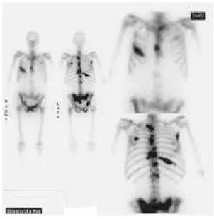
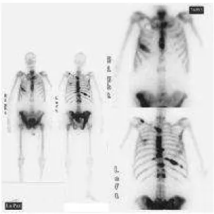
dose, the second dose was given between 13 weeks and 1 year (mean interval: 7 months and a half). Total dose number was between 3,145-5,550, and mean total dose number was 4,694. As established in the protocol, urine samples were collected 4-6 hours after treatment. Five patients needed a bladder catheter. Virtually all patients had a whole-body scintigraphy performed 4-6 hours after 153Sm-Lexidronam (Fig. 2).
Patients were followed up between 2 and 24 months (mean up: 14 months). On follow-up visits, clinical and laboratory (i.e., platelets, blood white cells, hemoglobin, creatinine, etc) data were collected to assess potential treatment-related toxicity. Pain severity was evaluated based on analgesia requirements according to WHO Analgesia Scale, and patients’ health status was evaluated according to the Karnofsky scale. However, the patients’ subjective assessment of pain was emphasized.
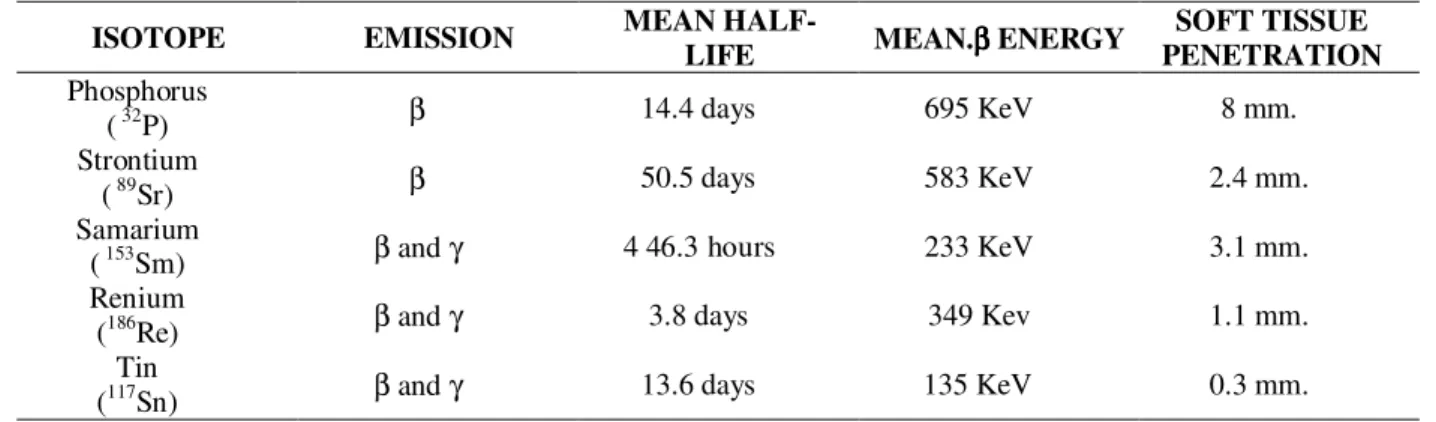
Table 1 - Radioactive isotopes used for metastatic bone pain management.
ISOTOPE EMISSION MEAN
HALF-LIFE MEAN.ββββ ENERGY
SOFT TISSUE PENETRATION
Phosphorus
( 32P) β 14.4 days 695 KeV 8 mm.
Strontium
( 89Sr) β 50.5 days 583 KeV 2.4 mm.
Samarium
( 153Sm) β and γ 4 46.3 hours 233 KeV 3.1 mm.
Renium
(186Re) β and γ 3.8 days 349 Kev 1.1 mm.
Tin
(117Sn) β and γ 13.6 days 135 KeV 0.3 mm.
Table 2: Results
Improved Unchanged Worsened
Subjetive 80(85%) 14(15%) 0
Karnofsky 51(54%) 43(46 %) 0
Analgesia 52(55,3%) 39(41,4%) (3%)
Table 3 - Results: Subjective response of pain.
PATIENTS
Worsened 0 0%
Unchanged 14 15 %
Slight relief 22 23 %
Moderate relief 38 40 %
RESULTS
No adverse effects from 153Sm-Lexidronam administration were noted during the study. Pain relief started between 3 and 30 days (mean: 7 days) after treatment, and it lasted between 1 and 12 months (mean: 3 months).
Both objective and subjective patients’ response to treatment, according to assessment based on WHO and Karnofsky scales are presented in Tables 2 and 3. As shown, no patients got worse, though three needed increased analgesia. Eighty-five per cent (85%) of the patients reported pain relief in different degrees, and analgesia could be reduced in 55% of them. Twenty-three per cent (23%) and 40% had mild or moderate, respectively, pain relief, and in 22% pain cessation was achieved. Four patients had been previously treated with
89
Sr. Three of them had moderate or complete response to 153Sm-Lexidronam, and the other, who had not responded to the previous treatment with
89
Sr, had a moderate response to 153 Sm-Lexidronam.
No relevant adverse effects were noted during the follow-up. In 5% of the patients, 48-72 hours posttreatment, an initial transient increment of pain was observed. Bone marrow toxicity was mild in all cases. Thrombocytopenia was noted in 3% of the patients, anemia in 8%, and leukopenia in 19%.
DISCUSSION
Several studies (including multicenter trials) support the efficacy of 153Sm-Lexidronam treatment for palliative metastatic bone pain, including its ability to improve patients’ life quality, at times in the terminal phase.
The findings reported herein are similar to those reported by earlier authors, with response rates ranging from 62 to 83%38,39,40,41,42.
According to criteria established by earlier authors, a dose of 37 MBq/kg, which seemed to be the most suitable, was administered. However, one can ask whether or not a higher dose would result in better clinical outcome, but data concerning the efficacy of greater doses are scanty in the literature, and it does not seem that better results have been be achieved when slightly higher doses are used42,43.
Clearance of 85% of the injected dose from the blood was achieved in the first 30 minutes, and aproximately 65% of the dose was taken up by the bone. Thirty-five per cent (35%) of the injected dose was excreted in the urine in the next six hours35,36. This, together with a short half-life and β energy from 153Sm, differs from other radioactive isotopes44, and enabled one to adjust the dose to the patient body weight, so that high activity, optimal biological radiation delivery and rapid release of a high radiation dose rate could be achieved in a short period of time.
Figure 2 - 153Sm-EDTMP
Thus, it is estimated that, for a dose of 2,590 MBq of 153Sm-Lexidronam, the equivalent effective dose is 796 mSv31. This would explain the rapid response of the pain to the radioactive isotope concerned41. Because of the rapid clearance and excretion of 153Sm-Lexidronam and its low exposure rate, patients can be treated on a day hospital-basis33.
153
Sm γ emission permits the patient to have a scintigraphy performed after the treatment, which can show the uptake of the isotope by the metastases visualized in the conventional scintigraphy carried out before initiation of treatment. At the beginning of the present study, the scintigraphy was performed 24 hours posttreatment33,42; however, because of the unsatisfactory clinical status of most patients, it was decided to perform the scintigraphy 4-6 hours before discharge, with results comparable to those achieved at 24 hours (Fig.2).
Duration of pain relief in this series was of approximately three months, which was slightly shorter than that reported by earlier authors37,40. There may be two reasons for such a difference. First, some of the patients in the series had a life expectancy as short as one month, and died pain-free two months after completing treatment. Secondly, a large number of patients with a life expectancy longer than three months died between 1 and 2 months after treatment. In contrast, some authors have reported higher mean survival rates (e.g., six months)42.
When the treatment was initiated with radioactive isotopes, specifically with 153Sm-Lexidronam, based on the results of a literature review40,42, the aforementioned protocol, including inclusión criteria, was developed. However, as clinical experience expanded, it became possible to more accurately determine the risk/benefit ratio for the patient to be treated. Thus, it was no longer necessary to adhere strictly the protocol. Knowing that pain palliation may be achieved within a few days, patients with a life expectancy shorter than four months were treated, bearing in mind that clinical benefits were to outweigh the risks taken. In three patients with a life expectancy of about one month, a significant pain palliation was achieved, and in one female patient with breast cancer, total pain cessation until her death was achieved. Also, we were not strict with patients undergoing hormotherapy. Most of these patients underwent hormonotherapy with no interference from 153Sm-Lexidronam.
153
response. As there were few tumors others than prostate and breast in our series, this topic warrants further investigation. Many authors report the best results for breast cancer37,38.
One of the problems encountered was the modality for pain assessment. It was believed that it would not be easy for patients with intemittent pain of varying location and with bad health status and deterioration to indicate the pain degree along a scale. Nevertheless, patients were thought to be able to indicate whether they felt better, unchanged or worse, as well as whether they had lowered or withheld the analgesic medication after treatment. Therefore, while taking the scores from Karnofsky and WHO into account, everything the patient or his/her relatives reported at follow-up visits was emphasized. Eighty-five per cent (85%) of the patients reported pain relief, and 54% reported improved life quality.
The only reported toxicity associated with 153 Sm-Lexidronam treatment reported in the literature is bone marrow toxicity, it being mild and reversible in most cases, and rarely severe37,38,39. In this series, bone marrow toxicity was mild in all cases. Therefore, 153Sm-Lexidronam treatment-associated toxicity is less severe than that associated with other treatment modalities for cancer patients. In the present study, patients receiving chemotherapy within less than eight weeks before treatment did not have increased bone marrow toxicity. At any rate, it is advisable to be cautious when adminstering 153Sm-Lexidronam to patients undergoing chemotherapy 4-8 weeks before treatment initiation.
In this series, 5% of the patients reported increased pain 48-72 hours after treatment, this finding being in keeping with earlier authors45,46. It may be due to radiation effects, because, although it is relatively low, it negatively affects the fluid circulating within the lesion. However, pain increment was transient and responded well to a slight increase in the usual analgesic.
153
Sm-Lexidronam permits multiple doses to be delivered, as required47,48. In the present study, nine patients needed a second dose, and a female patient with breast cancer received three doses. These 12 patients had a response as good as that with the first dose, with mild reversible bone marrow toxicity.
CONCLUSIONS
153
Sm-EDTMP administration is worthwhile for palliative metastatic bone pain management in patients with bone metastases from a number of primary malignant tumors because it provides relief and, is even able to stop the pain, thereby significantly improving patients’ life quality. Furthermore, 153Sm-EDTMP administration-associated adverse effects are mild and reversible.
ACKNOWLEDGEMENTS
I am in debt to Dr. L.M. Martín Curto and Dr. M. Coronado Poggio, Medical Head and Assistant Physician, respectively, of the Department of Nuclear Medicine of La Paz University Hospital (Madrid, Spain) for guidance and assistance. Without their kind collaboration, the present study would not have reached a satisfactory conclusión.
RESUMO
REFERENCES
Ackery, D. (1998), Principles of radionuclide therapy. In: Murray, I. P. C. and Ell, P. F. (Eds.). Nuclear medicine in clinical diagnosis and treatment. 2nd ed. Edinburgh : Churchill Livingstone. pp. 1039-1042. Alberts, A. S.; Smit, B. J.; Louw, W. K.; van Rensburg,
A. J.; van Beek, A.; Krizinger, V. and Nel, J. S. (1997), Dose-response relationship and multiple-dose efficacy and toxicity of samarium-153-EDTMP in metastatic cancer to bone. Radiother Oncol., 43, 175-179. Blake, G. M.; Zivanovic, M. A.; McEvan, A. J. and
Ackery, D. M. [19--], Sr-89 therapy: Strontium kinetics in disseminated carcinoma of the prostate.
Eur. J. Nucl. Med., 12, 447-454.
Centeno, C.; Sanz, A.; Vara, F. and Bruera, E. (2001), Metástasis óseas: manifestaciones clínicas y complicaciones. Un tratamiento multidisciplinar.
Medicina Paliativa, 8, 100-108.
CIS Bio International (1998), Metastatic bone pain.
International Report, 10-26.
De Camargo Etchebhere, E. C.; Pereira Neto, A. A.; Loes de Lima, M. C.; Oliveira Santos, A.; Ramos, C. M. and Camargo, E. E. (2004), Treatment of bone pain secondary to metastases using samarium -153-EDTPM. Sao Paolo Med. J., 22 : (5), 208-212. Doleys, D. and Doherty, D. (2000), Evaluación
psicológica y conductual. In: Abrahms, B.; Benzon, H.; Hanh, M.; Heaver, J.; Niv, D.; Parris, W. et al.
Tratamiento práctico del dolor. España : Harcout Brace. pp. 429-448.
Eary, J. F.; Collins, C.; Stabin, M.; Vernon, C.; Petersdorf, S.; Baker, M.; Hartnett, S.; Ferency, S.; Addisons, J.; Appelbaum, F. and Gordon, E. E. (1993), Samarium-153-EDTMP biodistribution and dosimetry estimation. J. Nucl. Med., 34 : (7), 1031-1036.
Fontana, A. and Delmas, P. (2000), Markers of bone turnover in bone metastases. Magazine supplement: skeletal complications of maligancy. Cancer, 88, 2952-2960.
Friedell, H. L. and Storaasly, J. P. (1950), The use of radiactive phosphorous in the treatment of the carcinoma of the breast with widespread metasatases to the bone.
Am. J. Roentgenol. Radat. Ther., 64, 559-575.
Friedlan, J. (1999), Local and systemic radiation for palliation of metastatic disease. Urol. Clin. N. Am.,
26, 391-402.
Goeckeller, W. F.; Trountner, D. E.; Volkert, W. A.; Edwards, B.; Simon, J. and Wilson, D. (1986), 153-Sm radiotherapeutic bone agents. Nuclear Medicine and Biology, 12 : (4), 479-482.
Goltzman, D. (1997), Mechanism of the Development of Osteoblastic Metastases. Magazine supplement: Skeletal complications of Malignancy. Cancer, 80, 1581-1587.
Han, S. H.; De Klerk, J. M.; Zonnenberg, B. A.; Tan, S. and Van Rijk, P. P. (2001), 186-rte-etidronate. Efficacy
of palliative radionuclide therapy for painful bone metasatases. Q. J. Nucl. Med., 45, 84-90.
Krishnamurthy, G. and Krishnamurthy, S. (2000), Radionuclides for metastatic bone pain palliation: a need for rational re-evaluation in the new millennium.
Journal of Nuclear Medicine, 41, 688-691.
Lovera, C.; Massardo, T.; Galleguillos, M. C.; González, P.; Comparini, B.; Yáñez, M.; Fodor, M.; Gil, M. C.; Araya, G. and Tomicic, M. (1998), Analgesic response and secondary effects in patients with osteoblastic metastasis treated with Samarium-153 ethylenediaminetetramethylenephos-phonate. Rev. Med. Chile, 126 : (8), 963-971.
Marsoni, S.; Hurson, S. and Eisenberger, M. (1985), Chemotherapy of bone metastases. In: Garranttini S., (Ed.). Bone resorption, metastases and diphosphonates. New York : Raven Press. pp. 181-195.
Mc Ewan, A. J. B. (1998), Pallation of bone pain. In: Murray, I. P. C. and Ell, P. F. (Eds.). Nuclear medicine in clinical diagnosis and treatment. 2nd ed. Edinburgh : Churchill Livingstone. pp. 1083-1099. Menda, Y.; Bushnell, D. L.; Williams, R. D.; Miller, S.
and Thomas, M. O. (2000), Efficacy and safety of repeated samarium-153-Lexidronam treatment in a patient with prostate cancer and metastatic bone pain.
Clin. Nucl. Med., 25, 698-700.
Meyer, R.; Campbell, J. and Raja, S. (1994), Peripheral neural mechanisms of nociception. In: Walla, P. and Melzack, R. (Ed.). Textbook of pain. Londres : Churchil Livinstone. pp. 13-14.
Nielsen, O.; Munro, A. J. and Tannock, I. F. (1991), Bone metastases: pathophysiology and management policy. J. Clin. Oncol., 9, 509-52.
Niv, D. and Devor, M. (2000), Analgesia preventiva: ¿Se puede prevenir el dolor postoperatorio agudo?. In: Abrahms, B.; Benzon, H.; Hanh, M.; Heaver, J.; Niv, D.; Parris, W. et al. (Eds.). Tratamiento práctico del dolor. España : Harcout Brace. pp. 1043-1044. Payne, R. (1997), Mechanisms and managemet of bone
pain. American Cancer Society. Magazine Supplement: Skeletal Complications of Malignancy,
80, 1608-1613.
Pons, F. and Fuster, D. (2002), Under-utilization of radionuclide therapy in metastatic bone pain pallation. Nuclear Medicine Communications, 23, 301-302.
Pons, F.; Herranz, R.; García, A.; Vidal-Sicart, S.; Conill, C.; Grau, J. J. et al. (1997), Strontium-89 for palliation of pain from bone metastases in patients with prostate and breast cancer. Eur. J. Nucl. Med.,
24, 1210-1214.
Pons, F.; Fuster, D. and Vidal-Sicart, S. (2003), Tratamiento paliativo del dolor óseo metastásico.
Rev. Esp. Med. Nuclear, 22 : (6), 429-438.
Resche, I.; Chatal, J. F.; Pecking, A.; Ell, P.; Duchesne, G.; Rubens, R.; Fogelman, I. et al. (1997), A dose-controled study of 153 Sm-Ethylenediaminetetrame-thylenephosphonate (EDTMP) in the treatment of patients with painful bone metastases. European Journal of Cancer, 33 : (10), 1583-1591.
Resche, I.; Chatal, J. F.; Pecking, A.; Ell, P. J.; Duchesne, G.; Rubens, R.; Fogelman, I.; Houston, S.; Fauser, A.; Fischer, M. and Wilkins, D. (1997), A dose-controlled study of 153 Sm-Ethylenediaminete-tramethylenephosphonate (EDTMP) in the treatment of patiens with painful bone metastases. Eur. J. Cancer, 33 : (10), 1583-1591.
Reyes, V. A. (2002), El manejo del dolor óseo intratable: perspectiva clínica y manejo con radionúclidos. In: Castro-Beiras, J. Avances en Medicina Nuclear y calidad científico-técnica. Consejería de Sanidad Comunidad de Madrid. pp. 649-669.
Robinson, R. G.; Preston, D. F.; Schiefelbein, M. and Baxter, K. G. (1995), Strontium-89 therapy for the palliation of pain due to osseous metastases. JAMA,
274, 420-424.
Rogers, M.; Gordon, S.; Benford, H.; Coxon, F.; Luckman, S.; Monkkonen, J. et al. (2000), Cellular and Molecular Mechanism of action of biphosphonates. American Cancer Society. Magazine Suplement: Skeletal Complications of Malignancy,
88, 2961-2978.
Sabino, M.; Ghilardi, J. and Feia, K. (2002), The involment of prostaglandins in tumorogenesis, tumor induced osteolysis and bone cancer pain. J. Musc. Skel. Neur. Interact.,2, 561-562.
Sartor, O.; Reid, R. H.; Hoskin, P. J.; Quick, D. P.; Ell, P. J.; Coleman, R. E.; Kotler, J. A.; Freeman, L. M. and Olivier, P. (2004), Samariun-153-Lexidronam complex for treatment of painful bone metastases in hormone-refractory prostate cancer. Urology, 63, 940-945.
Schimdt, R.; Scable, H.; Messlinger, K.; Heppelmann, B.; Hanesh, U. and Pawlak, M. (1994), Silent and active nociceptors: Structure, funtions and clinical implications. In: Gebhart, G.; Hammond, D. and Jensen, T. (Eds.). Progress in pain research abd management. Seattle : IASP. v. 2. pp. 213-150. Schlien, B. and Terpilak, M. S. (Eds.) (1984), The
health physics and radiological health handbook. Oincy, Maryland : Nucelon Lectern Associates Inc. Serafini, A. (2001), Therapy of metastatic bone pain.
J. Nucl. Med., 42, 895-906.
Serafini, A. N. (2000), Samarium SM-153 Lexidronam for the palliation of bone pain associated with metastases. Cancer, 88 : (12), 2934-2939.
Serafini, A. N. (2001), Systemic metabolic radiotherapy with samarium-153-EDTMP for the treatment of painful bone metastases. Q. J. Nucl. Med., 45, 91-99.
Serafini, A. N. (2002), Current status of bone pain palliation with systemic radioisotopes. In: Freeman, L. M. (Ed.). Nuclear Medicine Annual 2002. Philadelphia : Lippincott Wiliams & Wilkins. v. 2. pp. 53-74.
Serafini, A. N.; Houston, S. J.; Reche, I.; Quick, D. P.; Grund, F. M.; Ell, P. J. et al. (1998), Palliation of pain associated with metastatic bone cancer using Samarium
-153
Lexidronam: a double-bind placebo-control clinical trial. J. Clin. Oncol., 16 : (4), 1574-1581.
Silberstein, E. B. and Williams, C. (1989), Strontium-89 therapy for the pain of osseous metastases. J. Nucl. Med., 26, 345-348.
Singh, A.; Holmes, R. A.; Farhangi, M.; Volkert, A.; Williams, A.; Stringham, L. M. and Ketring, A. R. (1989), Human pharmacokinetics of Samarium -153-EDTMP in metastatic cancer. J. Nucl. Med., 30, 1814-1818.
Srivastava, S. C.; Atkins, H. L.; Krishnamurthy, G. T.; Zanzi, I.; Silberstein, E. B.; Meinkin, G. et al. (1998), Treatment of metastatic bone pain with tin-117m stannic diethylenetriaminepentaacetic acid: a phase I/II clinical study. Clin. Cancer, 4, 61-68.
Tian, J. H.; Zhang, J. M.; Hou, Q. T.; Oyang, O. H.; Wang, J. M.; Luan, Z. S.; Chuan, L. and He, I. J. (1999), Multicentre trial on the efficacy and toxicity of single-dose samarium-153 Ethylene diamine tetramethylene phosphonate as a palliative treatment for painful skeletal metastases in China. Eur. J. Nucl. Med., 26, 2-7.
Tong, D.; Gillck, L. and Hendrickson, F. R. (1982), The palliation of the symptomatic osseous metastases: final results of the study by the Radiation Therapy Oncology Group. Cancer, 50, 893-899.
Twycross, R. (1994), R. Meassurement of pain. In: Twycross, R. (Ed.). Pain relief in advanced cancer. Londres : Churchill Livingstone. pp. 129-147. World Health Organization (1990), Cancer pain relief
and palliative care: report of a WHO Expert
Committee. Genove, Switzerland : World Health Organization.