FERNANDO JORGE PEGO CRISTO
Molecular and functional analysis of DAND5 in
human Congenital Heart Disease (CHD)
UNIVERSIDADE DO ALGARVE
Departamento de Ciências Biomédicas e Medicina
2016
FERNANDO JORGE PEGO CRISTO
Molecular and functional analysis of DAND5 in
human Congenital Heart Disease (CHD)
Doutoramento em Ciências Biomédicas
Trabalho efetuado sob a orientação de:
Professor Doutor José António Henriques de Conde Belo
UNIVERSIDADE DO ALGARVE
Departamento de Ciências Biomédicas e Medicina
2016
iii
Molecular and functional analysis of DAND5 in human
Congenital Heart Disease (CHD)
Declaração de autoria de trabalho
Declaro ser o autor deste trabalho, que é original e inédito. Autores e trabalhos consultados estão devidamente citados no texto e constam da listagem de referências incluída.
Copyright – Fernando Jorge Pego Cristo. Universidade do Algarve.
Departamento de Ciências Biomédicas e Medicina.
A Universidade do Algarve reserva para si o direito, em conformidade com o disposto no Código do Direito de Autor e dos Direitos Conexos, de arquivar, reproduzir e publicar a obra, independentemente do meio utilizado, bem como de a divulgar através de repositórios científicos e de admitir a sua cópia e distribuição para fins meramente educacionais ou de investigação e não comerciais, conquanto seja dado o devido crédito ao autor e editor respetivos.
v
D
edicated to all the people who never stop believing
in me
………
“Your time is limited, so don‘t waste it living someone else‘s life. Don‘t be trapped by dogma – which is living with the results of other people‘s thinking. Don‘t let the noise of other‘s opinions drown out your own inner voice. And most important, have the courage to follow your heart and intuition. They somehow already know what you truly want to become. Everything else is secondary”
vii
Acknowledgements
Durante estes últimos anos, muitos foram aqueles com quem, trabalhei, troquei opiniões, partilhei ideias, aprendi e muitas vezes me diverti. Tive alguns amigos que sempre me apoiaram, ouviram as minhas tristezas e as minhas alegrias. A todos aqueles que ao longo deste período me ofereceram uma imutável e inestimável ajuda, permitiram a realização deste trabalho e de alguma forma contribuíram para o meu enriquecimento pessoal quero expressar os meus sinceros agradecimentos de forma especial, pois especial foi também o que me transmitiram.
Dito isto, desejo expressar os meus sinceros agradecimentos:
Ao Prof. José A. Belo, meu orientador, pela confiança em mim depositada para levar a cabo este projeto no seu laboratório, por todo o suporte e pela disponibilidade e generosidade reveladas ao longo destes anos que temos trabalhado juntos.
Ao José Inácio por todas as perguntas “parvas” que teve de me responder e todos os dias que teve de me aturar, por me ter ensinado quase todas as técnicas que sei até hoje e principalmente pela pessoa que é. Obrigado Zé por toda a paciência, disponibilidade e companheirismo.
À Salomé Almeida, por todo o suporte, disponibilidade, competência e boa disposição. Muito obrigado Salomé por acreditares em mim, no meu trabalho e dizeres sempre aquelas palavrinhas para levantar o animo.
Ao pessoal do laboratório, nomeadamente, Elizabeth, Sara, Marta, Marisa, Rita, Margaret, João Facucho, João Furtado (el pollo), Carolina (Brasileira), Ana Rubina (Rubi), Maria Silva, Oriol Bover e Paulo Pereira pela forma como me receberam e por toda a amizade, companheirismo e ensinamento durante este longo percurso. Um agradecimento especial ao Tiago Justo pelo excelente ano em que decidimos aventurar-nos por Lisboa, pelo companheirismo e ensinamentos tantos profissional como pessoal.
Um muito muito obrigado a todos os meus amigos de S.Tiago e claro aos da faculdade que estão sempre presentes, João Baptista e Nídia Cunha. Um agradecimento especial à Raquel Santa Maria por todo o suporte e acreditar em mim até mais do eu. Muito obrigado és uma pessoa espetacular, um verdadeiro exemplo, tanto profissionalmente como pessoalmente.
Quero também agradecer ao “pessoal do CEDOC” pela forma como me receberam no vosso grupo de amigos e pelos dias e noite bem passadas. Em especial quero agradecer à Inês Santarino por tudo o que fez por mim e pela pessoa espetacular que é, a tua amizade foi e é muito importante para mim. Uma palavra de apreço também á “Tati”, é muito difícil encontrar pessoas como tu e com o teu caracter. Temos uma amizade para uma vida. Obrigado por todos os conselhos.
Quero agradecer também ao CBMR e CEDOC pelas excelentes condições de trabalho.
Não podia deixar de agradecer também à Carla Machado por todo o suporte, por todos as noites que me stressava e me aturavas, por toda a compreensão, carinho e amizade. És uma pessoa única.
Por último, aos meus avós, tia, irmão e aos meus pais: a vocês o meu muito obrigado por todo o apoio incondicional, carinho, compreensão e por estarem sempre presentes nos momentos mais difíceis. Obrigada por tudo, Amo-vos!
ix
Resumo
Uma das questões fulcrais da biologia do desenvolvimento continua a ser como é que uma única célula, o oócito fecundado, se desenvolve num ser tão complexo e especializado, com os tecidos e órgãos diferenciados e organizados em torno de três eixos, o anterior-posterior (AP), dorso-ventral (DV) e esquerdo-direito (LR).
De um modo geral, o desenvolvimento embrionário humano pode ser dividido em três períodos: o primeiro, chamado de período zigótico, inicia-se com a fusão dos gametas masculino e feminino, formação do zigoto e termina com a implantação do blastocisto na parede do útero duas semanas após a fecundação. Durante o segundo período, ou período embrionário, que vai desde a terceira até à oitava semana de gestação, ocorre a gastrulação, com a formação e diferenciação dos folhetos embrionários (ectoderme, mesoderme e endoderme), e a formação dos órgãos e sistemas fisiológicos. O ultimo período, chamado fetal, vai desde o terceiro mês de gestação até ao nascimento e é caracterizado pelo crescimento do embrião, maturação e funcionalização das estruturas e órgãos.
Durante o desenvolvimento do embrião, o coração é o primeiro órgão interno a ser formado e a tornar-se funcional de modo a bombear sangue por volta das três semanas de gestação, suprindo as necessidades do embrião e garantindo a sua sobrevivência. Na maioria dos vertebrados o desenvolvimento cardíaco segue o mesmo padrão de formação. O coração forma-se a partir de duas regiões cardíacas bilaterais que advêm de uma região progenitora comum na gastrulação, a mesoderme cardíaca. A mesoderme cardíaca recebe informação posicional dos três eixos embrionários, porém o efeito do eixo Esquerda-Direita (ED) tem recebido particular atenção. Os dois “pools” bilaterais de células precursoras cardíacas mesodermais migram para a linha média onde se juntam e dão origem ao crescente cardíaco. Este crescente cardíaco vai formar posteriormente um tubo cardíaco linear primordial. De seguida, este tubo alonga e sofre um processo de “looping” para a direita levando à formação dos ventrículos e aurículas. Subsequentemente, ocorre a diferenciação, especialização e septação das quatro camaras cardíacas e válvulas
proporcionando as paredes necessárias para a separação física da circulação sistémica e pulmonar de sangue. Por último a formação do sistema de condução cardíaco e maturação dos cardiomiócitos, culminam na formação de um coração inteiramente funcional.
As doenças cardíacas congénitas (DCC) são a manifestação clínica de problemas durante o desenvolvimento embrionário do coração. Esta doença é a forma mais comum de defeito à nascença, ocorrendo em cerca de 8 em 1000 nados vivos, bem como a principal causa não-infeciosa de morte no primeiro ano de vida. Na maioria dos casos (80%), esta doença tem origem desconhecida e manifesta-se isoladamente em doentes não sindrómicos e não seguindo os padrões de hereditariedade definidos por Mendel, sendo considerada uma doença complexa com origem multifatorial. Esta complexidade advém do facto de a doença em muitos casos se apresentar geneticamente heterogenica, com penetrância reduzida e expressividade genética variável. Para além disso, fatores de risco ambientais foram também identificados como contribuído para o risco de desenvolver a doença. Nos dias de hoje, um modelo envolvendo tanto fatores genéticos como a interação genes-ambiente, é a explicação mais plausível para as altas frequências verificadas para a doença cardíaca congénita.
Nos últimos anos, vários esforços foram feitos de modo a se compreender os mecanismos moleculares que levam a uma má formação do coração. Estes estudos, usando principalmente animais modelo e genética molecular humana, da morfogénese do coração e dos primeiros passos da determinação do eixo de simetria esquerdo-direito durante a embriogénese, indicam que na maioria dos casos de distúrbios de lateralidade é observada uma malformação cardíaca complexa, sugerindo que as DCC podem dever-se a defeitos de lateralidade na morfogénese do coração.
Uma das redes regulatórias por detrás do estabelecimento do eixo esquerdo-direito é a via de sinalização Nodal, um fator de crescimento da família TGF-b, que é expresso assimetricamente do lado esquerdo do nó e placa lateral esquerda da mesoderme de ratinho. Os fenótipos cardíacos de ratinhos geneticamente modificados para desativar a sinalização de nodal ou a sua
xi regulação, demonstraram que o desenvolvimento cardíaco depende vivamente do estabelecimento do eixo E-D embrionário. Para além disso, foram identificadas variantes genéticas em genes envolvidos na via de sinalização Nodal em doentes com DCC.
O nosso laboratório identificou um membro da família Cerberus/DAN, o gene
Cerberus-like2 (Cerl2), que é assimetricamente expresso no lado direito do nó
em ratinho. O gene Cerl2 codifica para uma proteína secretada capaz de se liga diretamente a Nodal, inibindo a sua via de sinalização. Na presença de
Cerl2 a atividade da via de sinalização Nodal restringe-se apenas ao lado
esquerdo do embrião, o que culmina com uma morfogénese normal dos órgãos do abdómem. Ratinhos knock-out para o gene Cerl2, em que o gene Nodal passa a poder ser expresso e induzir a sua cascata de sinalização em ambos os lados da placa lateral da mesoderme, revelaram uma ampla gama de defeitos de lateralidade - situs inversus, isomerismos e heterotaxia - e malformações cardíacas - formação incompleta das aurículas, defeitos no septo ventricular, falha na rotação das principais artérias (transposição das grandes artérias, ventrículo direito de dupla saída), looping cardíaco anormal (randomizado), posicionamento do ápex cardíaco e hipertrofia ventricular - o que nos levou a suspeitar que alterações neste gene poderiam estar associadas a doenças de lateralidade e/ou casos isolados de doenças cardíacas congénitas.
Dado que as vias genéticas que regulam o desenvolvimento cardíaco em ratinhos e humanos são conservadas, o principal objetivo desta tese foi o estudo de alguns dos genes homólogos humanos envolvidos na via de sinalização Nodal, com principal foco no gene DAND5, homólogo humano de
Cerl2, mapeado no cromossoma 19, região 19p13.2, mas também verificando a
existência de variantes nos genes NODAL, PITX2C e CFC1 numa cohort de doentes com defeitos cardíacos congénitos com origem em perturbações do eixo esquerdo-direito.
A análise dos resultados da sequenciação de DNA do gene DAND5 permitiu-nos identificar dois doentes com a mesma variação. Clinicamente, um dos doentes apresenta um fenótipo de defeito no septo ventricular com aorta
conectada aos dois ventrículos, atresia pulmonar e isomerismo esquerdo. O outro doente apresenta um caso extremo de tetralogia de Fallot (defeito no septo ventricular com aorta conectada aos dois ventrículos, hipertrofia do ventrículo esquerdo e atresia pulmonar). A variante, identificada nos dois doentes como heterozigótica, leva à substituição de um nucleótido guanina por uma adenina na posição 455 do exão 2, resultando na substituição de um aminoácido arginina por uma histidina (p.R152H) numa região altamente conservada e funcionalmente importante da proteína DAND5. De modo a entendermos o potencial efeito da alteração p.R152H na proteína DAND5, fizemos um estudo in silico recorrendo a vários programas bioinformáticos. Os resultados dessas predições foram inconclusivos uma vez que 3 programas apontam para um possível efeito nefasto enquanto que outro programa sugere que a variante não tem qualquer efeito na proteína. Para clarificarmos a função desta proteína variante, avaliámos o seu efeito na regulação da via de sinalização Nodal recorrendo a um ensaio funcional de luciferase. O resultado obtido mostra uma diminuição substancial da função da proteína mutante comparada com a proteína wild-type.
Embora os fenótipos dos doentes sejam muito semelhantes, não podemos fazer uma clara correlação do genótipo com o fenótipo pois a variante c.455G>A foi reportada em bases de dados publicas como variante de nucleótido único (rs45513495). Além disso, a mãe de um dos doentes é portadora da variante sem apresentar indícios de doença, sugerindo uma penetrância incompleta, expressividade genética variável ou o efeito de fatores ambientais. Estas observações estão de acordo com a complexidade das doenças cardíacas congénitas e/ou defeitos de lateralidade e podem refletir a acção de variantes modificadoras ou outras variantes genéticas na via de sinalização que podem exacerbar ou atenuar o efeito da variante p.R152H na proteína DAND5 e nos fenótipos dos doentes. Portanto, nós propomos que esta variação no gene DAND5 pode ser um alelo de risco para o desenvolvimento de DCC e/ou defeitos de lateralidade.
Por esta razão, decidimos levar a cabo uma busca de possíveis alterações em genes que fazem parte desta cascata Nodal. Foram assim identificadas uma nova alteração na zona codificante do gene CFC1, sem efeito funcional
xiii aparente, e quatro polimorfismos, um na zona codificante e outro no intrão do gene NODAL e dois na zona não codificante do gene PITX2C em doentes com uma vasta gama de defeitos. Tendo em conta a análise levada a cabo por um programa bioinformático que permite analisar se alterações genéticas podem afetar o processo de splicing, nós verificamos que de facto, as variantes genómicas encontradas fora das zonas codificantes dos genes NODAL e
PITX2C podem resultar em moléculas de RNA mensageiro anormais,
principalmente devido à criação ou disrupção de locais para a ligação da maquinaria de splicing. Quanto à variante na zona codificante do gene NODAL, esta encontra-se no exão 2, e leva à substituição do aminoácido Histidina por uma Arginina na posição 165 (p.H165R). Esta variante já tinha sido reportada em dois estudos de 2008/2009 nos quais os autores verificaram, recorrendo a um ensaio funcional de lucifarese, que a variante leva à redução da atividade da proteína NODAL e parece atuar como variante modificadora e como fator de risco quando associada a outras possíbeis alterações em genes da via Nodal, com os quais este normalmente interage.
Para além disto, e com o objetivo de modelar a doença e estudar os mecanismos moleculares por de trás de uma simples variação num nucleótido, geramos células estaminais induzidas de um dos doentes com a variante no gene DAND5. Estas células foram caracterizadas, apresentado uma morfologia, expressão de marcadores pluripotentes e cariotipo normais.
Em conclusão, embora não possamos fazer uma correlação dos genótipo-fenótipo, nem classificar as alterações identificadas neste estudo como alelos associadas a doença por si, estas variantes, podem, no entanto, aumentar a suscetibilidade para o desenvolvimento de DCC e/ou defeitos de lateralidade. Uma vez que a maioria dos doentes apresenta mais do que uma das variantes no seu genoma, o efeito cumulativo de cada variante na via parece aumentar ainda mais o risco para desenvolver doença. Portanto, o desequilíbrio dos níveis adequados da via de sinalização Nodal, em ambos os lados da placa lateral da mesoderme, devido a uma ou várias variantes nos seus componentes, é um denominador comum para defeitos de lateralidade e/ou doenças cardíacas congénitas.
Palavras-chave: DAND5; Doenças cardíacas congénitas Defeitos de lateralidade, Via de sinalização Nodal, Variantes alélicas; Modelação de doença, Células estaminais induzidas
xv
Abstract
The majority of congenital heart disease (CHD) is sporadic, with a minority of cases associated with a known genetic abnormality. Combinations of genetic-environmental factors are implicated in the etiology of the disease. Recently, several studies, using mostly animal models, unraveled that perturbations in the molecular processes that precede the beginning of heart development might also be at the origin of CHD. In fact, some of the most complex CHDs are found associated with laterality defects, a disorder resulting from abnormal Left-Right axis formation. In our laboratory, the identified mouse Cerberus-like2 (Cerl2 – human DAND5), a protein that inhibit Nodal signaling, prompt us to study cardiac and laterality diseases, since the generated Cerl2 KO mice display a wide range of laterality defects and CHD. Considering the high conservation of genetic pathways regulating cardiac development in mouse and human, the main objective of the present thesis was the study of human genes involved in the Nodal pathway, focusing mostly in DAND5, in a CHD and/or laterality defects patients cohort. The sequence analysis of DAND5 revealed two patients displaying the same p.R152H variant, resulting in a substantial decreased in the function of the protein. We propose that p.R152H acts as a risk allele for CHD and/or laterality defects. In addition, we found two alterations in NODAL, two alterations in PITX2C and one alteration in CFC1. We hypothesized that the NODAL p.H165R variant can act as a common modifier and the intronic variants in NODAL and PITX2C might cause alterations in the splicing pattern of the mRNA molecules. Moreover, we generated patient-specific iPSCs to understand the molecular mechanisms of disease behind the DAND5 nucleotide variant. Although we cannot make a clearly genotype-phenotype correlation, the variants here identified probably increase the disease susceptibility due to the resulting abnormal Nodal signaling. Because most of the patients presented more than one alteration, the cumulative effect of each variant within the pathway seems to enhance even more these risk. Therefore, the imbalance in dosage-sensitive Nodal signaling is a common denominator for laterality defects and associated CHDs.
Keywords: DAND5, Congenital Heart Diseases, Laterality defects, Nodal
xvii
List of Contents
Acknowledgements ... vii
Resumo ... ix
Abstract ... xv
List of Figures ... xxi
List of abbreviations, acronyms and symbols ... xxiii
Chapter 1 – General Introduction ... 27
1. The primordium of life ... 3
1.1. Early embryogenesis ... 3
2. Patterning the embryo – Left-Right Axis Specification ... 7
2.1. Anterior-posterior and dorsal-ventral axis ... 7
2.2. Left – right asymmetry ... 7
2.2.1. The three main steps ... 8
2.2.1.1. Models for symmetry breaking (first step) ... 9
2.2.1.1.1. The leftward flow model ... 11
2.2.1.1.1.1. Cilia and the generation of the unidirectional leftward flow 12 2.2.1.1.1.2. Sensing the flow – morphogen/ chemosensor model 14 2.2.1.1.1.3. Sensing the flow – Two cilia/mechanosensor model 15 2.2.1.1.1.4. Readouts of the nodal flow – the role of ... 18
Cerl2/DAND5 ... 18
2.2.1.2. Transmission of a left–right-biased signal to LPM (second step) ... 22
2.2.1.2.1. The Signal ... 22
2.2.1.2.2. The Route of the signal to LPM ... 23
2.2.1.2.3. Patterning the LPM ... 25
2.2.1.3. Asymmetric position and morphogenesis of the internal organs .. 27
3.1. Embryonic origins of the heart ... 30
3.2. Heart fields and cardiac looping ... 32
3.3. Chamber morphogenesis and maturation of the heart ... 34
3.4. Other sources of cells in heart development ... 36
4. Congenital Heart Diseases ... 37
4.1. Epidemiology of Congenital Heart Diseases ... 37
4.2. Etiology of Congenital Heart Diseases ... 38
4.3. Laterality defects and Congenital Heart Disease ... 39
4.3.1. Cardiac left-right asymmetry ... 41
5. Disease modeling using patient-derived iPSCs ... 44
6. Objectives ... 46
Chapter 2 – Methods ... 47
1. Ethics ... 49
2. Study population ... 49
3. Clinical evaluation and inclusion criteria ... 49
4. Patient’s phenotypes ... 50
5. Genetic Screening of Nodal signaling pathway genes ... 52
5.1. Sample collection ... 52
5.2. DNA extraction and quantification of DNA concentration ... 52
5.3. Genes analyzed... 52
5.4. Oligonucleotide primers design ... 53
5.5. DNA amplification ... 55
5.6. Purification of PCR products ... 56
5.7. Sequencing and sequencing analysis ... 57
5.8. Sequence alignment of the variant proteins among vertebrate species ... 57
5.9. Characterization of the Causative Potential of the Variants ... 58
6. Functional analyses of the p.R152H DAND5 protein variant in the Nodal signaling... 58
xix
6.2. Nodal-dependent luciferase assay ... 60
7. In silico analysis of the intronic variants found in NODAL and PITX2C genes. ... 61
8. Generation and characterization of human iPSC line ... 62
8.1. Ethics statement and informed consent ... 62
8.2. Isolation of cells from urine ... 62
8.3. Non-integrative reprogramming and establishment of iPSC lines ... 63
8.4. Culture of iPSC ... 63
8.5. Sequencing analysis ... 64
8.6. Quantitative polymerase chain reaction (qPCR) analyses ... 64
8.7. Fluorescent immunocytochemistry... 65
8.8. Karyotyping ... 66
Chapter 3 – Results/Discussion ... 67
Part I – Functional study of DAND5 variant in patients with Congenital Heart Disease and laterality defects ... 69
1. Abstract ... 71
2. Background ... 72
3. Results and discussion ... 75
Part II – Genetic Screening of other Nodal signaling pathway genes ... 83
1. Abstract ... 85
2. Background ... 86
3. Results and discussion ... 87
3.1. NODAL H165R variant ... 88
3.2. NODAL intronic mutation ... 90
3.2.1. In silico analysis ... 91
3.3. PITX2C variants ... 93
3.3.1. PITX2C c.205+71A>G in silico analysis ... 94
3.3.2. PITX2C c.205+88 G>C in silico analysis ... 97
Part III – Disease Modelling using iPS derived cells ... 103
1. Abstract ... 105 2. Background ... 106 3. Results and discussion ... 108
3.1. Isolation and generation of Patient-Specific human iPSC lines from a Urine Sample. ... 108 3.2. Characterization of DAND5 allelic variant and healthy control human IPSCs lines. ... 111
3.2.1. Morphology... 111 3.2.2. Genotyping ... 112 3.2.3. Pluripotency analysis ... 113 3.2.4. Karyotype ... 115 Chapter 4 – General Discussion ... 117 Chapter 5 – References ... 131 Chapter 6 – Supplementary material ... 145 Supplementary Material 1 ... 147 Supplementary Material 2 ... 148 Supplementary Material 3 ... 149 Supplementary Material 4 ... 150 Supplementary Material 5 ... 151 Supplementary Material 6 ... 154 Supplementary Material 7 ... 155
xxi
List of Figures
Chapter 1
Figure 1.1 – Summary of the first three week of human development. ... 6 Figure 1.2 – General process of LR asymmetry generation ... 9 Figure 1.3 – “F” molecule model... 10 Figure 1.4 – Node structure and cilia microtubules organization ... 11 Figure 1.5 – Leftward nodal flow in the ventral node and posterior tilting of cilia ... 13
Figure 1.6 – The two versions of the “morphogen gradient”/chemosensor model ... 15
Figure 1.7 – “Two cilia” model. ... 17 Figure 1.8 - Nodal Signaling. ... 19
Figure 1.9 – Cerl2, Nodal and pSMAD2 activity in perinodal cells of the mouse node 20
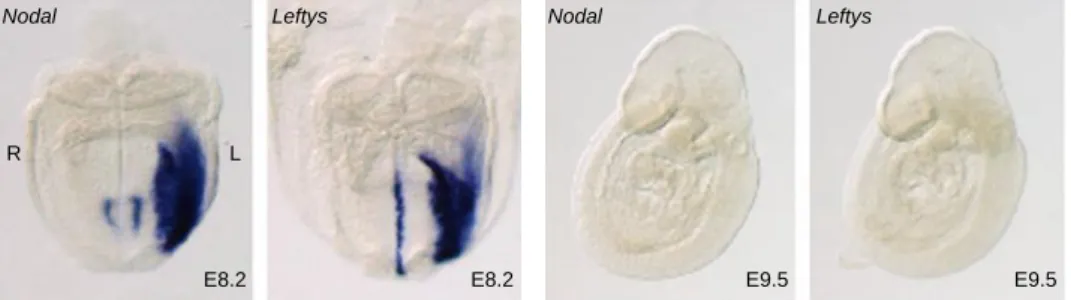
Figure 1.10 – Cerl2 mRNA and protein localization ... 22 Figure 1.11 – Nodal expression and the route of the signal to LPM ... 24 Figure 1.12 – Expression pattern of Nodal and Leftys in wilt-type mice. ... 26 Figure 1.13 - Mechanisms for the generation of morphological asymmetries and Pitx2 expression pattern. ... 28
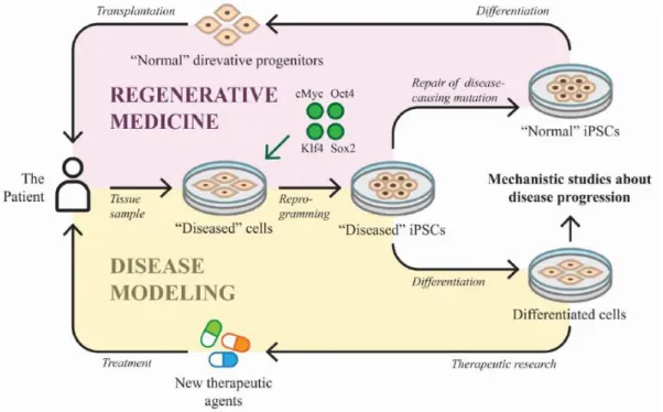
Figure 1.14 – Embryonic origins of the heart ... 31 Figure 1.15 – Heart fields regionalization of the heart ... 33 Figure 1.16 – Mature 4-chambered human heart ... 36 Figure 1.17 – Human laterality defects. ... 40 Figure 1.18 – Applications of iPSCs technology. ... 45
Chapter 2
Figure 2.1 – Genomic localization of DAND5; NODAL; CFC1 and PITX2C primers. . 54
Chapter 3
Part IFigure 3.1 - Genomic localization of DAND5 455 allelic variant ... 77 Figure 3.2 - Functional analysis of DAND5 variant. ... 80
Part II
Figure 3.3 - Sequencing and genomic localization of the NODAL 494 A>G allelic
variant. ... 88
Figure 3.4 - NODAL 494 G(C)>A(T) allele frequency according to 1000 genome
project. ... 89
Figure 3.5 - Relative activity of human NODAL p.H165R variant ... 89
Figure 3.6 – Conservation of the NODAL p.H165R variant between species ... 90
Figure 3.7 - Sequencing and allele frequency of the c.193+12C>T variant. ... 91 Figure 3.8 - In silico analysis of NODAL intronic using the Human Splicing Finder
online software... 92
Figure 3.9 - Sequencing and allele frequency of the PITX2C c.205+71A>G
substitution ... 93
Figure 3.10 - DNA Sequencing of PITX2C gene. ... 94 Figure 3.11 - In silico analysis of the PITX2C c.205+71 A>G variant. ... 95 Figure 3.12 - In silico analysis of the PITX2C c.205+88 G>C variant. ... 97 Figure 3.13 - CFC1 allelic variant. ... 99
Part III
Figure 3.14 - Genotype of patient and control samples used to generated iPSC
lines ... ….107
Figure 3.15 – Morphology of the proband urinary cells at different time points
after collection... ….109
Figure 3.16 - Morphology of the proband-derived cells at different time points
during transduction with Sendai virus carrying the Yamanaka reprogramming factors. ... ...110
Figure 3.17 - Morphology of proband iPSC-like cells at different time points after
expansion ... ...111
Figure 3.18 - Morphology of two picked clones of each iPS derived cell line ... ...112 Figure 3.19 - Confirmation of DAND5 allelic variant and Healthy control cell line
Genotype. ... ...112
Figure 3.20 - Pluripotency analysis at mRNA level. ... ...113 Figure 3.21 - Pluripotency analysis at protein level. ... ...114 Figure 3.22 - Karyotype of patient-derived iPSCs. ... ...116
xxiii
List of abbreviations, acronyms and symbols
°C – Degrees Celsius µg – Microgram µl – Microliter µM – Micro molar 3’ – 3 prime 5’ – 5 prime β-GAL – β-galactosidase μm – micrometer A A – Adenine ActRIIA – Activin receptor type IIA
ActRIIB – Activin receptor type IIB AKL7 – Activin receptor-like kinase 7
ALK4 – Activin receptor-like kinase 4
AP – Anterior-posterior
ARE – Activin responsive element ASD – Atrial septal defects ASEs – Asymmetric enhancers AVC – Atrioventricular canal AVSD – Atrioventricular septal defects
B
BLAST – Basic Local Alignment Search Tool BMP – Bone morphogenetic protein
Bp – Base pair
C
C – Cytosine Ca2+ – Calcium
Cerl2 – Cerberus like 2 CHD – Congenital heart disease/defects
CNC – Cardiac neural crest CNVs – Copy number variants
D
Da – Dalton
DAND5 – DAN Domain Family Member 5
DILV – Double inlet left ventricle
DNA – Deoxyribonucleic acid dNTP – Deoxyribonucleotide triphosphate DORV – Double outlet right ventricle DV – Dorso-ventral
Dvl – Dishevelled
E
E – Embryonic day ECM – Extracellular matrix EGF-CFC – epidermal growth factor-Cripto-FRL1-Cryptic EHF – Early head-fold
EMT – Epithelial-mesenchymal transition EPE – Entidade Pública Empresarial ESCs – Embryonic stem cells
F
FGF – Fibroblast growth factor FHF – First heart field
FoxH1 – Forkhead box protein H1
G
G – Guanine
GAGs – Glycosaminoglycans GATA4 – GATA Binding Protein 4 GDF1 – Growth differentiation factor 1
H
HAND2 – Heart- and neural crest derivatives-expressed protein 2 HEK – Human Embryonic Kidney
HTX – Heterotaxy
I
ICM – Inner cell mass
ICOs – Intraciliary calcium oscillations Information
iPSCs - induced pluripotent stem cells Isl1 – ISL LIM Homeobox 1
IV – Inversus viscerum IVS – Interventricular septum
K
Kif3a – Kinesin family member 3A Klf4 – Kruppel-like factor 4
KO – Knock-out
L
LA – Left atrium
Lefty - Left-right determination factor LHF – Late head-fold stage LPM – Lateral plate mesoderm LR – Left-right
LRO – Left-right organizer LV – Left ventricle
xxv M – Molar
MAPCAs – Major Aorto-Pulmonary Collateral Arteries Mef2c – Myocyte Enhancer Factor 2C
Mesp1 – Mesoderm posterior 1
Min – Minutes ml – Milliliters mRNA – Messenger ribonucleic acid
N
NCBI – National Center for Biotechnology
Nkx - Homeodomain factors NVPs – Nodal vesicular parcels
O
Oct – Octamer-binding transcription factor
OFT – Outflow tract OMIM – Online Mendelian Inheritance in Man
P
PA – Pulmonary atresia
PAM – Paraxial mesoderm PBS – Phosphate buffer saline
PCP – Planar cell polarity
PCR – Polymerase chain reaction PFP – Prospective floor plate
Pitx2 – Pituitary homeobox 2 PK – Proteinase K
Pkd1 – Polycystin-1
Pkd1l1 – Polycystin like 1 Pkd2 – Polycystin-2
PLSVC – Persistent left superior vena cava PS – Primitive streak
Q
qPCR – Quantitative polymerase chain reaction
R
RA – Retinoic Acid RA – Right atrium
RNA – Ribonucleic acid rpm – Rotations per minute RT – Room temperature
RT-PCR – Reverse transcription PCR RV – Right ventricle
S
SELI – Self-enhancement lateral inhibition SeV – Sendai virus
SHF – Second heart field Shh – Sonic Hedgehog
SI – Situs inversus
SMAD – Mothers against decapentaplegic homolog SNPs – Single nucleotide polymorphisms
Sox2 – Sex determining region Y-box 2
T
T – Thymine
TAPVC – Total anomalous pulmonary venous connection Tbx – T-box transcription factor TF – Transcription Factor
TGA – Transposition of great arteries TGF-β – Transforming growth factor beta
Tm – Temperature of melting
U
U – Unit
UTR – Untranslated region
V
Vangl1 – Van Gogh like 1 VSD – Ventricular septal defects
W
w/v – Weight/volume
Wnt – Wingless-Type MMTV Integration Site Family WT – Wild-type
Chapter 1
3
1.
The primordium of life
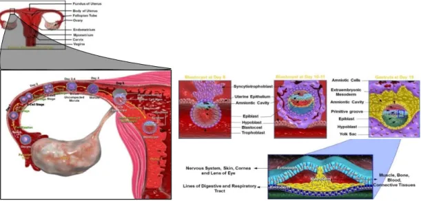
Human prenatal development starts at the time of fertilization and can be divided into three different periods, blastocyst or zygotic period; embryonic period and fetal period (Cochard & Netter, 2002). The zygotic period, which comprises the first two weeks of gestation, is a period of cell proliferation and leads to implantation of the blastocyst into the uterine wall. Through the embryonic period (third to the eighth week of gestation), the gastrulation occurs and most of the important organs and physiological systems develop. At this time, the embryo assumes a human appearance. The fetal period extends from the third month until birth. It is a period of growth, maturation and functionalization of the structures and organs. During these time course periods, the initial fertilized egg undergoes a systematic series of sequential changes to become increasingly complex, differentiated and ultimately giving rise to a fully developed human organism.
1.1. Early embryogenesis
Conception or fertilization is the fusion of a male spermatozoid with a female oocyte. This process, which normally occurs in the upper part of the uterine tube, marks the beginning of the human development and leads to the formation of a genetically unique and specialized cell, the zygote (Moore, Persaud, & Torchia). Immediately after fertilization, this newly formed totipotent cell makes its way to the uterus, a journey that takes five to seven days in humans. As it travels, the zygote undergoes a series of repeated mitotic divisions, called cleavages. Cleavage divisions result in a rapid increase in the number of cells that also become smaller with each division and are at this time called blastomeres. The blastomeres continue to divide and as the developing human enters in the uterus, by approximately three days after fertilization, it undergoes a compaction phenomenon leading to the formation of a 16-cell morula (Carlson, 2014; Moore et al.).
Posteriorly, by day four, a fluid starts to penetrate into the intercellular spaces of the morula and progressively forms a single blastocyst cavity. As the fluid
continues to increase into the blastocyst cavity, it separates the blastomeres and we can differentiate two groups of cells (Moore et al.). At this time, the embryo is a hollow ball of hundreds of cells, called blastocyst, with an internal mass of cells, the inner cell mass (ICM) or embryoblast, at one pole of the blastocyst, and an outer cell layer that forms the epithelial wall of the blastocyst, the trophoblast. The trophoblast will form the extraembryonic structures and the fetal component of the placenta whereas the cells of the ICM are pluripotent stem cells that can give rise to all cell types of the three embryonic germ layers, i.e., Ectoderm, Mesoderm, and Endoderm, and the Germ Cell Lineage, as well as to the non-trophoblast tissues (Yolk sac, Allantois, and Amnion) that support the developing embryo (Carlson, 2014; Moore et al.; Schoenwolf et al.).
Around day six of development, the 150 micrometers sized embryo starts to invade the endometrial epithelium and by the end of the second week of human gestation the blastocyst is completely implanted in the endometrium (Cochard & Netter, 2002; Moore et al.). As implantation proceeds, the trophoblast proliferates quickly and differentiates into the cytotrophoblast, an inner actively proliferating layer, and into the syncytiotrophoblast, which erodes the endometrial tissues, whereas the blastocyst changes morphologically resulting in a flat bilaminar plate of epithelial cells, the embryonic disc. This bilaminar embryonic disc consists of an upper layer of columnar cells, the epiblast, and a lower layer of cuboidal cell, the hypoblast or primitive endoderm. The epiblast contains the pluripotent cells that will form the three germ layers and the hypoblast will give rise to the yolk sac (Carlson, 2014; Moore et al.; Schoenwolf et al.). The formation of the bilaminar embryonic disc marks the appearance of the first axis in the embryo, the primitive dorsal-ventral axis with the epiblast defining the dorsal side and the hypoblast defining the ventral side of the embryo (Carlson, 2014; Moore et al.; Schoenwolf et al.).
At the start of the third week of human development, the cells of the embryonic epiblast begin to differentiate in a process known as gastrulation – formation of the three germ layers. This process, which marks the beginning of morphogenesis (creation of body shape), initiates between days fourteen and sixteen after fertilization and at this stage the embryo is known as gastrula (Schoenwolf et al.). The first morphological sign of gastrulation is the formation
5 of a longitudinal midline structure, the primitive streak (PS), on the surface of the epiblast, near the caudal end of the bilaminar embryonic disc (Moore et al.). This structure results from the proliferation and movement of the epiblast cells to the median plane of the embryonic disc. As the primitive streak elongates by the addition of cells to its caudal end, its cranial end proliferates to form the primitive node or Hensen’s node (Moore et al.). The primitive node contains a circular depression called the primitive pit, which is continuous caudally down the midline of the primitive streak with a trough like depression called the primitive groove (Carlson, 2014). This groove results from the invagination movement (inward movement) of cells from the epiblast through the primitive streak, a process called ingression (Schoenwolf et al.).
Shortly after the primitive streak appears, cells from the epiblast move toward the primitive streak, ingress in this structure and then migrates, in a temporal and spatial well-defined sequence, into the interior of the embryonic disc, to form two new germ layers. During this process, the cells change their structure and organization, undergoing an epithelial-mesenchymal transition (EMT). While in the epiblast they present the typical properties of epithelial cells, i.e. closely connected, well-defined apical and basal surfaces, expression of E-cadherin, when they become free of the epiblastic layer in the primitive groove, they assume the morphology and characteristics of mesenchymal cells (spindle-shaped morphology, expression of N-cadherin instead of E-cadherin), which are able to migrate as individual cells if provided with the proper extracellular environment (Carlson, 2014).
The first epiblast cells to ingress in the primitive streak invade the hypoblast, displacing its cells and replacing them with a layer of definitive endoderm. Others epiblast ingressing cells remain there and organize into the space between the epiblast and definitive endoderm to form a third germ layer – the intraembryonic mesoderm. As soon as the definitive endoderm and intraembryonic mesoderm are formed, epiblast cells no longer migrate toward the PS nor ingress through it. This remaining epiblast cells now constitutes the last germ layer, the ectoderm (Carlson, 2014; Moore et al.; Schoenwolf et al.). The embryonic ectoderm gives rise to peripheral and central nervous systems, the epidermis, hair, nails, sensory epithelia of eyes, ears and nose, many
Figure 1.1 – Summary of the first three week of human development.
During the first two weeks of gestation, the initial zygote embraces a period of cell proliferation that culminates with the implantation in the uterine wall and appearance of a bilaminar embryonic disc consisting of the epiblast and hypoblast. During the third week, gastrulation occurs and the bilaminar embryonic disc was converted in a three-layered disc, composed of ectoderm, endoderm and mesoderm.
connective tissues of the head, some glands and neural crest cells, which contribute to the formation of many organic systems (Moore et al.; Schoenwolf et al.). Embryonic endoderm is the source of the epithelial linings of the digestive and respiratory tracts, auditory tube, urinary bladder and most of the urethra. It also contributes to the parenchyma of the liver, pancreas thymus, thyroid and parathyroid glands (Carlson, 2014; Gilbert, 2010; Moore et al.; Schoenwolf et al.). Embryonic mesoderm gives rise to striated and smooth muscles, blood cells, blood vessels lineage, serosal linings of all body cavities, ducts and organs of the reproductive and excretory systems, and most of the cardiovascular system. It is the source of all connective tissues, including cartilage, bones, tendons, ligaments, dermis, and stroma (connective tissue) of internal organs, excluding the head and limbs (Carlson, 2014; Gilbert, 2010; Moore et al.; Schoenwolf et al.). By the end of the third week, the process of gastrulation is completed and the bilaminar embryonic disc was converted into a flat three-layered disc, composed of ectoderm, endoderm and mesoderm that will give rise to all the tissues and organs of the embryo. Furthermore, gastrulation is responsible for the definition of all major body axes through the formation of a temporary structure called the primitive streak. The primitive streak establishes a visible longitudinal axis of bilateral symmetry around which all embryonic structures will organize and align.
7
2.
Patterning the embryo – Left-Right Axis Specification
2.1. Anterior-posterior and dorsal-ventral axis
As three-dimensional objects, all vertebrate and invertebrate organisms present three different body axes, the anterior-posterior (AP) axis, the dorso-ventral (DV) axis and the left-right (LR) axis. The formation of these axes however varies from species to species. Whereas in fish and frog the dorso-ventral and anterior-posterior axes are determined at fertilization by the distribution of the yolk and the entry position of the sperm (Gilbert, 2010), in mouse and human, when the embryo implants into the wall of the uterus it is still symmetric without any axe. In these embryos, as embryogenesis proceeds, the DV axis is specified as the proximal-distal axis from the implantation site. Subsequently, the AP axis is arbitrarily determined in the plane perpendicular to the DV axis (Alarcon & Marikawa, 2003; Beddington & Robertson, 1999). So, after implantation, the future AP axis is marked anteriorly by the polarization of the head primordium and posteriorly (tail) by the appearance of the primitive streak, while the prospective DV axis is defined by the formation of the three germ layers, the endoderm marking its ventral side and the ectoderm being dorsal (Hamada & Tam, 2014; Hirokawa et al., 2009).
2.2. Left – right asymmetry
Theoretically, in a three-dimensional structure, once the orientation of two orthogonal axes is acquired, the orientation of a third axis would be spatially delineated by default (Hamada & Tam, 2014). This axiom of geometry seems to fit to the formation of the three body axes.
During development, once the AP and DV axes are established, it is argued that the LR axis is consistently oriented relative to the dorsal–ventral and anterior– posterior axes (Hashimoto & Hamada, 2010). Although many insights had been made regarding the generation of the anterior-posterior and dorsal-ventral axes, much remains to be understood about how the LR axis is consistently oriented with respect to the other two and what are the molecular mechanisms
underlying the process of LR asymmetry generation (Vandenberg & Levin, 2012). In the past 25 years, this process has been extensively studied and many genes and pathways were uncovered. Nonetheless, the precise mechanisms have not been determined making difficult to conceptualize therapeutic or preventative approaches (Amack, 2014).
In the next pages, I will provide a review about the current knowledge of how left-right symmetry is established in animal models. Resuming the agreements and disagreements in the field and the proposed models to explain how by breaking the bilateral symmetry and creating LR differences in the body plan, the initial symmetric embryo is transformed into a complex organism, externally bilateral symmetric and internal asymmetric.
2.2.1. The three main steps
The establishment of left-right (LR) asymmetry during early embryogenesis is critical for the organization of the embryo body plan and for the correct positioning and morphogenesis of the internal organs (Shiratori & Hamada, 2014). There is a general agreement in the field that the process by which this LR asymmetry is generated requires and can be divided in three different main phases/steps (Figure 1.2): In the first step, there is a breaking of the initial bilateral symmetry, allowing that the LR axis is consistently oriented relative to the dorsal-ventral and anterior-posterior axes. The second step consists in the transference of an LR-biased signal (or signals) to the lateral plate mesoderm (LPM), which leads to differential gene expression of signaling molecules in cell fields on either side of the midline. In the third step, this asymmetric gene expression of signaling molecules drives changes in cell behavior, such as proliferation and migration rate, which lead to the LR asymmetric position and morphogenesis of the internal organs (Shiratori & Hamada, 2006, 2014; Vandenberg et al., 2013; Vandenberg & Levin, 2013). In the last years, this process has been extensively studied to decipher the molecular mechanisms underlying these three steps of left-right asymmetry establishment. These studies revealed a general agreement between researchers regarding the main molecular pathways involved in steps two and three, decoding that these
9
Figure 1.2 – General process of LR asymmetry generation.
Generation of LR asymmetry comprises 3 steps. In the first one, LR axis is oriented relative to the DV and AP axes due to the action of an asymmetric signal. The second steps involve the transference of this signal to the LPM and differential genetic expression on either side of LPM. The last step relay on the LR asymmetric position and morphogenesis of the internal organs.
(Adapted from: (Vandenberg, Lemire, & Levin, 2013))
Step 1 – Symmetry breaking LR axis consistently oriented relative
to the dorsal-ventral and anterior-posterior axes.
Step 2 – Amplification and transmission of a left–right-biased signal to LPM Differential genetic expression on either
side of LPM.
Step 3 – situs-specific morphogenesis LR asymmetric position and morphogenesis of the internal
organs. Step 2
Step 3
cascades are highly conserved from sea urchin to mouse (Nakamura & Hamada, 2012). In contrast, the origin of the symmetry-breaking event (first step) is controversial because no unifying mechanism between model organisms has been identified to date and it seems that this process has diverged during evolution (Nakamura & Hamada, 2012; Sauer & Klar, 2012).
2.2.1.1. Models for symmetry breaking (first step)
The mechanism beyond the process of symmetry breaking was put in debate with the conceptual F-molecule model (Figure 1.3) proposed by Brown and Wolpert in the nineties (Brown & Wolpert, 1990). In their analysis, they postulated that a symmetric embryo must distinguish is left from its right side after the establishment of the anterior-posterior and dorsal-ventral axis, by consistent orientation of an intracellular component that is chiral by virtue of its biochemical structure (Vandenberg & Levin, 2013). This “F-molecule” would
have three arms and when two arms are aligned with the preexisting axis, the third arm would be automatically aligned along the future LR axis (Brown & Wolpert, 1990; Hashimoto & Hamada, 2010; Vandenberg & Levin, 2013). Therefore, in the field, there is an agreement about the need of an “F” molecule for the origin of asymmetry prior asymmetric transcriptional events. However, there is considerable debate on some major issues: what is the chiral element that first breaks symmetry? When during embryogenesis is asymmetry initiated? How conserved across diverse species are these mechanisms, and which model systems best represent the “general case”?(Vandenberg et al., 2013; Vandenberg & Levin, 2013). In recent years, two models, which are cytoskeleton-dependent and whose mechanism of action fulfill the chiral “F” molecule hypothesis proposed by Brown and Wolpert, were identified and debated. One model predicts that unidirectional cilia-driven extracellular fluid flow (nodal flow), produced on the LR organizer, during gastrulation, is the origin of LR asymmetry, whereas the second model – intracellular model – postulates that LR asymmetry originates during very early developmental stages due to the chiral nature of the cytoskeleton (Levin, 2003, 2004; Vandenberg et al., 2013; Vandenberg & Levin, 2013). Although both mechanisms implicate the cytoskeleton, as the cilium is effectively a microtubular extension of the cytoskeleton, and lead to asymmetric Nodal activity (May-Simera & Kelley, 2012), they act at two very different developmental stages and seem to have less in common. Here, I will introduce only the first model.
Figure 1.3 – “F” molecule model.
(A) The chirality of a molecule (schematized by an F) tethered with respect to the other axes can orient the direction of the LR axis. (B, C) Three-dimension representation of how an “F” molecule allows each cell to know which direction is left and which is right.
(Adapted from: (Levin, 2004))
R L
11
2.2.1.1.1. The leftward flow model
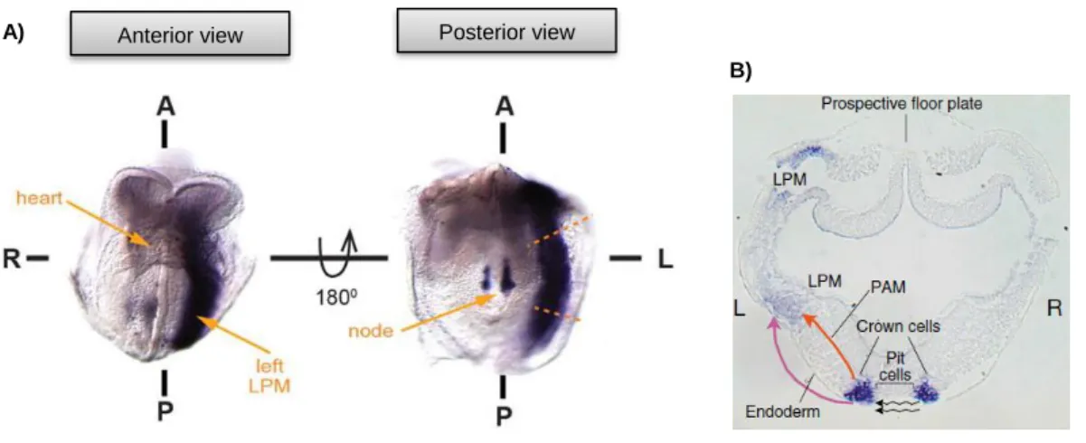
In some mammals (mouse, rabbit and humans), fish and amphibians the LR symmetry seems to be broken by the action of rotating cilia in a pit like structure called the “embryonic node” in mouse, “Kupffer’s vesicle’’ in zebrafish, and ‘‘gastrocoel roof plate’’ in frogs (Shiratori & Hamada, 2006; Yoshiba & Hamada, 2014; S. Yuan, Zhao, Brueckner, & Sun, 2015). The mouse node (Figure 1.4A), formed during gastrulation, is a transient small triangular structure with 50–100 μm in width and 10–20 μm in depth, located between the anterior notochord and the primitive streak in the ventral midline of the embryo (Hamada & Tam, 2014; Hirokawa et al., 2009). There are two types of cells in this cavity, the node pit cells, which are columnar epithelial cells located in the central region of the ventral node and the crown cells, squamous epithelial cells located on the edge of the node (Yoshiba & Hamada, 2014). Although both cell types present monocilia that project into the extraembryonic space, the crown cells have in most of the cases (90%) immotile cilia that are believed to sense the nodal flow, whereas, most of pit cells possess motile rotating cilia which are responsible for the generation of the asymmetric left-directed flow of extracellular fluid (nodal flow) in the node cavity (Figure 1.4B) (Hirokawa, Tanaka, & Okada, 2012).
Figure 1.4 – Node structure and cilia microtubules organization.
A) Scanning electron microscope of a mouse node at 7.5 days postcoitum. B) Direction of the leftward
fluid flow across the ventral node. Motile cilia are located at the central region of the node, while immotile cilia are present at the periphery of the node cavity (marked by dashed lines). C)Normal cilia with nine pairs of 9+2 arrangement microtubules connected with dynein motors. D) The central pair of microtubules is missing in immotile primary cilia and nodal cilia. In nodal cilia, the dynein motors remain in a chiral arrangement and produce a rotation-like movement (arrows). (Adapted from: (Hamada & Tam, 2014; Hirokawa, Tanaka, & Okada, 2009))
A) B)
C) D)
Left Right
2.2.1.1.1.1. Cilia
and
the
generation
of
the
unidirectional leftward flow
Normal motile ciliated cells present an axoneme composed of nine pairs of double microtubules arranged longitudinally along the axes and a pair of microtubules, referred as central pair, in the center of the cilia (9+2 arrangement) (Figure 1.4C) (Hirokawa et al., 2009). This central pair of microtubules, which defines the direction of the beating plane, is absent in the axoneme of immotile cilia cells and nodal cells. Thus, based on the microtubule arrangement (9+0) (Figure 1.4D), the node monociliated cells were thought to lack motility (Hirokawa et al., 2009). However, in 1998, Nonoka and co-workers examined the behavior of these monocilia using video-light microscopy. They discovered, for the first time, that the monocilia presented in the mouse node are motile but instead of move back and forth, like most motile cilia or flagella, they rotate vigorously at approximately 600rpm (Nonaka et al., 1998). Furthermore, when they added fluorescent beads in the extraembryonic fluid at the node region of wild-type embryos, they verify that the cilia of these cells rotate in a clockwise direction, generating a leftward (right-to-left) flow of fluid in the node cavity, which they designated nodal flow (Takeda et al., 1999) (Figure 1.5A). Okada also supported this hypothesis in 1999, by the analysis of node monocilia motility of inversus viscerum (iv) mutant mice, which harbors a mutation in the axonemal dynein protein Dnah11 (Lrd) gene. These results confirmed that the monocilia of heterozygous iv mutant embryos also rotates as fast as the wild-type embryo (~600rpm) and this movement can also generate a leftward flow of extraembryonic fluid in the node cavity. In contrast, in the iv mutant homozygous mice, the cilia are almost immotile and the flow absent (Okada et al., 1999), suggesting that in fact cilia are the responsible for the generation of the nodal flow.
But how and why is the flow generated in a leftward direction by the rotational movement of the motile monocilia? The answers to these questions rely in the posterior position of the basal body of each cilium, which lead to a posterior tilting of the cilia in the node cells. High-speed video microscopy observation revealed that the node cilia do not project vertically from the node cells, but they
13 are tilted posteriorly at an average angle of 30°- 40º (Nonaka et al., 2005), which allows the cilia to orient along the anterior‐posterior and dorsal‐ventral axes, as the F-molecule model proposed by Brown and Wolpert. These observations also revealed that a rightward rotational movement of the cilium occurs close to the cell surface, whereas a leftward motion is away from the surface (Hirokawa et al., 2009). Based on this, hydrodynamic principles predict that the shear resistance of the cell surface slows the rightward swing, but leaves the leftward stroke unimpeded to generate a leftward fluid flow, leading to a more efficient sweep of the cilia towards the left as opposed to the right (Okada, Takeda, Tanaka, Izpisua Belmonte, & Hirokawa, 2005). However, these observations do not explain the posterior tilting of the cilia. This tilting could be explained by the convex curvature of the apical membrane of the node cells and the shifting in the position of the basal body (Figure 1.5B), which seems to be related with planar cell polarity (PCP). Indeed, some core PCP components, like Dishevelled (Dvl), Van Gogh like 1 (Vangl1) and Prickle2 have polarized subcellular localizations (Figure 1.5B) in the node cells and knockout mice for these components result in disrupted posterior position of nodal cilia, abnormal nodal flow and LR defects (Hirokawa et al., 2012; Yoshiba & Hamada, 2014). Thus, the posterior positioning of the basal body, ensued from a mechanism of PCP, results in the posterior tilting of the cilia, which in turn is responsible for the generation of a unidirectional leftward flow that disrupts the initial LR symmetry in the mouse embryo.
A)
B)
2.2.1.1.1.2. Sensing
the
flow
–
morphogen/
chemosensor model
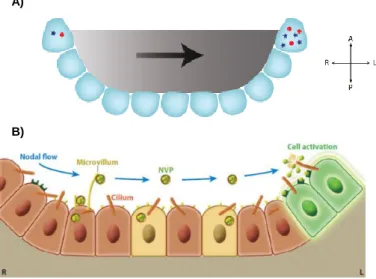
It is widely accepted that while central cilia of pit cells are mostly motile and generate the nodal flow, peripheral cilia of crown cells are mostly immotile and act as sensors of the fluid flow. However, how these cells at the edge of the node sense the flow is still debated. Two main model hypotheses have evolved to answer to this question. One hypothesis, termed “morphogen gradient” or chemosensor model, was firstly proposed by Nonaka and colleagues in 1988 (Figure 1.6A) (Nonaka et al., 1998). This model assumes that signaling morphogens, with sizes between 20-40 kDa, like Sonic Hedgehog (Shh), Fibroblast growth factor (FGF), Retinoic Acid (Ra) or Nodal are secreted into the node cavity and transported, due to the action of the unidirectional nodal flow, towards the left side of the embryo, where they bind to cell surface receptors and initiate a signaling cascade (Hirokawa, Tanaka, Okada, & Takeda, 2006). This stationary asymmetric accumulation of a determinant morphogen triggers signaling events that cement the asymmetry in the developing embryo. Although simple and straightforward, this hypothesis is questionable whether nodal flow can generate a chemical gradient in a closed cavity of node, where the leftward flow is balanced by the rightward counterflow (Hirokawa et al., 2006). Furthermore, what is the identity of the morphogen itself?
In a more sophisticated version of the chemical model (Figure 1.6B), the authors observed flowing materials, named nodal vesicular parcels (NVPs) (Hirokawa et al., 2012) that are released from slowly growing filopodia-like processes in all regions of the ventral node into the extracellular fluid flow, fragmented by contacting rotational cilia and finally absorbed by left-sided node cells (Hirokawa et al., 2012). Furthermore, pharmacological studies suggest that FGF, Shh and RA regulate the release of NVPs and that NVPs seem to carry
Figure 1.5 – Leftward nodal flow in the ventral node and posterior tilting of cilia.
A) Unidirectional leftward flow (blue) is generated by the clockwise rotation of nodal cilia due to the
posteriorly tilting of these cilia. B) Posterior tilting of cilia result from the posterior position of the basal body, which seems to be related with the subcellular localization of some core planar cell polarity molecules, Dishevelled, Van Gogh like 1 and Prickle2. Adapted from:(Yoshiba & Hamada, 2014).
15
Figure 1.6 – The two versions of the “morphogen gradient”/chemosensor model.
A) First “morphogen gradient” model proposed by Okada and colleagues. In this model, a morphogen
secreted into the node becomes asymmetrically accumulated on the left side due to the action of the nodal flow. Resulting thereby in symmetry breaking. B) Recent version of the “morphogen gradient” model propose that nodal vesicular parcels (NVPs) containing a morphogen are carried to the left side of the node by nodal the flow. These NVPs are then fragmented and absolved by left-sided node cells, resulting therefore in the breaking of symmetry. Adapted from: (Norris, 2012).
the signal to elevate the calcium (Ca2+) concentration on the left side of the
node, which in turn is thought to influence the asymmetric gene expression in adjacent cells (Hirokawa et al., 2006). Indeed, Shh and RA are unsheathed and transported in the NVPs but they are unlikely to be the molecular determinant of LR polarity because asymmetric expression of downstream Shh or retinoic acid response genes has not been detected in or near the mouse node.
2.2.1.1.1.3. Sensing
the
flow
–
Two
cilia/mechanosensor model
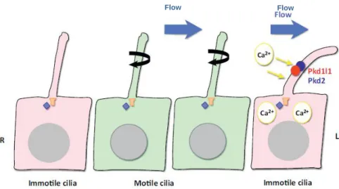
An alternative to the chemosensor hypothesis is that the hydrodynamic force generated by the flow is mechanosensed in the responding cells, most likely the crown cells, in the peripheral region of the ventral node (McGrath, Somlo, Makova, Tian, & Brueckner, 2003). This model, named “two-cilia” model (Figure 1.7), postulates that motile cilia at the center of the node produce an asymmetric left-directed flow of extracellular fluids detected by mechanical bending of a second non-motile group of cilia, which contain the Ca2+
permeable cation channel Polycystin-2 (Pkd2) exclusively on the left edge of the
A)
node. This results in the elevation of intracellular Ca2+ and leads to subsequent
asymmetric patterns of gene expression (Hamada & Tam, 2014; McGrath et al., 2003; Yoshiba & Hamada, 2014).
Some evidences are in agreement with this idea, the first comes from kidney cells in which immotile cilia respond to flow-induced bending mechanical stress by initiating an influx of extracellular Ca2+ through mechanosensory complex
channels (Pkd1/Pkd2 (Praetorius & Spring, 2001). Surprisingly, nodal cilia cells (motile and immotile) do not express Pkd1 and in its absence LR asymmetry is not disrupted (Karcher et al., 2005), suggesting that in nodal cells, Pkd2, which is thought not to be a sensory protein per se, will form a sensory complex with another partner. In fact, in mouse and fish cilia, Pkd2 physically interact and form a complex with a paralogue of Pkd1, Pkd1l1. Moreover, PKd1l1 is expressed in a pattern that corresponds spatially and temporally to the establishment of LR asymmetry and both proteins localize in motile and immotile cilia of the ventral node (Field et al., 2011; Kamura et al., 2011). So, in nodal cells, the same process of sensing the flow may occur via the flow-sensing complex Pkd1l1/Pkd2, being Pkd1l1 the sensor and Pkd2 the effector. Second, asymmetric elevation of intracellular Ca2+ became randomized in iv
mice (flow absent) and absent when Pkd2 was deleted (Babu & Roy, 2013). Moreover, rescues assays of the Pkd2 gene in specific regions of the Pkd2 deficient mice revealed that Pkd2 expression is not necessary in pit cells, composed mostly of motile cilia, for correct LR development. However, correct LR development requires Pkd2 expression specifically in crown cells, where most immotile cilia are located, suggesting that Pkd2 function is needed in the immotile cilia to sense and respond to the flow (Norris & Grimes, 2012). Lastly, an elegant work by Hamada’s lab in Japan has provided proof of the sensory function of cilia in Nodal cascade induction. First, the authors verified that only two rotating cilia were required to establish normal sidedness. Second, they verified that artificially applied fluid flow was able to rescue the Nodal cascade in a mouse model of Kartagener syndrome, i.e. in embryos without flow due to immotile cilia, in a mechanism dependent on the calcium channel Pkd2.
17 Furthermore, rescue of Kinesin family member 3A (Kif3a) expression only in the crown cells of these animals, results in the generation of a weak leftward flow and Nodal expression in the LPM. These observations suggest that a weak and transient local flow produced by the few motile cilia crown cells and sensed by the neighboring immotile cilia cells is sufficient to initiate LR asymmetric gene expression.
Although all the evidences mentioned above support the two cilia hypothesis, the bending of sensory cilia has not been verified by in in vivo observations and all the cilia in Kupffer’s vesicle of Medaka fish were directed visualized as motile cilia, expressing Lrd, Pkd1l1 and Pkd2 (Babu & Roy, 2013). Furthermore, inhibition of FGF signaling, which is involved in the release of NVP in the morphogen hypothesis, suppresses Ca2+ asymmetric intracellular elevation
without disturbing the flow, suggesting that FGF signaling and NVPs are needed for the asymmetric distribution and consequent normal LR patterning (Tanaka, Okada, & Hirokawa, 2005). Therefore, these two mechanisms may not be mutually exclusive, but rather may run in parallel or work synergistically with each other.
Figure 1.7 – “Two cilia” model.
Motile cilia at the center of the node generate a leftward nodal flow that is detected by mechanical bending of immotile cilia cells in the left edge of the node. These cells contain the Ca2+ permeable channel Pkd2 and Pkd1l1 exclusively on the left edge of the node resulting in the elevation of intra cellular Ca2+ and subsequent asymmetric patterns of gene expression. Adapted from: (Yoshiba & Hamada, 2014).