Universidade de Lisboa
Faculdade de Medicina de Lisboa
T
HE
C
LINICAL
R
ELEVANCE OF
D
RUG
I
MMUNOGENICITY
Sandra Pinheiro Garcês da Gama
Doutoramento Medicina
Reumatologia
Universidade de Lisboa
Faculdade de Medicina de Lisboa
T
HE
C
LINICAL
R
ELEVANCE OF
D
RUG
I
MMUNOGENICITY
Sandra Pinheiro Garcês da Gama
Orientador: Professora Doutora Jocelyn Demengeot
Co-Orientador: Professor Doutor Lucien Aarden
Doutoramento Medicina
Reumatologia
Todas as afirmações efetuadas no presente documento são da exclusiva responsabilidade do seu autor
.
A impressão desta dissertação foi aprovada pelo Conselho
Científico da Fculadade de Medicina de Lisboa em reunião de
28 de Outubro de 2014.
A
BSTRACT
The introduction of biologic therapies into clinical practice has greatly improved the
treatment of chronic disabling inflammatory diseases, such as Rheumatoid Arthritis,
Spondylarthritis or Inflammatory Bowel Diseases, among others. However, a sizeable
fraction of patients never achieve therapeutic response or, more often, cannot maintain
therapeutic response over time. Among the pitfalls of biologicals is their potential
immunogenicity and the associated anti-drug antibodies (ADAb) produced by the patients,
which promote faster clearance/neutralization of the drug in circulation and thus interfere
with drug efficacy. Moreover, ADAb have also been associated with adverse events.
In this work we aimed at formally document the impact of ADAb in clinical outcomes, and
use this analysis to construct and test an algorithm for therapeutic decisions based on explicit
biomarkers of immunogenicity.
To evaluate the clinical relevance of biological drug’s immunogenicity, we first performed
a systematic review of the literature coupled with a meta-analysis. We evidenced that in the
presence of detectable ADAb therapeutic response may be reduced by as much as 80%. Such
impact is attenuated, although not abrogated, by concomitant immunosuppression,
particularly with methotrexate that associates with reduced ADAb production. Differences
in the immunogenic profile of specific biologics were also verified, with monoclonal
antibodies exhibiting higher immunogenicity than fusion proteins. We next assessed the impact of immunogenicity on drug’s safety profile by following a cohort of patients receiving intravenous infliximab, a TNF-inhibitor. Infusion-related adverse events occurred
developed an acute reaction during or immediately after the infusion, requiring medical
intervention.
To evaluate the relevance of drug immunogenicity assessment for therapeutic decisions, we
first defined a convenient method to assess immunogenicity on a routine basis. We verified
that a newly developed Bridging ELISA performed as well as antigen-binding radio-immuno
assay, currently considered by many as the “gold-standard” to assess ADAb. Next, we
designed an algorithm for the management of patients receiving biologic therapies, which
combines the usual clinical evaluation with immunogenicity assessment at every three
months. This algorithm was tested in a cohort of RA patients treated with one of the three
most commonly used biologics. We evidenced that patients who followed therapeutic
strategies concordant with the proposed algorithm had close to 10-times higher probability
of achieving low disease activity, when compared to those who followed other strategies
commonly adopted in current clinical practice.
Our work demonstrates that a personalized, evidence-based approach for the management
of patients receiving biologic therapies will lead to safer and most cost-effective strategies.
R
ESUMO
As terapêuticas biológicas revolucionaram o prognóstico de doenças inflamatórias crónicas
e incapacitantes como a Artrite Reumatóide, as Espondilartrites, as Doenças Inflamatórias
do Intestino, entre outras. Ainda assim, um número significativo de doentes não responde a
esses fármacos ou, mais frequentemente, perde a resposta inicial ao longo do tempo de
tratamento. Uma das principais limitações destas terapêuticas é o seu potencial imunogénico
e a consequente formação de anticorpos anti-fármaco (AAF) por parte dos doentes a quem
são administrados. Os AAF promovem uma rápida eliminação/neutralização do fármaco em
circulação e podem também interferir com o seu perfil de segurança, associando-se a um
maior risco de efeitos adversos.
Este trabalho pretende documentar formalmente o impacto clínico dos AAF, utilizando essa
informação na construção e validação de um novo algoritmo de apoio à decisão terapêutica
baseado em biomarcadores de imunogenicidade.
Para avaliar a relevância clínica da imunogenicidade foi inicialmente realizada uma revisão
sistemática da literatura e uma meta-análise. Evidenciámos que na presença de AAF
detetáveis há uma redução de até 80% da resposta à terapêutica. Este impacto é atenuado,
mas não eliminado, pelo tratamento concomitante com imunossupressores, particularmente
com metotrexato, que se associa a uma redução da produção de AAF. Foram também
verificadas diferenças importantes no perfil imunogénico entre os fármacos, com os
anticorpos monoclonais a exibirem maior imunogenicidade que as proteinas de fusão.
Seguidamente foi avaliado o impacto da imunogenicidade no perfil de segurança destes
fármacos, avaliando no tempo uma cohort de doentes tratados com infliximab, um inibidor
do TNF-alfa. Reações adversas agudas associadas à infusão do fármaco ocorreram
com AAF tiveram uma reação aguda durante ou imediatamente após a infusão do biológico,
requerendo intervenção médica imediata.
No sentido de avaliar a relevância da avaliação da imunogenicidade para a melhoria das
decisões terapêuticas, começámos por definir o método laboratorial mais conveniente para
monitorização da imunogenicidade na prática clínica de rotina. Constatámos uma boa
concordância entre um novo método de ELISA desenvolvido (“Bridging ELISA”) e o
método de radioimmunoensaio (“RIA-ABT”), considerado por muitos como “gold
standard” na deteção dos indivíduos AAF-positivos. Seguidamente, desenhámos um
algoritmo de apoio à decisão terapêutica para doentes medicados com terapêuticas
biológicas, que combina a atual avaliação clínica com a monitorização da imunogenicidade
a cada três meses. Este algoritmo foi testado numa cohort de doentes com Artrite Reumatóide
inicialmente medicados com um dos três agentes biológicos mais utilizados no tratamento
desta doença. Evidenciámos que os doentes que tinham seguido estratégias terapêuticas
concordantes com o algoritmo proposto tiveram cerca de 10 vezes maior probabilidade de
alcançarem uma baixa atividade de doença, quando comparados com os doentes que
seguiram outras estratégias, frequentemente adotadas na atual prática clínica.
O nosso trabalho demonstra que uma abordagem personalizada e cientificamente orientada
aos doentes medicados com terapêuticas biológicas permite desenhar estratégias mais
seguras e custo-efetivas. Os nossos resultados têm não só uma elevada relevância clínica,
A
GRADECIMENTOS
Este trabalho marca e descreve uma etapa que para sempre viria a mudar a minha forma de
ser e de pensar. Muitos foram aqueles que contribuiram para esta mudança e tornaram
possível este trabalho, aos quais me sinto profundamente grata.
Primeiramente, o meu agradecimento à Professora Doutora Jocelyne Demengeot pelos
ensinamentos, orientação, encorajamento e amizade que sempre demonstrou. É hoje uma
das minhas grandes referências, a nível professional mas também pessoal. A sua
generosidade foi determinante na minha chegada até aqui e poder trabalhar com ela é para
mim um enorme privilégio.
Os meus agradecimentos a toda a comunidade do Instituto Gulbenkian de Ciência (IGC),
um instituto de características científicas e humanas de excelência, onde as palavras rigor e
cooperação assumem uma dimensão maior. Um profundo agradecimento a todos os
elementos do grupo Fisiologia dos Linfócitos que me acolheram ao longo de três anos,
sempre disponíveis a ensinar, a ajudar e a discutir, a qualquer hora, resultados e tantas dúvidas… Um agradecimento especial ao Professor Doutor Constantin Fesel pelos seus ensinamentos e disponibilidade, bem como À Professora Doutora Ana Regalado, pelo seu
apoio e contribuição técnica laboratorial. Com todos eles adquiri uma nova perspectiva do
trabalho em equipa e formei amigos para a vida.
Agradeço profundamente ao Programa Gulbenkian de Formação Médica Avançada
(PGFMA) onde, na verdade, tudo começou. Um especial agradecimento à Professora
Doutora Leonor Parreira, ao Professor Doutor João Ferreira e ao Professor Doutor António
Coutinho, pelos ensinamentos, pelo apoio, por toda a amizade e inspiração. A todos os
conhecimentos e experiências de vida, o meu muito obrigada. Aos meus colegas do PGFMA
com quem partilhei diariamente, ao longo de seis meses, uma experiência única de formação
científica que para sempre viria a mudar a nossa forma de ser Médicos. Uma palavra de
sentimento especial para com o nosso colega e amigo Jaime Almeida, infelizmente já não
entre nós, que nos deixa um sentimento de vazio e perda irreperáveis.
O meu profundo agradecimento ao Professor Doutor Lucien Aarden que me introduziu na
área da Imunogenicidade, pelos seus ensinamentos, pelo apoio e generosidade
demonstradas. Ao Sanquin Research Institute, diretamente aos elementos do Departamento
de Investigação em Imunopatologia, o meu agadecimento por todo o apoio e ensimentos
técnicos na execução dos testes laboratoriais de avaliação de Imunogenicidade.
À Dra. Elizabeth Benito-Garcia, reumatologista e epidemiologista, pela orientação e
ensinamentos na condução da meta-análise e desenho dos estudos clínicos.
À Professora Doutora Marília Antunes, estatista, pela orientação e apoio em determinadas
análises estatísticas.
Ao Hospital Garcia de Orta, em especial ao Serviço de Reumatologia, o meu agradecimento
pelo apoio prestado, em particular à Enfermeira Lurdes Barbosa pela preciosa colaboração
na colheita das amostras biológicas. Um agradecimento muito especial aos meus doentes
que sempre de forma tão generosa atenderam a todas as minhas solicitações. Muito obrigada
por participarem neste trabalho, feito para vocês.
Aos meus familiares e amigos que sempre me incentivaram e apoiaram. Entre eles estão os
amigos de sempre e para sempre. Aos meus sogros, por me ajudarem a manter uma estrutura
Aos meus pais, Dário, Maria e Cipriano, agradeço tudo. Obrigada pela educação, pela
inspiração e pelos valores que me transmitiram ao longo de uma vida. Agradeço o seu apoio
incondicional, o suporte imenso e constante, a mim e à minha família.
Por fim agradeço e peço desculpa aos meus filhos, Simão e Afonso pelas horas de brincadeira
perdidas. Espero que, mesmo quando ausente, lhes possa ter ensinado o valor do trabalho e
da preserverança para atingir os nossos sonhos. Ao Gama, meu marido, meu companheiro e
amigo incondicional, muito obrigada pelo apoio, motivação e inspiração. Obrigada por
T
ABLE OF
C
ONTENTS
Abstract ... vii
Resumo ... ix
Agradecimentos ... xi
Table of Contents ... xv
List of Figures ... xix
List of Tables ... xxi
Acronyms ... xxiii
1 General Introduction ... 1
1.1 Biologic Therapies ... 3
1.2 Imunoglobulins Structure and Function ... 3
1.3 Antibody Production: from hybridoma technique to advanced biothechnology... 6
1.4 Drug Immunogenicity ... 9
1.4.1 Drug-related Factors Influencing Immunogenicity ... 11
1.4.2 Patient-related Factors ... 13
1.4.3 Immunogenicity Assessment... 16
1.5 Rheumatoid Arthritis ... 23
1.6 Tumor Necrosis Factor Alpha ... 26
1.7 TNF Inhibitors in RA ... 28
1.8 Non-TNFi Biologic Therapies in RA ... 31
1.9 The Current Management of RA And The Treat-To-Target Strategy ... 33
2 Objectives/Scope of the Thesis ... 39
3 Results ... 41
3.1 The Immunogenicity of TNFi Therapies in Immune-mediated Inflammatory Diseases – a systematic review of the literature with a meta-analysis ... 41
3.1.2 Methods ... 42
3.1.3 Results ... 43
3.1.4 Discussion ... 46
3.2 The impact of Immunogenicity on Drug Safety Profile ... 74
3.2.1 Introduction ... 74
3.2.2 Objectives ... 75
3.2.3 Methods ... 75
3.2.4 Results ... 76
3.2.5 Discussion ... 78
3.3 Immunogenicity Assessment in Routine Clinical Practice ... 84
3.3.1 Introduction ... 84
3.3.2 Objectives: ... 85
3.3.3 Methods: ... 85
3.3.4 Results: ... 89
3.3.5 Discussion ... 90
3.4 A Preliminary Algorithm Introducing Immunogenicity Assessment in the Management of RA Patients Receiving Tumor Necrosis Factor Inhibitors Therapies ... 97 3.4.1 Introduction ... 97 3.4.2 Objectives ... 98 3.4.3 Methods ... 99 3.4.4 Results ... 102 3.4.5 Discussion ... 105 4 General Discussion ... 115
4.1 The Impact of ADAb on Therapeutic Responses ... 116
4.2 The Influence of Concomitant Immunosuppression and Other Possible Modulator Factors on ADAb Detection ... 117
4.3 Etanercept (fusion protein): Biologic Free of Significant Immunogenicity . 119 4.4 The Impact of ADAb on Drug’s Safety Profile ... 121
4.5 Suitable Assays to Monitor Immunogenicity in Routine Clinical Practice .. 123
4.6 Integrating Immunogenicity Information in the Management of Patients Receiving Biologic Therapies ... 125
4.8 Biologic Dose Reduction Programs Based on Serum Drug Levels ... 135
5 Conclusions ... 139
5.1 Relevance (and perspective) of This Work for Clinical Practice ... 139
5.2 Relevance for Economic and Societal Values ... 140
6 References ... 143
L
IST OF
F
IGURES
Figure 1 – Immunoglobulins Structure ... 4
Figure 2 – Complementary-Determining Regions (CDR1, CDR2 and CDR3) ... 5
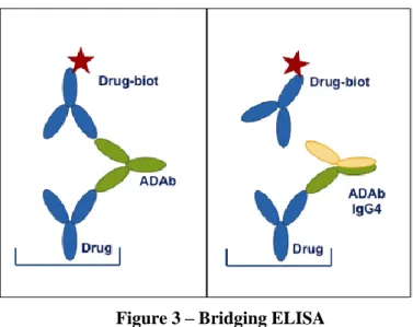
Figure 3 – Bridging ELISA ... 21
Figure 4 – Radioimmunoassay (RIA) – Antigen Binding Test (ABT) ... 21
Figure 5 – Flow of studies through the systematic review (SR) process ... 56
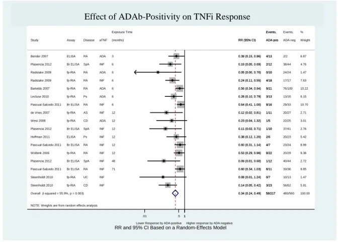
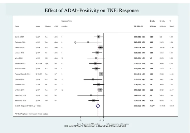
Figure 6 – Effect of ADAb positivity on TNFi response (excluding the study “Bartelds 2011”) ... 57
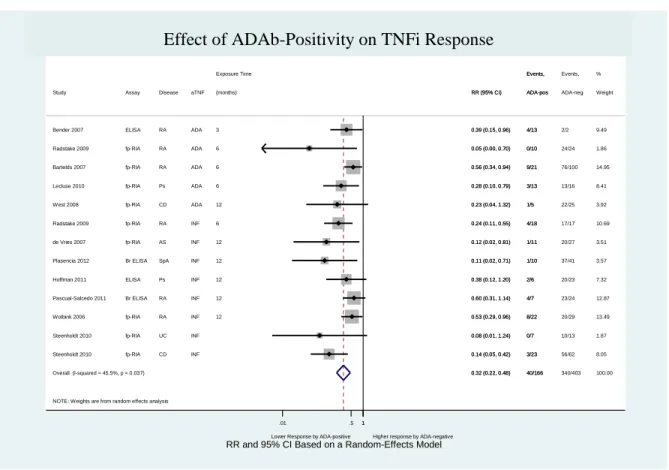
Figure 7 – Effect of ADAb positivity on TNF responsei (excluding the study “Bartelds 2007”) ... 58
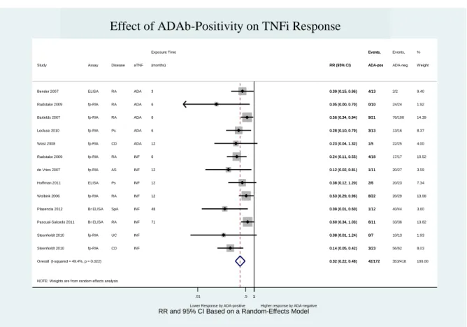
Figure 8 – Effect of ADAb positivity on TNFi response (including “Pascual-Salcedo 2011” and “Plasencia 2012” at 6 months of follow-up time)... 59
Figure 9 – Effect of ADAb positivity on TNFi response (including “Pascual-Salcedo 2011” and “Plasencia 2012” at 12 months of follow-up time)... 60
Figure 10 – Effect of ADA positivity on TNFi response (including “Pascual-Salcedo 2011” and “Plasencia 2012” at > 48 M follow-up time) ... 61
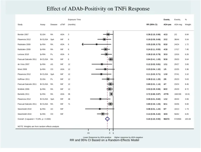
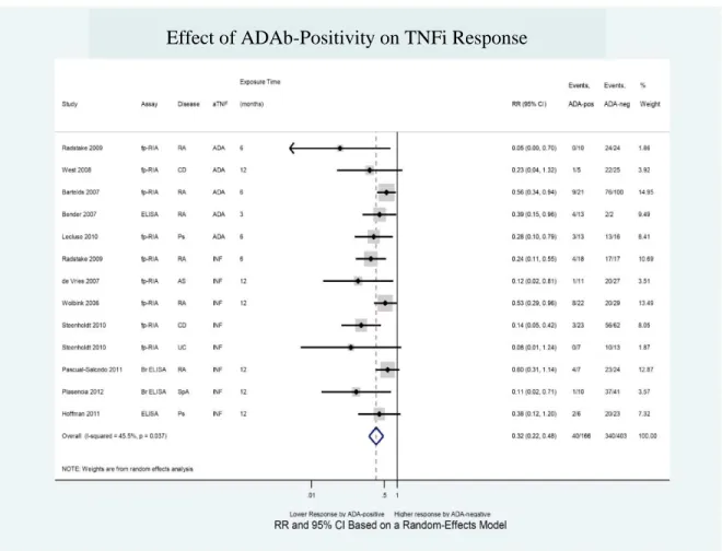
Figure 11 – Effect of ADAb positivity on TNFi response ... 62
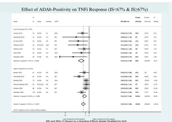
Figure 12 – Effect of ADAb positivity on TNFi response (IS<67% and IS≥67%) ... 63
Figure 13 – Effect of ADAb positivity on TNFi response (MTX<74% and MTX≥74%) .. 64
Figure 14 – Effect of ADAb positivity on TNFi response (by diagnosis) ... 65
Figure 15 – Effect of ADAb positivity on TNFi response (by diagnosis) ... 66
Figure 16 – Effect of ADAb positivity on TNFi response according to initial biologic dose ... 67
Figure 17 – Egger’s publication bias plot for the effect of ADAb positivity on TNFi response ... 68
Figure 18 – Effect of immunosuppression on ADAb production (by excluding “Bartelds 2010” and “Bartelds 2011”) ... 68
Figure 19 – Effect of immunosuppression on ADAb production (by excluding “Bartelds 2007” and “Bartelds 2011”) ... 69
Figure 20 – Effect of immunosuppression on ADAb production (by excluding “Bartelds
2007” and “Bartelds 2010”) ... 70
Figure 21 – Effect of immunosuppression on ADAb production ... 71
Figure 22 – Effect of immunosuppression on ADAb production (by assay) ... 72
Figure 23 – Egger’s publication bias plot for the effect of IS on ADAb production ... 73
Figure 24 – Patients with infusion-related adverse events (IrAE) ... 82
Figure 25 – Patients with detectable ADAb ... 82
Figure 26 – Proportion of patients with IrAE among ADAb-positives and ADAb-negatives ... 83
Figure 27 – Serum drug levels at every week between two infliximab infusions in 4 IBD patients who had IrAE ... 83
Figure 28 – ELISA to assess serum drug levels ... 95
Figure 29 – Assays used to measure ADAb ... 95
Figure 30 – Proportion of ADAb-positive patients detected in each assay. ... 96
Figure 31 – ADAb titres assessed by IgG-RIA-ABT and specific IgG4-RIA-ABT ... 96
Figure 32 – Preliminary Algorithm to Approach RA Patients receiving TNF Inhibitor Therapies, introducing immunogenicity assessment ... 113
Figure 33 – C-Reactive Protein (CRP) concentration among negative and ADAb-positive patients, at study beginning ... 114
L
IST OF
T
ABLES
Table 1 – Eligibility criteria for studies included in the systematic review ... 50 Table 2– Study and baseline patient characteristics // 1. Quantitative Evidence ... 51 Table 3 – Meta-Regression stratified by clinical characteristics to address the effect of ADAb on drug response and the effect of IS on ADAb detection ... 55 Table 4 – Patient’s Baseline Characteristics... 110 Table 5 – Response and Low Disease Activity in patients from Group A and Group B, over one year after therapeutic decision ... 111 Table 6 – Therapeutic Response and Low Disease Activity among ADAb-negative and ADAb-positive patients, at study beginning ... 111 Table 7 – Patient’s Baseline Characteristics among ADAb+ e ADAb- ... 112
A
CRONYMS
Abs – AntibodiesACPA – Anticitrullinated Protein Antibodies
ACR – American College of Rheumatology
ADAb – Anti-drug Antibodies
ADCC – Cell-mediated Cytotosicity
ADCP – Antibody-dependent Cellular Phagocytosis
AE – Adverse Events
AE – Ankylosing Spondylitis
AIRE – Autoimmune Regulator Protein
APC – Antigen Presenting Cell
ASAS – Assessment of Spondyloarthritis International Society
ASDAS - Ankylosing Spondylitis Disease Activity Score
AZA – Azathioprine
BCR – B Cell Receptor
C – Constant
CD – Crohn’s Disease
cDNA – Complementary Deoxyribonucleic Acid
CDR – Complementary-Determining Regions
CI – Confidence Intervale
CNS – Central Nervous System
CRP – C-Reactive Protein
CTLA-4 – Cytotoxic T-Lymphocyte-associated Antigen 4
DAS – Disease Activity Score
DC – Dendritic Cells
DMARDs – Disease-modifying Antirheumatic Drugs
DNA – Deoxyribonucleic Acid
EAE – Experimental autoimmune Encephalomyelitis
EMA – European Medicines Agency
ESR – Erythrocyte Sedimentation Rate
EULAR – European League Against Rheumatism
FADD – Fas-Associated Death Domain
FDA – Food & Drug Administration
GEE – Gneralized Estimation Equation
GM-CSF – Granulocyte-Macrophage Colony Stimulating Factor
HCQ – Hydroxycloroquine
HLA – Human Leucocyte Antigen
IBD – Inflammatory Bowel Diseases
IFN – Interferon
Igs – Immunoglobulins
IL – Interleukin
IQR – Interquartile Range
IrAE – Infusion-Related Adverse Events
IS – Immunosuppression
IVIG – Intravenous Immunoglobulin
LFN – Leflunomide
LPS – Lypopolysaccharide
LTa – Lymphotoxin Alpha
mAb – Monoclonal Antibody
MCP – 6-Mercaptopurine
MHC – Major Histocompatibility Complex
MS – Multiple Sclerosis
mTEC – Medullary Thymic Epithelial Cells
MTX – Metothrexate
Nab – Neutralizing Antibody
NF-kB – Nuclear Factor kB
NK – Natural Killer
OMERACT – Outcomes Measures in Rheumatology Initiative
OR – Odds Ratio
PAD – Peptidyl Arginine Deaminase
PCR – Polymerase Chain Reaction
PD – Pharmacodynamics
PK – Pharmacokinetics
Ps – Psoriasis
PsA – Psoriatic Arthritis
RA – Rheumatoid Arthritis
RCTs – Randomized Clinical Trials
REM – Random Effect Models
RF – Rheumatoid Factor
RIA – Radioimmunoassay
RR – Risk Ratio
SD – Standard Deviation
SDAI – Simplified Disease Activity Index
SLZ – Salazopirine
SpA – Spondylarthritis
TACE – TNF Alpha Converting Enzyme
TCR – T Cell Receptor
TEC – Thymic Epithelial Cells
TLR – Toll-like Receptor
TNFa – Tumor Necrosis Factor alpha
TNFi – Tumor Necrosis Factor inhibitors
TRAF – TNF Receptor-Associated Factor
Tregs – T Regulatory Cells
TSA – Tissue-Specific Antigens
UC – Ulcerative Colitis
1 G
ENERAL
I
NTRODUCTION
Chronic inflammatory immune-mediated diseases are a heterogeneous group of poorly understood disorders, which aetiology remains largely unknown. They have been conceived as a result of a combination of genetic variants, acquired environmental triggers and stochastic events.
Rheumatoid Arthritis (RA), Spondylarthritis (SpA), Psoriasis (Ps) and Inflammatory Bowel Diseases (IBD) are among the most prevalent chronic inflammatory immune-mediated diseases that affect predominantly young people at productive age of life, inducing significant morbidity and mortality. Therefore, their social and economic impact on society is extremely high.
Despite the poorly understood aetiology of those diseases, their physiopathology ends in a chronic inflammatory response against self-tissues with the release of inflammatory mediators, production of autoantibodies and activation of leukocytes that will perpetuate immune response, leading to an extensive tissue damage and malfunction of the corresponding target organs.
Cytokines are potent-rate limiting extracellular molecules that specifically regulate the inflammatory response, the tissue damage and the repair mechanisms. An increasing body of evidence has revealed the critical role that cytokines play in the initiation and perpetuation of autoimmunity [1, 2]. The important role of cytokines in immune-mediated inflammatory mechanisms had lead to the idea that cytokine-based manipulation could offer a possibility to interfere with autoimmune process.
Such idea has revolutionized the treatment approach of the above-mentioned diseases, which the most well known example has been RA.
RA is a chronic disabling disease that affects primarily the joints, inducing an irreversible joint damage with significant loss of functionality. In the beginning of 90’s a paradigm shift occurred in the treatment approach of RA. In addition to small chemical molecules, such as methotrexate (MTX) or corticosteroids, large therapeutic proteins, also known as biologics, were introduced in the arsenal of therapeutic options for RA treatment. Biologics represent a distinct therapeutic class, which are produced through living organisms using
biotechnology and genetic engineering instead of being simply chemically synthesized. There are today a wide variety of biotechnologic-derived therapeutic proteins. In the context of chronic inflammatory immune-mediated diseases, biologics are mainly represented by monoclonal antibodies, and to a less extend by fusion proteins, which target several cytokines or cells that play critical role on immune-mediated inflammation. The first biologics used in the treatment of RA targeted the Tumor Necrosis Factor (TNF). Given the successful results other targets emerged, such as CD-20, co-stimulatory molecules B7.1/B7.2 and interleukin (IL)-6. These therapies provide better control of inflammation, increasing patient’s quality of life and, most importantly, their functionality.
The clinical benefit that biologics have brought to RA patients has been extensively demonstrated in randomized clinical trials (RCTs). However, biologics have now been used in clinical practice for more than a decade and a general pattern seemed to emerge, best analysed for Tumor Necrosis Factor inhibitors (TNFi) in RA: there is evidence that i) about one third of the patients would not respond to the biological therapy (primary non-responders); ii) one third would show clinical response, but the beneficial effect would fade away within the first 6-12 months of continuous therapy (secondary non-responders); and iii) the remaining third would maintain a clinical response beyond a year.
Over the last years increasing evidence has revealed the production of anti-drug antibodies (ADAb), naturally produced by the patients upon administration of biologics, as one of the main factors interfering with drug efficacy and safety profile. Although there were good indications that immunogenicity of TNFi is one of the main mechanisms behind treatment failure, this notion did not permeate the clinical practice.
The lack of suitable assays to assess immunogenicity in clinical practice has also prevented the expansion of the field. Immunogenicity assessment is technically challenging and only recently optimized assays, specifically tailored to detect ADAb, have emerged though little experience still exists.
Despite the great improvement in overall clinical responses afforded by biologicals, therapeutic failures to these drugs are frequent. Therapeutic decisions in these cases, such as whether to increase the dose or to switch to another biological of similar or different mechanism of action, would benefit to be guided by reliable biomarkers. Moreover, in responding patients, the same lack of guiding biomarkers prevents an educated and desirable
dose reduction program, as these therapies are supposed to be maintained for life. Monitoring drug levels and potential immunogenicity should help optimize the use of biological therapies. However, the way such information might be integrated in clinical practice towards more cost-effective strategies remains to be defined.
The high costs of biologic therapies represent a big concern for societies. Monitoring drug levels and ADAb might represent a very promising tool for an optimized and personalized use of biological therapies, as it allows revision of the costs engaged in these therapies while keeping as a priority the welfare of patients.
1.1
B
IOLOGICT
HERAPIESThe critical role of cytokines in all stages of the immune-mediated inflammatory process, lead to the idea that the manipulations of cytokine network could modulate immune responses and autoimmune diseases [1, 2]. The easiest and more efficient way to block cytokines is through monoclonal antibodies (mAbs), although other type of molecules such as decoy receptors can also bind cytokines with specificity and high affinity. The therapeutic potential of mAbs has revolutionized the pharmaceutical industry over the last years, which has lead to important refinements in the antibody manufacturing techniques.
Biotechnology-derived therapeutic proteins represent a group of medicines that are produced through live organisms and not simply chemically synthetized. Hence, they are often mentioned as biologics. There are a wide variety of biotechnology-derived therapeutic proteins available today for the treatment of several diseases. Beyond mAb and soluble receptors, enzymes, clotting factors, hormones, or cellular growth factors also exist, namely for the treatment of some genetic diseases where there is an absence or pathologic modification of the endogenous protein. Nonetheless, mAbs represent the great bulk of biologics today produced with therapeutic purposes.
1.2
I
MUNOGLOBULINSS
TRUCTURE ANDF
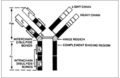
UNCTIONAntibodies (Abs) are a family of structurally related glycoproteins, also known as Immunoglobulins (Igs). Antibodies represent the secreted form of Igs that can also be produced in a membrane form (B cell receptor, BCR). All Abs have a common symmetric core structure of two identical covalently linked heavy chains and two identical light chains, each linked to one of the heavy chains – Figure 1.
Abs are classified into different isotypes and subtypes on the basis of amino-acid differences in the heavy chain constant (C) regions, which consist of three or four C domains [3]. The antibody classes or isotypes are called IgM, IgD, IgE, IgA and IgG. IgG isotype consist of four subtypes, numbered according to their frequency in peripheral blood: IgG1, IgG2, IgG3 and IgG4. Each Ig molecule contains 2 light chains of the same isotype, kappa (κ) or lambda (λ), which differ in their single C domain. Even within the same isotype, slight differences in the amino-acid sequences of the constant heavy or light chain also exist among different individuals, designated as allotypes [4].
Figure 1 – Immunoglobulins Structure
Allotypes expressed on the constant region of IgG heavy chain are referred as Gm (genetic markers) together with the isotype. With the exception of IgG4, different allotypes have been described for IgG1 (G1m), IgG2 (G2m) and IgG3 (G3m). Allotypes expressed on the constant region of κ light chain are referred as Km. No allotypes have been described for λ light chains. Specific Gm haplotypes exist in different populations. In a Caucasian population the G1m1,17 allotype is much less frequent than G1m3 [5]. Even within the same population group, inter-individual variations may also occur [4].
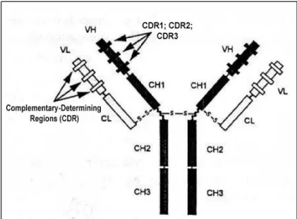
The N-terminal domains of heavy and light chains form the variable (V) regions of Ab molecules, which differ among antibodies of different specificities. The V regions of heavy and light chains each contain three separate hypervariable regions of about 10 a.a that are spatially assembled to form the antigen-combining site of the antibody molecule, known as complementary-determining regions (CDR) – Figure 2.
Figure 2 – Complementary-Determining Regions (CDR1, CDR2 and CDR3)
Antibody binding to antigen can be highly specific, but cross-reactions may also occur and the same antibody may bind different antigens. Antibodies can bind to two, or in the case of IgM, up to ten identical epitopes simultaneously, leading to enhanced avidity of the antibody-antigen interaction. The relative concentrations of antigens and antibodies may favour the formation of immune complexes.
The antibodies’ role does not extinguish in the antigen recognition. Antibodies also have effector functions, which are mainly mediated by the C regions of the heavy chains [6-8]. IgG1, IgG3 and to a lesser extend IgG2 are able to activate the complement system, by binding to the C1 complex via their Fc terminals [9]. Activation of the complement system may lead to target cell lysis, phagocytosis of pathogens, release of anaphylatoxins, among other pro-inflammatory effects [10]. Antibodies may also interact with Fc receptors, displayed at surface of immune cells such as macrophages, monocytes, dendritic cells or platelets, leading to their activation. This interaction may result in antibody-dependent cellular phagocytosis (ADCP), antibody-dependent cell-mediated cytotoxicity (ADCC) and cytokine release [11]. The interaction of Fc regions with neonatal Fc receptor protects IgG from degradation, increasing their half-live and mediates IgG transfer from mother-to-fetus [12]. In contrast, IgG4 has limited effector functions due to its low affinity for complement and Fc receptors [13, 14]. IgG4 represents a small fraction of total IgG content and it has been described in cases of prolonged or repeated antigenic stimulation [15]. It has been thought that IgG4 production may have regulatory or protective effect against chronic inflammatory reactions [16, 17]. Human IgG4 has the particular ability to exchange half of
the molecule with another IgG4 molecule of different specificity, resulting in a bispecific immunoglobulin molecule unable to cross-link identical antigens [17-21]. This dynamic Fab arm exchange introduces unpredictability on the binding ability of IgG4, which may be unwanted in case of human immunotherapy.
1.3
A
NTIBODYP
RODUCTION:
FROM HYBRIDOMA TECHNIQUE TO ADVANCED BIOTHECHNOLOGYThe production of antibodies at a large scale, in a reliable manner, started in 1975, with the development of mouse hybridoma technology by Cesar Milstein and Georges Köhler [22]. The contribution of such invention for science and medicine conceived to its inventors the Nobel Prize of 1984 for Medicine and Physiology, shared with Niels Jerne, who also made critical contributions to immunology.
The hybridoma technique consists in the fusion of a mouse myeloma and mouse spleen cells from an immunized donor, able to secrete antibodies of a single specificity against a predetermined antigen. Such hybrid cells can be grown in vitro in massive cultures to provide specific antibodies. Mouse hybridomas were developed for a number of research and therapeutic applications. However, clinical studies with murine mAbs had been disappointed due to their weak effector functions, short half-life and ability to evoke immune reactions in the human organism [23-25]. An intensive work was subsequently conducted in order to refine the production and the clinical use of therapeutic antibodies. To that, much had certainly contributed the easy access to hybridoma technology, which was never patented by its inventors, deliberatively.
In 1986, the US Food and Drug Administration (FDA) approved OKT3 (muromonab), the first therapeutic monoclonal antibody for human use. OKT3 is a murine monoclonal antibody of the IgG2a isotype, which targets CD3, a molecule that is part of a multimolecular complex found in association of T-cell receptor (TCR) for antigen. OKT3 acts by blocking T-cell function and it was shown to effectively treat acute allograft rejection [26, 27]. Subsequently, many other monoclonal antibodies emerged, providing great advantages for the treatment of several disabling diseases. However, the use of mouse monoclonal antibodies in humans was also accompanied by important immune reactions, limiting its clinical use [28-30]. This fact has propelled the pharmaceutical companies to refine the production techniques of biologics, creating products which are increasingly identical to
human constituents. Even though, immune reactions to biologics have been difficult to overcome [31, 32].
One of the major challenges in applying biopharmaceuticals for medical purposes has been to ensure that they circumvent recognition by the immune system, being accepted as self-molecules and do not elicit a specific or non-specific immune response against themselves.
Improved techniques have been developed to reduce non-human sequences in biologics. One of the first techniques developed with that aim was the antibody chimerization, a process in which mouse variable domain regions are fused to human constant regions [33]. This process generates chimeric antibodies, which have reduced immunogenicity when compared to fully mouse antibodies [34-37].
Later on, humanized antibodies were developed by grafting mouse CDRs into human antibody backbone, restricting even more the non-human sequences [38, 39]. The exclusive replacement of the CDRs without framework regions may affect the antibody affinity to the antigen, which led to the replacement of human amino acids in the framework region by their corresponding mouse sequences. This replacement is then followed by “resurfacing” approach, where the exposed mouse residues at surface are replaced by human amino acids [40, 41].
The clinical success obtained with chimeric and humanized antibodies have motivated continuous innovation at the production level, and currently an increasing number of fully human antibodies exist [42]. These human antibodies are typically derived from large phage display libraries expressing human antibodies fragments or from transgenic mice engineered with human immunoglobulin genes [43].
Phage display platforms started in 1985 with the discovery that foreign deoxyribonucleic acid (DNA) sequences could be cloned into filamentous bacteriophages, being displayed at the surface of phage particles as fusion proteins [44]. This discovery was combined with polymerase chain reaction (PCR) amplification methods developed in the late 1980s for cloning expressed immunoglobulin variable region complementary DNA (cDNA) repertoires derived from B cells, to create diverse libraries of antibody variable regions that reflect the natural B cell repertoire [45]. The library is then used to select against the target protein to capture the phage that binds specifically to the protein. These libraries allow a
rapid screen of large number of antibodies against the target protein, in contrast to the laborious generation of hybridomas. Libraries started to be built from immune fragments isolated from immunized animals or infected humans, resulting in biased libraries toward certain specificities [46, 47]. Then naïve libraries emerged, built from a pool of healthy donors, followed by a synthetic repertoire rearranged in vitro, which is not biased and therefore can be used for selecting specificities against a wide range of targets [48, 49]. Large and diverse semi-synthetic libraries represent a valuable source of antibodies against a large number of target proteins.
In 1994, by genetic engineering, it was possible to generate mice expressing fully human antibody repertoires [50, 51]. These engineered animals comprised target disruptions of the endogenous mouse heavy and κ light chain genes together with introduced transgenes of unrearranged human heavy and κ light chain gene segments. The observation that a limited germline repertoire can be used by the mouse immune system to construct high specificity and high affinity antibodies to a wide variety of antigens reflects the relevance of somatic recombination and affinity maturation processes in generating diversity [52]. Subsequent progress has included the expression of more V segments by the transgenic mice, thereby expanding the potential repertoire of recovered antibodies [53].
There are no major advantages for the use of one technique over another. Transgenic mice-derived antibodies undergo affinity maturation in vivo, which obviates the requirement for subsequent in vitro affinity maturation, thereby reducing timelines. However, the reported affinities for antibodies derived from the two types of platforms fall into the same range, with no overall significant difference between affinity-optimized phage displays derived mAbs and un-optimized transgenic derived mAbs [43]. It is often desirable to obtain species-crossreactive antibodies, which allow the evaluation of their biologic function in animal models, which are often mice. Typically self-reactive mice-derived antibodies are not selected, due to processes of immune tolerance induction. This constitutes a particular strength of phage display libraries, which in contrast to transgenic mice platforms and also hybridoma technology, allows direct selection for exquisitely specific binding properties, such as species cross-reactivity [54].
Despite all the above mentioned advances in mAbs manufacturing, drug immunogenicity still represents one of the major drawbacks in the usage of these biologics in clinical practice.
1.4
D
RUGI
MMUNOGENICITYImmunogenicity is the ability that virtually all therapeutic proteins have to elicit and unwanted immune response against themselves, with the production of anti-drug antibodies (ADAb).
The theoretical basis for biologic’s immunogenicity relies on two main principles: on the foreign nature of biopharmaceutical (neo-antigens or non-self antigens) or on their similarity to self-molecules in cases of protein replacement, where little or no endogenous protein is expressed. In the later case, immune tolerance was never developed for the defective or absent protein and the biologic will be recognized as a foreign molecule by the recipient's immune system.
To elicit an antibody response, therapeutic protein must interact with three major types of cells: professional antigen presenting cells (APCs), T helper (Th) cells and B cells.
The first step involves the uptake of therapeutic protein by APCs, through pinocytosis, receptor-mediated endocytosis or phagocytosis. The efficiency of this process varies according several factors, such as the aggregation state of the therapeutic protein, its receptor-binding affinity, the route of administration, the capacity of binding membrane receptors that will lead to internalization and effective protein processing [55-58]. Receptor-mediated endocytosis provides a more efficient antigen uptake than non-specific endocytosis, enabling presentation of antigens that are present at very small concentrations [59]. Inside the APCs, therapeutic protein is cleaved in a mixture of small peptides. Each peptide will bind human leukocyte antigen class II (HLAII) molecules, and those who bind with “strongly enough” affinity will be displayed at the cell surface. As such, the concentration of a particular epitope that is being presented is a function of the amount loaded and, crucially, of its affinity for HLAII receptors. HLA polymorphism and its impact on the binding of specific peptides (HLA restriction) are primary mechanisms by which patients genetics contributes to immune responses to particular protein therapeutics [60-63]. Antigen-specific T cells, through its T cell receptor (TCR), will recognize a specific complex peptide-HLAII. This interaction, together with co-stimulatory signals provided by the APC through CD80 and CD86 molecules, fully activates the specific Th cell. In the absence of this co-stimulation, T cells become inactive. Once fully activated, T cells divide and produce an array of pro-inflammatory cytokines. Naive B cells that will recognize, through IgM and
IgD surface receptors, a cognate antigen bound via a specific T cell epitope-HLA/TCR will be activated, proliferate and mature toward a plasma cell. This interaction results in the engagement of CD40 and CD40L between T and B cell, which will provide a further signal to B cells that will lead to B cell clonal expansion and differentiation into antibody-secreting plasma cells and memory B cells. In the absence of activated Th cells, naïve B cells do not fully mature, and are rendered anergic or undergo apoptosis.
T-cell independent activation of B cells may also occur, usually induced by highly repetitive structures, where the co-stimulatory signal can be mediated via alternative signalling pathways, such as Toll-like receptors (TLR) engagement [64]. Antibodies produced by direct B cell activation are mostly IgM or low affinity IgG isotypes. However, the great bulk of ADAb are high affinity IgG antibodies, denoting T cell help to antigen-specific B cells [65].
The great dynamic diversity of TCRs and BCRs ensures the recognition of a wide variety of sequences and structures, which is important for the defence role of the immune system. On the other hand, such diversity implies the generation of new hypervariable sequences to which immune system is not tolerant. This explains, in part, why even fully human therapeutic mAbs can be immunogenic.
Tolerance is ensured by complex mechanisms with origin at the thymus. Promiscuous gene expression by thymic epithelial cells (TEC) of tissue-specific antigens (TSA) is highly relevant to ensure tolerance, as demonstrated in autoimmune regulator protein (AIRE)-deficient mice [66-68]. It has been argued that T cells carrying TCRs with “too much high” affinity for self HLA-peptide complex will be subjected to negative selection mediated by medullary TECs (mTECs) [69, 70]. However, clonal deletion at the polyclonal level is incomplete, and autoreactive T cells often escapes into periphery [71-76]. Those autoreactive T cells are controlled at the periphery by a particular subset of lymphocytes, which although less frequent than effector cells, are able to regulate auto-reactive T cells, as elegantly demonstrated by Coutinho et al in the beginning of 90’s [77]. Those cells are known today as T regulatory cells (Tregs), which are phenotypically characterized as CD4 cells that express the high affinity receptor for IL-2 (CD25) and also Foxp3, a transcriptional factor codified by X chromosome that is essential for these cells’ development and function [78-81]. It has been suggested that Foxp3+ Tregs are selected on high-affinity self-reactive TCRs, having higher resistance to negative selection than conventional T cells [82-87].
Therefore, Tregs cells will be selected in an antigen-specific manner, according to the antigens presented in the thymus during T cell development. It is thus conceivable to postulate that newly variable sequences as those continuously generated through V(D)J recombination might represent foreign sequences, not represented in the thymus, thereby able to elicit an immune response. Recent evidence has, however, demonstrated that the antigens may also reach the thymus via the blood stream or immigrating antigen-loaded dendritic cells [88-90]. This, together with the possibility of antigen co-presentation by thymic APCs and the existence of inducible Tregs at periphery, may explain tolerance for antigens not expressed by TEC [91-96].
Taken together, this highlights the complexity of immune response to biologics, which represents a highly complex phenomenon resulting from the interaction of drug- and patient-related factors not fully elucidated.
1.4.1 Drug-related Factors Influencing Immunogenicity
Perhaps the most understandable factor responsible for an immune response to biopharmaceuticals is the proportion of non-self sequences in the biopharmaceutical. Non-human sequences represent a primary target of Non-human immune system. However, even fully human sequences may elicit the production of ADAb. T cell epitope content represents one of the major aspects influencing immunogenicity. Fab regions represent the highly immunogenic part of mAbs [60]. As mentioned before, the hypervariable CDR regions may represent new determinants unlikely to be present in the thymus during T cell development.
That notion has propelled the development of immunoinformatic tools, able to easily identify T-cell epitopes present in a biologic [97-100]. Despite the good correlation that has been found between the in silico prediction and in vivo immunogenicity, often in silico assays leads to an overestimation of the potential immunogenic T cell epitopes, as not all peptides that fit into the HLAII groove are generated by protein processing in vivo. Therefore, a wide array of in vitro and in vivo methodologies exist to further validate the ability of those peptides to elicit immune responses. Those methodologies include HLA binding assays, antigen and presentation assays, T cell proliferation or cytokine assays, T cell phenotyping (effector versus regulatory T cells), naïve blood assays, or humanized mouse models [101].
In the course of searching for T effector epitopes, regulatory T-cell epitopes (Tregitopes) were also identified in the structure of biologic proteins [102]. Tregitopes are specific, highly conserved and promiscuous epitopes from conserved regions of human immunoglobulins, able to activate Tregs, with the phenotypic properties of “natural” Tregs that supress immune responses in vitro and in vivo [102, 103]. In contrast to T effector epitopes, which mainly resides in the CDR regions of immunoglobulins, Tregitopes have been found mainly at the Fc portion of the human immunoglobulins [102]. There is a close correlation between the presence of highly promiscuous HLAII Tregitopes and the absence of HLA-binding T effector epitopes, with lack of immunogenicity in published clinical studies [104]. Therefore, validated Tregitopes have been integrated in the immunogenicity prediction, greatly improving the accuracy of the in silico analysis [104]. Hence, tolerization represents today an emerging approach to reduce unwanted immune responses to therapeutic mAb and biologic proteins. With the introduction of tolerogenic sequences in the biologic, which are thought to induce T regulatory cell expansion, a tolerogenic immune response to biologic is expected [103, 105-108]. Preliminary studies have demonstrated that co-administration of antigens with Tregitopes in vivo and in vitro leads to the induction of antigen-specific tolerance and suppression of both humoral and cellular immune responses to co-administered antigens [102, 105, 106, 109, 110]. Other strategy that has been adopted to induce tolerance has been the design of ‘stealth’ antibodies. These ‘stealth’ antibodies are composed of a peptide “mimotope” linked to the antibody via a flexible linker and can enable the host to develop tolerance to a non-binding conformation of the therapeutic protein. This approach has been achieved for alemtuzumab in which tolerance was induced to a soluble non-binding (single mutation) alemtuzumab variant [58].
One of the highest risk factor for drug immunogenicity is still protein aggregation despite all the advances in drug formulation over the last years. Aggregates are one of the major concerns of regulatory agencies, as their presence may results in quantitative and qualitative changes in T cell epitope presentation and cellular activation [64, 111, 112]. Certain aggregates, such as fibrils, which are large and highly ordered proteins aggregates can be presented in an “array” format, such is the case of viral capsids and bacterial cell walls. Such arrays may act as potent immunostimulators of innate defence system, which in turn leads to an adaptive, more specific immune response [64, 113, 114]. Multiple sources of protein aggregation, particles and leachates exist, namely at the stages of product manufacture, storage, shipping and drug infusion [115-119]. Patients’ serum characteristics (e.g.
hyperlipidemia) may also influence protein aggregation, an issue that needs to be further explored. Product mishandling by patients or health care professionals can also contribute to protein aggregation, although the extent of this problem remains unknown [120]. Methods for predicting aggregation are not currently available.
Glycosylation pattern is also a key aspect in the biologic’s structure. Non-human carbohydrate residues might be highly immunogenic [121-123]. The challenge of engineering and analysing glycosylation is a cornerstone of biopharmaceutical drug-design in nowadays [124]. Therapeutic proteins are produced in cell lines that arederived from a variety of sources, including mamals (human and non-human), bacteria, plants, yeast and viruses. Small differences in the protein sequence and/or post-translational modifications, such as glycosylation, oxidation, damination, acylation and alkylation, may influence the immunogenicity profile of the therapeutic protein.
Other important sources of immunogenicity in a biopharmaceutical are the so-called product-related factors. These factors represent the additional compounds other than the active ingredient found in the final product, such as degradation products, process- or product-related impurities and additives. These impurities, even in small quantities, have the potential to stimulate an unwanted immune response [125].
Even though, despite all the advances in protein engineering technologies, chemistry and manufacturing techniques, which have lead to the production of highly pure fully human or humanized mAbs, immunogenicity is still a limitation of biologic therapies. However, the fact that the same biological may be highly immunogenic in some patients, but not in all, strongly suggests that also patient-related factors are determinant to immunogenicity.
1.4.2 Patient-related Factors
Genetic differences among individuals might certainly influence the ability to produce and maintain a significant immune response against a biopharmaceutical.
Allelic differences in HLAII molecules are known to account to the inter-individual susceptibility to autoimmune diseases, specific infectious diseases and efficacy of some vaccines [126-129]. Similarly, those individual allelic differences may also affect drug immunogenicity, as demonstrated for factor VIII or Interferon-alpha (IFNα) [130, 131].
Detailed in silico studies have also confirmed the link between HLA and immunogenicity [132].
Interindividual variation of T cell repertoire may also contribute to differences in immunogenicity between individuals. T-cell repertoire is though to be the result of previous exposures to related epitopes, vaccination and gut microbiome [133, 134]. Such individual specificity, in addition to antigen processing may explain why some HLA binding peptide sequences do not induce T cell activation in some individuals [135].
Recently, a correlation between Il-10 gene polymorphisms and antibody production against adalimumab (fully human TNFi) has been described, but a causal relation has not been investigated [136].
It has been recently proposed that drug immunogenicity may possibly be affected by pre-existing antibodies, namely by the natural antibody repertoire. Pre-pre-existing antibody responses can regulate immune responses following subsequent antigen challenge [137]. It was demonstrated that the presence of antibodies specific for Galα1-3Galβ1-4GlcNAc-R (αGal), encoded in the natural antibody repertoire, increases T and B cell responses to poorly immunogenic antigens that have been modified to express αGal epitopes [121]. Although the exact mechanism is not fully elucidated, it has been shown that the presence of αGal-specific antibodies may allow immune complex formation or efficient antigen presentation by B cells, which increases the efficiency of priming immune response to the antigen [121, 138, 139]. Anti-hapten responses have shown to be either increased or suppressed depending on the titre of pre-existing antibodies [140, 141]. Similarly, in IBD patients, pre-treatment infliximab-Fab reactive IgGs were significantly higher in patients developing infusion-reaction to infliximab than in remaining patients, while were significantly lower among those who were in remission one year after infliximab treatment [142]. The role of antibodies as immune regulators has first described more than one century ago [143], and may explain the effectiveness of intravenous immunoglobulin (IVIG) therapy in many autoimmune conditions. IVIG exerts its effect through a wide array of mechanisms, including immune complexes formation and immuno-modulation via anti-idiotypic interactions [144, 145].
The evidence that our immune system contains a large variety of antigenic structures in the form of the V-regions of immunoglobulins and TCR, able to interact even in the absence of nominal antigens, lead Niels Jerne, in early 70’s, to postulate that idiotypic interactions
would be on the basis of the selection of “pre-immune repertoires” and in the establishment of natural tolerance [146]. Natural antibodies are critical in providing early protection against pathogens, although they may also participate in T-cell dependent immune responses [147-150]. A huge fraction of natural antibodies are conserved among different individuals [151]. Computer simulations of the immune network show that the greater the degree of connectivity of a clone, the greater is its degree of tolerance to chronic antigenic stimulation [152]. The study of those specificities might integrate further models of drug design, aiming to select V-regions of therapeutic antibodies that show the wider connectivity profile with pre-existing clones.
The type of the disease and the immune status of the patient might also influence immunogenicity. We might expect to verify higher immune responses in patients with autoimmune diseases that are more prone to antibody production. High disease activity and baseline inflammatory markers (C-reactive protein) have been detected among RA patients who become ADAb-positives, maybe reflecting a highly active immune system [153]. Neutralizing ADAb to a Granulocyte-Macrophage Colony Stimulating Factor (GM-CSF) product was described in 95% of immune-competent cancer patients but only in 10% of immune-compromised cancer patients [154]. Similarly, rituximab, a chimeric anti-CD20 mAb, elicited no immune response from B cell chronic lymphocytic leukemia patients, but was immunogenic in 27% of Sjögren syndrome and in 65% of Systemic Lupus Erythematous [155-158].
Drug immunogenicity may also be age related, as protein turnover is different in children compared to adults. However, there is a lack of studies specifically comparing the immunogenicity of biologics in these two populations.
The use of concomitant immunosuppressive therapies has also been associated with decreased immunogenicity. Co-treatment with methotrexate, azathioprine, 6-mercaptopurine and hydrocortisone has been associated with lower antibody responses to TNFi in RA or Crohn’s Disease patients [159, 160]. Similarly, pre-treatment with methotrexate plus rituximab ± IVIG in children receiving alglucosidase for Pompe disease or pre-treatment with IVIG in hemophilic patients receiving recombinant human FVIII, was associated with reduced immunogenicity and better clinical outcomes [161, 162].
Factors such as administration route, drug regimen and dosage may also affect drug immunogenicity. Administration of a biologic into sites with a high prevalence of dendritic cells (DCs), such as subcutaneous tissue, seems to increase the risk of immunogenicity due to prolonged exposure to and uptake by DCs, as compared with intravenous administration [163-165]. In a recent study, intravenous abatacept (a fusion protein consisting of CTLA4-Fcγ1) was revealed to be less immunogenic than subcutaneous abatacept [166]. Moreover, the immune complexes formation may lead to local inflammation at injection site, and may also reduce the absorption of the biopharmaceutical, preventing them to reach the target sites.
Intuitively, we would say that frequent drug administration increase the potential drug immunogenicity. However, regular, scheduled administrations have been associated with lower incidence of ADAb, in comparison with episodic or on-demand regimens [160, 167, 168]. Similarly, it is conceivable to assume that higher doses of biologic would increase immunogenicity, as higher amounts of drug (antigen) and product-related factors would be administered. However, it has been shown that in contrast to initial lower-doses of antigen, initial higher doses lead to small numbers of memory T cells and less efficient immune responses upon re-challenge with the same antigen [169, 170]. This point is of major relevance, since induction dosages are preconized only for some biologics in the treatment of specific diseases [171, 172].
In comparison to the extensive knowledge that has emerged about the drug-related factors, very few are still known about the patient-related factors implicated in drug immunogenicity. Further research is warranted to better understand the inter-individual variability on immune responses, aiming to predict the patients at risk to develop significant immune responses to a given biologic and to modulate immunogenicity below to its clinical significance.
1.4.3 Immunogenicity Assessment
As drug immunogenicity may have dramatic impact on product safety and efficacy, the assessment of immunogenicity starts at the very early phases of drug development. This notion have led the regulatory agencies to develop risk-based guidelines for immunogenicity screening, which is mandatory for the approval of biopharmaceuticals [173, 174].
Currently, combined in silico, in vitro and in vivo strategies are adopted by drug developers in order to rapidly screen therapeutic proteins for potential immunogenicity. Different bioinformatics tools are today available to screen T cell epitopes that bind HLAII with high affinity [97-101]. However, those in silico methods cannot be used alone to evaluate immunogenicity, as they cannot evaluate aspects such as processing and peptide presentation, affinity or peptide-HLA stability, TCR affinity or post-translational modifications that may influence the immune response against therapeutic proteins. Therefore, further in vitro and in vivo validation is warranted to confirm the potential of predicted epitopes to induce an immune response.
A wide array of in vitro assays can be used: 1) HLA binding assays to evaluate the ability of peptides to bind HLA class II proteins; 2) Dendritic Cell (DC) assays to detect potential endogenous and exogenous DC stimuli, either related to the drug (aggregates, misfolding, denaturation) or related to the formulation (impurities and excipients); 3) T cell assays to evaluate cellular proliferation, phenotype and cytokine release, which also provides the information about the nature of the T cell response. However, in vitro testing may not reflect differences in how the protein is processed and presented in vivo or the inter-individual variability in DC responses or T cell repertoire, which may lead to some discrepancies in the results.
In vivo methods represent another mean of evaluating the potential immunogenicity of a protein or peptide. Most commonly, NOD scid gamma (NSG) mice transplanted with human immune systems and/or transgenic ‘immune tolerant’ mice that express the protein therapeutic have been used to predict immunogenicity in humans [175]. They also allow us to modulate factors such as dosing, drug concentration and route of administration. Transgenic animal models can be used to study the immune response as a consequence of a break in tolerance [176-178]. One example of such an approach is the use of a transgenic mouse model expressing human IFNβ which was used to model the development of ADAs against various recombinant IFNβ products. These transgenic mice were more sensitive than previous ‘hybrid’ transgenic mice models, in that they were not only suitable for studying factors that break immunological tolerance but could also be used to dissect the effects of protein structure, formulation and aggregation on the induction of ADAs [175, 179, 180]. However, important limitations exist in each model because the mechanisms underlying
immunogenicity are still unknown and there may be important differences between the human and animal responses.
Such combined approach for immunogenicity prediction at very early phases of drug development allows the rapid identification of potentially high immunogenic drugs before entering in clinical phase, saving time, costs and efforts. Aditionally, the identification of T effector epitopes in therapeutic proteins also allow the identification of the best candidates for de-immunization with the aim to eliminate or camouflage those epitopes. Such strategy reduces the interaction of therapeutic proteins with immune effector cells, thereby reducing their immunogenicity [29, 181-186]. Point mutations, pegylation or glycosylation are examples of techniques that have been used to mask the immunogenic epitopes [187-189]. However, de-imunization techniques have also some limitations, as those changes may alter the structural and functional properties of therapeutic proteins, which may render them non-functional or even more immunogenic.
Still, it should be also stressed that despite the good correlation that has been verified between the T cell epitope content predicted by in silico tools and the development of significant immunogenicity in further clinical trials, immunogenicity at population level can be quite different from immunogenicity at individual level [61, 132, 190]. Inter-individual variability, such as HLA haplotype, TCR repertoire, immune status or concomitant therapies may influence immunogenicity at individual level. Therefore, there is a need to monitor immunogenicity beyond the approval phase, as recently recommended by the European Medicines Agency (EMA) [191].
Because ADAb production is the end-result of immune response to therapeutic proteins, circulating ADAb has been the chief criterion for defining an immune response to biologics. However, the detection of ADAb is technically challenging.
Different types of ADAb may be defined according to the target epitope region in the therapeutic antibodies: anti- idiotypic, anti-isotypic and anti-allotypic ADAb.
Anti-idiotypic antibodies, represent the bulk of antibody response against therapeutic mAbs [60, 192-194]. Those ADAb will compete with the endogenous ligand for the binding to the drug and are designated as neutralizing antibodies because they can immediately inhibit the working mechanism of the drug [153, 195]. Antibodies targeting different regions of the