UNIVERSIDADE DE LISBOA
FACULDADE DE CIÊNCIAS
DEPARTAMENTO DE QUÍMICA E BIOQUÍMICA
Study of the self-assembly of the pro-inflammatory S100A9
protein driven by metal ion binding
Mestrado em Bioquímica
Especialização em Bioquímica
Gonçalo Raimundo Nogueira
Dissertação orientada por:
Professor Doutor Cláudio M. Gomes
iii
Acknowledgments
I would like to express my gratitude to Professor Cláudio M. Gomes, for welcoming me in his laboratory, for the learning opportunity and mentorship, for his support, patience, criticism and motivation.
A special thank you to my friends and colleagues at the Proteins Folding and Misfolding Lab: To Joana Cristovão, Tânia Lucas, Bárbara Henriques and Sónia Leal, for their patience, constant ideas and guidance during my first steps as a scientist.
To thank Rodrigo David and Javier Fernandez for being the boys from the group and also Mariana Romão for their friendship inside and outside the lab.
Words cannot express how grateful I am for the good moments that we shared and for providing a happy and comfortable working environment.
I would like to thank Prof. Margarida Godinho, Prof. Mário Rodrigues and Dr. Carlos Marcuello from the BioISI Magnetic Nanosystems Group and FCUL Department of Physics for the AFM measurements and analysis.
Finalmente, quero agradecer à minha família, amigos e, em especial, à Nocas, pelo amor e apoio incondicional. A pessoa que sou hoje devo-o a vocês.
v
Abstract
S100A9 is a Ca2+ and Zn2+-binding protein which has been increasingly associated with
Alzheimer’s disease due to its dual roles as a pro-inflammatory and amyloidogenic agent. This neurodegenerative condition is characterized by neuroinflammation, amyloidogenesis and also disturbance of metal homeostasis. Previous studies have shown that S100A9 is capable to undergo self-assembly into dimer, tetramer and larger oligomers, including formation of amyloid fibrils as a result of its inherent amyloidogenicity. Interestingly, the formation of these different conformational states is thought to be regulated by Ca2+ and Zn2+ binding. Herein, we provide
insights of how binding of these metal ions influences S100A9 self-assembly reactions using a set of complementary techniques, including fluorescence spectroscopy with different conformational dyes, conformational antibodies, SEC analysis, turbidimetry assays, and AFM imaging.
The results obtained suggest that Zn2+ binding induces the formation of S100A9
assemblies and precipitates; albeit these exhibited ThT-reactivity, AFM imaging elicited mostly amorphous aggregates rather than amyloidogenic fibril structures. SEC analysis of the formed oligomers indicated a size corresponding to that of the S100A9 tetramer, a finding corroborated by AFM measurements. Regarding Ca2+-binding effects, thioflavin-T (ThT)-binding kinetics indicate the occurrence if a polymerization reaction, which leads to the formations of string-like structures as noted by AFM. Interestingly, in control experiments using apo-S100A9, we observed that these string-like structures are also formed upon reaction in the same conditions with no added metal ions. When exposed to both Zn2+ and Ca2+, we noted that S100A9 forms heterogeneous self-assemblies, as inferred from reactivity with different fluorophores including luminescent conjugated oligothiophene dyes which are able to detect a wide range of amyloidogenic protein aggregates. Interestingly, we observed that metal ion chelation using EDTA fully reverts the self-assembly reaction, as shown by disappearance of turbidity, decrease in ThT emission and a decrease on AFM-observable oligomers/structures.
Altogether, the results from this work contribute to unveil possible mechanisms through which Zn2+ and Ca2+ binding influences S100A9 self-assembly reaction and will open new avenues for investigations on the roles of such assemblies in pathophysiological conditions.
vii
Resumo
A causa de doenças neurodegenerativas está muitas vezes associada à formação de agregados proteicos e estruturas amiloides. Um exemplo mais representativo desta situação é a doença de Alzheimer, que se caracteriza pela existência de um cenário de neuro-inflamação e amiloidogénese. Neste contexto biológico, ocorre a acumulação do péptido β-amiloide (Aβ) em placas extracelulares e a deposição da proteína tau na forma híper-fosforilada em emaranhados neurofibrilares intracelulares. Como consequência deste fenómeno, o principal sintoma da doença é a deterioração das capacidades cognitivas, porém, os mecanismos subjacentes a estes sintomas não são ainda totalmente compreendidos. Além disso, a desregulação da homeostase dos metais é também observada em pacientes que sofrem desta patologia.
A proteína S100A9 tem vindo a ser frequentemente associada com a doença de Alzheimer devido ao seu papel tanto como agente pro-inflamatório e amiloidogénico. A S100A9, também conhecida como Mrp14, pertence à grande família das proteínas S100, as quais possuem dois domínios EF-hand de ligação a cálcio (Ca2+), ligando também zinco (Zn2+) e cobre (Cu2+) em locais distintos dos locais de ligação do Ca2+. Esta proteína é uma das mais potentes proteínas pro-inflamatórias da família S100, sendo sobrexpressa em cenários inflamatórios, incluindo a inflamação decorrente na doença de Alzheimer. Estudos prévios demonstraram que a proteína S100A9 tem a capacidade de se reorganizar-se (self-assembly) em dímero, tetrâmero e também em estruturas oligoméricas maiores, onde se inclui a formação de fibras amiloides, como resultado da sua amiloidogenicidade inerente. Deste modo, sabendo que esta proteína tem a capacidade de ligar Ca2+ e Zn2+, e que, por sua vez, a ligação dos metais a proteínas é um fenómeno que induz alterações conformacionais na estrutura das mesmas, foi proposto que a interacção entre a proteína S100A9 e os iões metálicos pode ser a causa da agregação amilóide e da citotoxidade que advem deste fenómeno. Assim, neste estudo são dadas evidências de como os metais influenciam a reacção de self-assembly da proteína S100A9, através do uso de técnicas complementares, nomeadamente espectroscopia de fluorescência recorrendo ao uso de diferentes sondas conformacionais (ThT, ANS, LCOs: p-FTAA e h-FTAA), ensaios de turbidimetria, uso de anticorpos conformacionais (OC e A11), analise por cromatografia de exclusão molecular e também microscopia de força atómica (AFM).
Numa primeira parte deste estudo, foi necessário expressar e purificar o homodímero da proteína S100A9 de modo a serem obtidas quantidades significantes de proteína pura, para que os subsequentes ensaios pudessem ser efectuados. Para tal, a proteína recombinante humana foi expressa em E.coli, seguindo-se uma sequência de etapas, que permitiram isolar os corpos de inclusão e extrair as proteínas contidas nestes, incluindo a proteína em estudo. O extracto proteico foi após submetido a uma serie cromatografias: cromatografia de dessalinização, para remover o cloreto de guanidina (agente desnaturante), cromatografia de exclusão molecular e cromatografia de troca-iónica. Finalmente, e dado que o principal objectivo deste estudo era avaliar o efeito dos iões metálicos (Ca2+ e Zn2+) na proteína S100A9, foi necessário desmetalar a proteína pura obtida. Para tal, o extracto foi incubado com DTT e EDTA, seguindo-se nova cromatografia de exclusão molecular, de modo a obter a forma pura final da proteína desmetalada.
Neste estudo, demonstramos que o Zn2+ tem a capacidade de induzir a agregação da proteína S100A9, quando presente em concentrações superiores à capacidade da sua ligação à proteína S100A9 e quando esta última está presente numa concentração superior a 10µM (concentração critica para agregar). Os agregados formados apresentaram reactividade para com a sonda ThT (usada para detetar estruturas amiloides) e foram visíveis, macroscopicamente, sob a forma de um precipitado branco, conferindo um aspecto turvo, que possibilitou seguimento
viii
deste fenómeno por ensaios onde monitorizou a turbidez da solução a 360 nm. A formação deste precipitado ocorreu numa escala de tempo de minutos. Além disso, observou-se que o aumento da quantidade de Zn2+ se correlacionou com um potenciamento do processo de agregação, onde
se observou um aumento do sinal em ambos os ensaios (fluorescência e turbidez) e diminuição da fase de inicial (lag phase). Os resultados com as outras sondas conformacionais mostraram estar de acordo com a existência desta agregação. No entanto, apesar de os agregados formados levarem à formação de espécies reactivas à ThT e aos LCOs, os resultados obtidos através de ensaios de seeding, análise por AFM e detecção com os anticorpos conformacionais, OC e A11, sugerem que os agregados formados não são de origem amilóide, mas por outro lado que parecem ser maioritariamente de natureza amorfa. A divergência de resultados pode ser devida ao facto da sonda ThT ter já ter mostrado capacidade de se ligar não só a estruturas amiloides mas também de outra natureza. A possibilidade de estes agregados serem induzidos por interações electrostáticas que afectam a solubilidade da proteína foi excluída, uma vez que as cinéticas de S100A9 em presença de excesso de NaCl não conseguiram reproduzir o mesmo efeito que o Zn2+.
Por último, a análise das amostras de S100A9 incubadas com Zn2+ (razão 4:1), por cromatografia
de exclusão molecular, foi possível observar a presença de espécies com um tamanho semelhante ao da proteína S100A9 na sua forma tetramérica, o que está concordante com o ensaio de AFM. Estes resultados sugerem que o Zn2+ induz a formação de agregados não amiloides, que precipitam, com um possível papel na quelação do Zn2+.
Relativamente ao efeito do Ca2+, sendo este um ligando natural da proteína S100A9, seria expectável que a ligação deste ião metálico não induzisse a agregação amilóide da proteína. De facto, os ensaios de cinética de ligação das várias sondas conformacionais e de imunodetecção pelos anticorpos OC e A11, excluíram a existência de formação de estruturas amiloides. Em concordância, as imagens obtidas por AFM indicam a ocorrência de uma reacção de polimerização do tipo não-amilóide, onde a proteina S100A9 adquire uma aparência semelhante longas “cordas”. Curiosamente, um ensaio controlo usando a forma apo da proteína S100A9, revelou a formação de estruturas semelhantes, mesmo sem a adição de iões metálicos. Estas evidências sugerem uma possível função biológica destes agregados.
Curiosamente, quando combinada a ligação de Zn2+ e Ca2+ à S100A9 observou-se um efeito aditivo que se refletiu numa agregação heterogénea, com presença de fibras amiloides e outros agregados intermediários passiveis de serem observados por AFM e de serem detectados pelos anticorpos conformacionais OC e A11. Além disso, todas as sondas mostram reactividade para com a S100A9, mas de uma forma sequenciada, demostrando a complexidade deste fenómeno. Neste caso, à semelhança da agregação induzida apenas pelo Zn2+, a solução tornou-se túrbida com formação de precipitado branco.
Por fim, é de salientar que foram efectuados ensaios com EDTA, um agente quelante, para remover os iões metálicos ligados à S100A9 com o objectivo de verificar a dependência dos agregados formados. Assim, foi observada a reversão da reacção de self-assembly pelo desaparecimento da turbidez, diminuição da fluorescência da ThT e diminuição dos oligómeros/estruturas observadas por AFM.
Em suma, este estudo contribuiu para revelar possíveis mecanismos, pelos quais a ligação de Zn2+ e/ou Ca2+ à proteína S100A9 influencia a sua reacção de self-assembly. Abrindo, assim, caminho para investigação dos papéis dos vários agregados em condições patológicas, nomeadamente na doença de Alzheimer, e para evidenciar o papel e a relevância da proteína S100A9 no despoletar da doença.
ix
Contents
List of Figures ... xi
List of Tables ... xiv
Abbreviations ... xv
I. Introduction... 1
1.1. Protein Folding and Misfolding... 3
1.1.1. Protein folding ... 3
1.1.2. Protein folding regulation ... 4
1.1.3. Metal ions and protein folding ... 6
1.1.4. Protein aggregation and amyloid formation ... 7
1.2. S100 proteins ... 11
1.2.1. Function and structural characteristics of S100 proteins ... 11
1.2.2. Metal binding to S100 ... 12
1.2.3. S100A9 ... 14
1.2.4. S100A9 in Alzheimer’s disease ... 16
1.3. Methods for structural analysis and protein folding monitoring ... 18
1.3.1. Fluorescence spectroscopy ... 18
1.3.2. Conformation studies by Imunodetection ... 19
1.3.3. Atomic Force Microscopy ... 19
1.4. Objectives ... 20
II. Materials and Methods... 21
2.1. Expression and purification ... 23
2.1.1. S100A9 homodimer expression and purification ... 23
2.1.2. S100A9 demetalation ... 23
2.1.3. Determination of S100A9’s concentration... 24
2.1.4. Electrophoresis ... 24
2.2. Morphology, structural and kinetic studies ... 24
2.2.1. Aggregation Assays ... 24
2.2.3. Dot-blot Analysis ... 25
2.2.4. Atomic force microscopy (AFM) assay... 25
2.2.5. Analytical size exclusion chromatography (SEC) ... 26
III. Results and Discussion ... 27
3.1. Purification of S100A9 homodimer ... 29
x
3.2.1. Analysis of the formation of zinc-dependent aggregates ... 32
3.2.2. Analysis of the formation of Ca-dependent aggregates and the effect
combined of Zn and Ca ... 38
3.2.3. Imunodetection and SEC analysis discriminate metal-induced structures .. 42
IV. Conclusions ... 45
xi
List of Figures
Figure 1.1.1 – The free energy landscape for protein folding. Folding occurs through the progressive organization of ensembles of structures on a funnel-shaped free energy landscape. Conformational entropy loss during folding is compensated by the free energy gained as more native interactions are formed. Kinetics is determined by the local roughness of the landscape,
relative to thermal energy. Adapted from [10]. ... 4
Figure 1.1.2 - Regulation of protein folding in the ER. Many newly synthesized proteins are translocated into the ER, where they fold into their three-dimensional structures with the help of a series of molecular chaperones and folding catalysts (not shown). Correctly folded proteins are then transported to the Golgi complex and then delivered to the extracellular environment. However, incorrectly folded proteins are detected by a quality-control mechanism and sent along another pathway in which they are ubiquitinated and then degraded in the cytoplasm by proteasomes. Adapted from [19]. ... 5
Figure 1.1.3 – States accessible to a protein molecule. Adapted from [28]. ... 7
Figure 1.1.4 - Structure of an amyloid fibril at atomic resolution. Adapted from [36]. ... 8
Figure 1.1.5 – Thermodynamics of the amyloid state. Adapted from [40]. ... 8
Figure 1.1.6 – General mechanism of aggregation to form amyloid fibrils. The earliest species generally resemble bead-like structures (D), thereafter transform into structures called protofibrils or protofilaments (A) which in turn assemble into mature fibrils (B). Adapted from [3]. ... 9
Figure 1.1.7 - Kinetics of amyloid formation. Adapted from [42]... 10
Figure 1.2.1 – Structural features of S100 proteins. Ribbon diagrams of (A) and EF-hand motif (B) and EF-hand domain and (C) the integration of two EF-hand domains into a S100A9 dimer. Adapted from [82]. ... 11
Figure 1.2.2 – Comparison of single sub-units of S100A12 to emphasize the differences in the packing of Helix III induced by Ca2+ and transition metals. This reveals that the consequences of binding Ca2+ are much greater than those of binding transition metals. Adapted from [82]. 12 Figure 1.2.3 – Alignments of S100 proteins containing transition metal binding sites. Structure based sequence alignment of S100 proteins from the (A) His-rich and (B) Cys-rich categories. Conserved residues in His-rich sites are highlighted with blue background, and those in Cys-rich sites in red background. Adapted from [82]. ... 13
Figure 1.2.4 – Structural similarity of tetrahedral zinc and copper binding sites in S100 proteins. (A) Overlay of the structures of (Ca2+)4, (Zn2+)2-S100A7 (light green), (Ca2+)4, (Zn2+)2 -S100B (blue) and (Ca2+)4, (Cu2+)2-S100A12 (purple) showing that the transitions metal ions are chelated in similar manner by side chains in the same position in the sequence. (B) Zoom in on the tetrahedral Zn2+ and Cu2+ sites showing the similar spatial disposition of the 3 His and 1 Asp chelating side chains. The Zn2+ and Cu2+ ions are colored gray and orange, respectively. Adapted from [82]. ... 13
Figure 1.2.5 – S100A9 homodimer crystal structure (CHAPS removed). The Ca2+ ions are colored green. Images generated in pymol using PDB entry 1IRJ. ... 14 Figure 1.2.6 – Amino acid sequence alignment of the human S100 family of proteins. Proteins whose three-dimensional structures have already been analyzed are marked by an asterisk. Secondary structure elements of S100A9 are given. Residues that coordinate calcium ions are marked as follows: m, main-chain carbonyl group; s, mono-dentate side-chain of Asp or Asn; b, bi-dentate side-chain of Glu. The residues highlighted in blue are well-conserved residues the side-chains of which coordinate calcium ions. The residues highlighted in yellow represent highly conserved hydrophobic residues forming an intra-monomer hydrophobic cluster.
xii
are other conserved hydrophobic residues that form an inter-monomer hydrophobic cluster. All sequences were obtained from the SWISS-PROT protein sequence database [110]. Adapted from [108]. ... 15 Figure 1.2.7 – A comprehensive model of amyloid fibril formation enhancement. Local
inflamation by Aβ amyloid deposition induces S100A9 production by activated phagocytes and up-regulation of the inflamatory condition. Thereafter, S100A9 induces increased formation of Aβ amyloid fibril and futher inflamation. Moreover S100A9 drives microglia into proinflamtory state thereby compromising microglial phagocytosis. Adapted from [136]. ... 17 Figure 1.3.1 – ThT, ANS, p-FTAA and h-FTAA chemical structures and respective excitation emission wavelengths. ... 18 Figure 1.3.2 - Dotblot resulted from detection of fibrils and oligomers, from different proteins, by the conformation-dependent antibodies A11 and OC [148]. ... 19 Figure 1.3.3 - The operating principle of AFM and AFM images of amyloid fibrils. a)
Schematic representation of AFM measurement. AFM image of b) twisted ribbons, c) helical ribbon d) multi-stranded helical fibril. Adapted from [149]... 19
Figure 2.2.1 - Calibration curve used for S100A9 self-assembly analysis by SEC. ... 26
Figure 3.1.1 – Diagram of purification steps for S100A9 homodimer.... 29
Figure 3.1.2 – Desalting chromatography purification step. (A) Chromatogram with Abs280nm vs
elution volume (Ve). (B) SDS-PAGE: 1-standarts; 2-protein extract injected; 3-fractions from 2nd peak; 4-fraction from between peaks; 5-fraction from 1st peak. ... 29 Figure 3.1.3 - Size-exclusion chromatography purification step. (A) Chromatogram with
Abs280nm vs elution volume (Ve). (B) Calibration curve for the size-exclusion chromatography
column used in purification of S100A9. (C) SDS-PAGE with different fractions of this step: 1-standarts; 2,3-fractions from 2nd peak (red square); 5,6,7- fractions from 1st peak (green square). ... 30 Figure 3.1.4 – Ionic-exchange chromatography purification step. (A) Chromatogram with Abs280nm (black line) and buffer B % (red line) vs elution volume (Ve). (B) SDS-PAGE showing
purified S100A9 homodimer. ... 30
Figure 3.2.1 – Zn2+ enhances apo-S100A9 aggregation. (A) apo-S100A9 concentration
gradient. (B) effect of 4Zn on the same S100A9 concentration gradient. (C) Zoom of red section marked in B ... 32
Figure 3.2.2 – Zn2+ gradient induces further aggregation. (A) S100A9 submitted to a Zn2+
gradient from 0 to 8-fold protein’s concentration. (B) ThT fluorescence variation represented for each Zn/S100A9 ratio. ... 33
Figure 3.2.3 – Zn2+-dependent aggregates formation and its reversion by EDTA. (A) Zn2+
gradient over S100A9. (B) Effect of 4Zn/S100A9 for different protein concentrations. (C) Native-PAGE of the S100A9 incubated samples with different Zn2+ concentrations. Each pair of
wells has the samples before (wells 3, 5, 7 and 9) and after (4, 6, 8 and 10) addition of EDTA. Black arrows highlights smear. Apo-S100A9 was also loaded without (well 1) and with incubation (well 2) conditions. (D) Plate wells display solution’s macroscopic appearance and the effect of EDTA... 34
Figure 3.2.4 – ThT-kinetics show EDTA effect (added at 5 hours of incubation). ... 34
Figure 3.2.5 - Correlation between ThT kinetics and AFM imaging of apo S100A9 and
4Zn/S100A9 incubated 25h at 37ºC with 250rpm agitation. Panel (A) and (B) display the effect of Zn2+ on S100A9. Scan size of 2 and 1 µm, respectively. Panel (C) and (D) display apo S100A9 self-assembly. Scan size of 1 µm. ... 35
Figure 3.2.6 - AFM imaging of EDTA effect on Zn2+-induced aggregates. Scan size of 5 µm. 35
Figure 3.2.7 – Zn2+-induced aggregates seed aggregation of 4Zn:S100A9. (A) 50 hour
monitoring of seeding effect by adding 20%, 10% and 5% of aggregates. (B) Zoom of the first 3 hours (red section in A). ... 36
xiii
Figure 3.2.8 – No seeding effect on apo-S100A9. (A) Different amounts of Zn2+-induced
aggregates added to S100A9 homodimer. (B) Control - aggregates alone. ... 36 Figure 3.2.9 – Comparison of kinetic profiles obtained for S100A9 alone, 4:1 and 6:1 ratio of Zn/S100A9, with different probes. ... 37
Figure 3.2.10 – Binding of both Ca2+ and a transition metal (in our case, Zn2+) to S100 protein.
... 38
Figure 3.2.11 – Zn2+ and Ca2+ together induced S100A9 aggregation. (A) Ca2+ gradient over a
non-aggregator Zn2+ concentration. (B) Zn2+ gradient over a full Ca2+-loaded S100A9
homodimer. ... 38 Figure 3.2.12 – Correlation between ThT kinetics and AFM imaging of S100A9 4Ca samples incubated 25h at 37ºC with 250rpm agitation. Images with different scan sizes: (A) 2 µm, (B) and (C) 1 µm ... 39
Figure 3.2.13 – AFM imaging shows polymerization reaction induced by Ca2+. Images taken at
5, 15 and 25 hours of incubation... 39
Figure 3.2.14 – AFM imaging of EDTA effect on metal-dependent S100A9 structures. ... 40
Figure 3.2.15 - Comparison of kinetic profiles obtained for S100A9 alone, 4:1 Ca/S100A9 ratio and 4:4:1 Ca/Zn/S100A9 ratio, with different probes. ... 41 Figure 3.2.16 – Dotblot of the 25h end-points of metal-induced aggregates. (A) OC reactivity to amyloid fibrils (B) A11 reactivity to prefibrillar oligomers. ... 42
Figure 3.2.17 – Aggregation analysis by SEC reveals S100A9 species with different sizes. .... 43
Figure 3.2.18 – Changes in quantities of soluble protein induced by metal binding and EDTA. (A) Bar chart representing bradford quantification. (B) SDS-PAGE of the respective conditions in chart above. ... 44
xiv
List of Tables
Table 1.1.1 – Typical coordination environments of selected metal cations in proteins. Adapted from [8]. ... 6 Table 1.1.2 – Representative protein folding diseases. Adapted from [2]. ... 7
xv
Abbreviations
°C Celsius degrees
µmol Micro mole
Abs280 Absorbance at 280nm
AD Alzheimer’s disease
AFM Atomic Force Microscopy
ANS 8-anilino-1-naphthalenesulfonic acid
Asn Asparagine
Asp Aspartic Acid
Aβ amyloid-β protein
Ca Calcium
Cu Copper
Cys Cysteine
DNA Deoxyribonucleic acid
DTT Dithiothreitol
E. Coli Escherichia coli
EDTA ethylenediaminetetraacetic acid
EGTA Triethylene glycol diamine tetraacetic acid
ER Endoplasmic Reticulum
Fe Iron
G Free energy
g Gram
Gln Glutamine
Glu Glutamic Acid
H Enthalpy
h Hour
His Histidine
Hsp Heat Shock Protein
IPTG Isopropyl β-D-1-thiogalactopyranoside
kDA Kilo Dalton
L Litre
LCOs luminescent conjugated oligothiophenes
M Molar
min Minutes
mL Mili liters
mM mili Molar
mAu milli Absorbance Unit
N Nitrogen
NaCl Sodium Chloride
nm Nanometers
nmol Nano mole
OD600 Optical density at 600 nm
PAGE Polyacrylamide gel electrophoresis
PDB Protein Data Bank
Phe Phenylalanine
xvi
PMSF phenylmethane sulfonyl fluoride
s second
SDS sodium dodecyl sulfate
SEC Size-exclusion chromatography
TCEP tris(2-carboxyethyl)phosphine
ThT Thioflavin-T
TLR Toll Like Receptor
Trp Tryptophan
Tyr Tyrosine
µM micro Molar
1
3
1.1. Protein Folding and Misfolding
1.1.1. Protein folding
Proteins are essential in a variety of biological functions and, to work properly, following their synthesis on the ribosome, they must undergo through a complex process named folding. This process consists on the acquisition of a specific three-dimensional structure, native conformational state, which is encoded in each protein’s own amino acid sequence [1, 2].
Uncovering protein folding pathways and the principles underlying this phenomenon is considered to be one of the most challenging problems in all structural biology [1-3]. Protein folding is a high efficiency and tightly regulated physical process that originates a wide range of defined structures which vastly contributed for biological living systems to develop diversity and selectivity in their biochemical processes [3].
Indeed, only the proteins that have folded correctly will acquire long-term stability in crowded biological environments and will be able to selectively interact with their natural partners. Consistently, unsuccessful protein folding is frequently associated with pathological scenarios [2-6].
The folding mechanism results from a set of interactions formed between a specific group of amino acid residues that comprises de core of the folding which in turn triggers the folding reaction, into the final conformation [7]. The latter corresponds to the so-called native state, which tends to be the most stable structure under physiological conditions [8].
Proteins are structurally organized in a hierarchical manner. The first level of this hierarchy is the primary structure, which correspond to the sequence of amino acids that composes the protein chain. Within this chain, certain segments tend to fold into simple shapes, such as helices and loops, etc, that together constitute the secondary structure. The overall chain tends to fold further into a three-dimensional compact tertiary structure, the most stable form of the protein, since it optimizes the various attraction forces between the different amino acids residues. Moreover, the tertiary structure correspond to active form of the protein, thus it is often referred to as the native structure of the protein. These levels of structure exist in all proteins, although sometimes deviations from the classical “rules” occur, namely in the formation of fibrous proteins [9]. Some proteins are composed of more than one chain, in which cases the tertiary structure is not the final hierarchical level of structure. In these cases, each chain must fold separately into a tertiary structure and then join together to form a biologically active complex, referred to as quaternary structure [9].
In order to achieve the native state, proteins have to find their way to this unique conformation rather than one of the countless alternatives. Preferable folding pathways are determined by the folding landscape of protein which can be conceptually described as a funnel [10]. This concept allows the correlation between free energy changes and the protein’s three-dimensional structure (Figure 1.1.1) [7]. The top of the funnel contains a wide variety of unfolded conformations characterized by high energy levels resulting from a large conformational entropy [10]. Contrarily, the bottom of the funnel presents an energetic minimum corresponding to the most stable structure under physiological conditions [10].
The energetics of protein folding can be described by the central thermodynamic equation (equation 1):
∆𝐺 = 𝛥𝐻 − 𝑇∆𝑆 (Equation 1.1.1)
In this equation, ΔH represents the additive contributions of chemical bonds and interactions, while −TΔS correspond to the variations in the degree of order within the system [8].
4
Figure 1.1.1 – The free energy landscape for protein folding. Folding occurs through the progressive organization of
ensembles of structures on a funnel-shaped free energy landscape. Conformational entropy loss during folding is compensated by the free energy gained as more native interactions are formed. Kinetics is determined by the local roughness of the landscape, relative to thermal energy. Adapted from [10].
Folding in vivo can occur in different occasions and cellular locations. On the one hand, it can happen co-translationally, when the nascent chain is still attached to the ribosome [11]. On the other hand, in most case protein folding occurs in the cytoplasm after translation. Additionally, folding can take place in subcellular compartments, such as the mitochondria or the endoplasmic reticulum (ER), after the translocation of the polypeptide chain through the organelle membrane occurs [12, 13]. The particular environment in which folding takes place makes a difference in many details, however the fundamental principles of folding are indeed universal [3].
1.1.2. Protein folding regulation
Considering the above-mentioned complexity of the protein folding process, misfolding events can happen, therefore leading to unsuitable interactions with other molecules inside of the cell [14]. As such, living systems have strategically evolved to prevent such phenomena [12-14]. The folding process takes place in a complex and highly crowded environment, where it is aided by auxiliary proteins, termed molecular chaperones, whose role is to secure the protein’s proper folding [12-16]. Chaperones act both in an early stage of protein synthesis, interacting with the chain as it emerges from the ribosome, and also at later stages, guiding the protein folding after the dissociation from the ribosome [12, 13].
The role of molecular chaperones in protein folding is to increase the efficiency of the overall process by reducing the probability of competing reactions, particularly misfolding and ultimately aggregation. Thus it is often observed the tandem work of different molecular chaperones to guarantee the success of the various stages of the protein folding [3].
One of main classes of molecular chaperones are the heat shock proteins (Hsps). Hsps are especially induced in conditions of cell stress, which is consistent with their role in preventing misfolding. Hsps are classified according to their molecular weight (Hsp40, Hsp60, Hsp70, Hsp90, Hsp100…) and are involved not only in protecting proteins as they fold but also in rescuing misfolded and even aggregated proteins, allowing them to fold correctly [12, 13].
5
Potentially slow steps in the folding process can be accelerated by a group proteins that act as folding catalysts, such as peptidylprolyl isomerases and protein disulphide isomerases. Their role is to increase the rate of the cis–trans isomerization of peptide bonds comprised in proline residues, and to increase the rate of formation and reorganization of disulphide bonds, respectively [17].
As mentioned above, folding can take place in different cellular locations. In this context, newly synthesized proteins, meant for secretion, are translocated to the ER, where the folding process happens. In the ER, proteins are subjected to a “quality-control” check that involves a series of glycosylation and deglycosylation reactions that tags proteins, allowing the distinction between the correctly folded from misfolded ones (Figure 1.1.2) [18, 19]. Misfolded proteins are targeted for degradation [20]. This mechanism is also upregulated in stress conditions [21].
Figure 1.1.2 - Regulation of protein folding in the ER. Many newly synthesized proteins are translocated into the ER,
where they fold into their three-dimensional structures with the help of a series of molecular chaperones and folding catalysts (not shown). Correctly folded proteins are then transported to the Golgi complex and then delivered to the extracellular environment. However, incorrectly folded proteins are detected by a quality-control mechanism and sent along another pathway in which they are ubiquitinated and then degraded in the cytoplasm by proteasomes. Adapted from [19].
6
1.1.3. Metal ions and protein folding
Metal ions are essential for virtually all organisms and metal-binding proteins constitute around one third of the proteome [8]. Among others, metal cations such as iron (Fe3+/Fe2+), zinc
(Zn2+), copper (Cu2+/Cu+), calcium (Ca2+), magnesium (Mg2+) or manganese (Mn2+/Mn3+) are
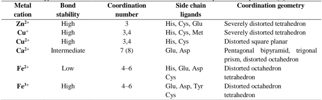
extremely important in biological processes such as electron transfer reactions, catalysis and stabilization of the protein structure [22]. The protein-metal association leads to a series of adjustments on the protein fold that results from a compromise on coordination numbers, bond lengths and angles which are imposed both on the metal and on the protein fold [8]. These changes alter protein energetics, thus influencing its stability and dynamic properties [22]. There are amino acid residues, such as histidine, cysteine, aspartate and glutamate, that have high affinity to certain metals resulting in a selectivity pattern [22-24]. Hence, there are preferable combinations of amino acids residues, which constitute metal coordination motifs, whose interaction with the metal ion ensures its proper insertion in a catalytic or structural site (Table 1.1.1) [8].
Table 1.1.1 – Typical coordination environments of selected metal cations in proteins. Adapted from [8]. Metal cation Bond stability Coordination number Side chain ligands Coordination geometry
Zn2+ High 3 His, Cys, Glu Severely distorted tetrahedron
Cu+ High 3,4 His, Cys, Met Severely distorted tetrahedron
Cu2+ High 3,4 His, Cys Distorted square planar
Ca2+ Intermediate 7 (8) Glu, Asp Pentagonal bipyramid, trigonal
prism, distorted octahedron
Fe2+ Low 4–6 His, Glu, Asp
Cys
Distorted octahedron tetrahedron
Fe3+ High 4–6 Glu, Asp, Tyr
Cys
Distorted octahedron tetrahedron
Generally, the protein-metal association involves both electrostatic and coordinative interactions, but in around one third of metalloproteins metal binding is essentially coordinative [8, 24]. Alternatively, this interaction can be established indirectly via a metal cluster or a larger chemical group [8].
When multiple binding sites are involved, the protein fold energetics become more complex due to cooperative events. In this cases, when a metal binds or dissociates from one site, conformational adjustments occur that affects the coordination sphere and binding energetics of another site, either by increasing (positive cooperativity) or decreasing (negative cooperativity) its affinity [8].
It is now clear that metal binding modulates both the protein folding landscape and folding trajectories [25, 26]. Thus, living systems have created a complex protein machinery whose function is the maintenance of metal ion homeostasis [8]. The metal delivery to polypeptides is assured by metallo-chaperones, whose role is to deliver the metal ion to its target hollo protein [27]. There are three generic scenarios of the mechanisms that mediate metal insertion into newly folded proteins: co-translational metal ion binding; post-translational metal ion binding to incompletely folded proteins; and post-translational metal ion binding to folded apo proteins [24].
7
1.1.4. Protein aggregation and amyloid formation
In addition to the native globular structure, proteins can populate other states, including disordered and partially ordered conformations, and different aggregated assemblies, which are important to cell homeostasis including growth, development and proliferation (Figure 1.1.3) [3]. The state a protein adopts under specific conditions depends on the relative thermodynamic stabilities of the many accessible conformations and on the kinetics of their interconversion (Figure 1.1.3) [2, 28]. The transitions between the different states are highly regulated by the environment and by the presence of molecular chaperones and degradation mechanisms. Therefore, aberrant behavior of the chaperones and other intervenients can be a key factor for development of misfolding and aggregation diseases (Table 1.1.2) [21, 29].
Table 1.1.2 – Representative protein folding diseases. Adapted from [2].
Figure 1.1.3 – States accessible to a protein molecule. Adapted from [28].
Disease Protein Site of folding
Hypercholesterolemia low-density lipoprotein receptor ER
Cystic fibrosis Cystic fibrosis trans-membrane regulator ER
Phenylketonuria Phenylalanine cytosol
Huntington’s disease Huntingtin cytosol
Marfan syndrome Fibrillin ER
Osteogenesis imperfecta procollagen ER
Sickle cell anaemia hemoglobin cytosol
αl-antitrypsin deficiency αl-antitrypsin ER
Tay-Sachs disease β -hexosaminidase ER
Scurvy Collagen ER
Alzheimer’s disease β-amyloid/presenilin ER
Parkinson’s disease α-synuclein cytosol
Scrapie/Creutzfeldt-Jakob disease prion protein ER
Familial amyloidoses transthyretin/lysozyme ER
Retinitis pigmentosa rhodopsin ER
Cataracts crystallins cytosol
8
Amyloid fibrils are the most well studied type of protein aggregate, given its unique kinetic and thermodynamic stability that culminates a progressive build-up of deposits in tissues. As a consequence, these deposits can physically disrupt specific biological components such as organs and tissues, thereby leading to a pathological behavior [5].
Several peptides and proteins that are involved in the most common misfolding diseases, such as the amyloid-β peptide in Alzheimer’s disease [30, 31] and α-synuclein in Parkinson’s disease [32, 33], form amyloid fibrils. Although their soluble forms have distinct characteristics, the aggregated forms are typically similar, characterized by their cross-b structure, in which β-strands form effectively continuous hydrogen-bonded β-sheets that run along the length of the fibril [34, 35]. Amyloid fibrils tend to appear as unbranched filamentous structures with a few nanometers in diameter but often micrometers in length (Figure 1.1.4) [36].
Figure 1.1.4 - Structure of an amyloid fibril at atomic resolution. Adapted from [36].
This phenomenon is not restricted to a small number of proteins, but instead it seems to be a generic feature of polypeptide chains, since it has been shown that fibrils can be formed in vitro by many other peptides and proteins, under specific conditions [2, 37, 38]. However, the propensity to form amyloid fibrils under given circumstances can differ between different amino acid sequences. The amyloidogenic propensity of a given peptide or protein correlates with its physicochemical features, such as charge, secondary-structure propensities and hydrophobicity [39].
The highly ordered and compact structure, provides an exceptional high level of kinetic and thermodynamic stability to the fibrils, which led to the assumption that the amyloid state might be more stable than the functional native state of a protein, even under physiological conditions [40, 41]. However, in the amyloid state intramolecular interactions are predominant and essential, which means that its thermodynamic stability (ΔG) is dependent on the protein concentration [42]. Thus, the amyloid state in only more stable than the native state when the critical concentration is achieved and the free energy (G) of the peptide or protein is lower than its native state. This evidence suggests that proteins might occasionally function at concentrations that exceed their conventional thermodynamic solubility [41, 43].
9
The formation of amyloid fibrils has been characterized, by in vitro studies, as a typical nucleated process composed by a lag phase, followed by a period of rapid growth [44, 45] and if the total amount of protein is limited, this process ends with a plateau phase as a result of the depletion of all soluble species and its conversion into fibrils (Figure 1.1.7). The self-assembly into amyloid fibrils begins with the formation of soluble oligomers resulting from nonspecific interactions, however, in some cases, specific structural transitions are a key factor [46]. The earliest species generally are characterized as small bead-like structures, sometimes linked together, often described as amorphous aggregates or micelles. Sequentially, the “prefibrillar aggregates” give rise to short, thin and sometimes curly fibrillary species termed “protofilaments” or “protofibrils”. Finally, the latter species are thought to assemble into long mature fibrils (Figure 1.1.6) [3].
Figure 1.1.6 – General mechanism of aggregation to form amyloid fibrils. The earliest species generally resemble
bead-like structures (D), thereafter transform into structures called protofibrils or protofilaments (A) which in turn assemble into mature fibrils (B). Adapted from [3].
Besides classic nucleation, the amyloid formation can be triggered by templating or seeding [47, 48] from existing aggregates. Templating is the mechanism by which structured aggregates promote the conversion of soluble protein species into similar aggregates [42]. On the other hand, the seeding process occurs when aggregates are used to promote the formation of larger aggregates, eliminating the lag phase [42].
The nucleation process can also involve secondary steps that depend on the behavior of the aggregates being formed [49, 50] (Figure 1.1.7). In this cases, formation of new aggregates arises from existing fibrils through fragmentation or from a combination of both monomeric and aggregated species through secondary nucleation [49, 50].
10
Figure 1.1.7 - Kinetics of amyloid formation. Adapted from [42]
The formation of these fibrils is known to be associated with both protein loss of function and generation of toxic intermediates in the process of self-assembly, which in turn are the basis of many misfolding diseases [30, 31, 49, 51-57]. Thus, living systems have evolved to avoid the formation of such fibrils by alternating amino acid residues with polar and hydrophobic characteristics [58, 59] that favor b-sheet structure seen in amyloid fibrils or by inserting highly aggregation-resistant residues, known as “gatekeepers” [58, 60], and by controlling proteins structure by means of molecular chaperones and degradation mechanisms [61-63].
11
1.2. S100 proteins
1.2.1. Function and structural characteristics of S100 proteins
S100 proteins belong to the EF-hand calcium-binding protein superfamily constituted by 24 members of low molecular weight (10–13 kDa) [64]. These proteins were named S100 by Moore, due to their solubility in 100% saturated ammonium sulfate [65].
Importantly, S100 proteins are expressed exclusively in vertebrates, exhibit tissue, cell, and subcellular-specific expression patterns [64, 66], and are induced by specific growth factors, cytokines and toll-like receptor (TLR) ligands [67-72]. In pathological conditions, however, the expression of a particular S100 protein can be triggered in a cell type that does not express it in normal physiological conditions [73-78]. The majority of the human S100 genes, are located within chromosome 1q21, except S100A11P, S100B, S100G, S100P and S100Z which map to chromosome 7q22-q3, 21q22, Xp22, 4p16 and 5q13, respectively [64].
All S100 proteins possess highly conserved overall structural architectures, although their sequences share no more than 25–65% homology [79]. Like other EF-hand Ca2+ binding proteins,
the S100 proteins contain two calcium-binding EF-hand motifs bridged by a so called hinge region, that are highly conserved among the S100 family members (Figure 1.2.1) [80, 81]. A single monomer is composed of two calcium-binding helix-loop-helix motifs, containing four helices (helix I, II, III, and IV) (Figure 1.2.3) [80, 81].
These proteins are distinguished from other EF-hand proteins by their both intracellular and extracellular functions, their tendency to form homodimers, and the ability to bind transition metals at the dimer interface [82]. Some members of the S100 protein family are also able to form heterodimers, for example S100A8 and S100A9 or S100A1 and S100P, suggesting different functions for homo- and heterodimers [82]. Moreover, in many situations, biological functions and signalling are carried out by S100 proteins that form higher order non-covalent oligomers, which can be promoted through binding of metal ions, such as Ca2+ and transition metals
(particularly Zn2+) [82, 83]. These oligomers include tetramers (S100B [84] , S100A2 [85], and
S100A8/A9 [86]), hexamers (S100B [84], S100A12 [87, 88]), and octamers (S100B [84]). Regarding intracellular functions, S100 proteins are involved in the regulation of several biological processes such as proliferation, differentiation, apoptosis, Ca2+ homeostasis, energy metabolism, inflammation and migration/invasion, through the interaction with a variety of targets, namely enzymes, receptors, transcription factors and nucleic acids [89]. On the other hand, at extracellular level, S100 can regulate functions of target cells in an autocrine and paracrine manner via activation of surface receptors [89].
It has been shown that several S100 proteins have the ability to undergo β-aggregation, as a cause or consequence of pathophysiological states [90]. Indeed, an example is the S100A8/A9 heterodimer that was found in amyloid deposits from prostate cancer patients, in inclusions called copora amylacea [91]. Moreover, there is the assumption that S100 proteins have intrinsic disordered regions [92] and the propensity to form amyloid-like structures [91].
Figure 1.2.1 – Structural features of S100 proteins. Ribbon diagrams of (A) and EF-hand motif (B) and EF-hand
12
1.2.2. Metal binding to S100
The ability of the S100 proteins to bind different metals, such as Ca2+, Zn2+ and Cu2+, is
a matter of great interest since it is known that these metals can have an effect on the structure, function and biochemical properties of the protein [82]. Both Ca2+ and transition metals (particularly Zn2+) have been shown to stimulate oligomerization in vitro, and crystal structures have revealed a range of oligomeric states [82].
As mentioned above, the S100 proteins bind Ca2+ ions through their EF-hand domains
[80, 81]. Typically, EF-hand Ca2+-binding motifs are arranged in pairs of EF-hands held together
by a very short anti-parallel β-strand and numerous hydrophobic interactions between the four helices [82]. S100 family members are an unique set of hand family, because one of the EF-hand motifs in the pair (designated as a pseudo- or S100-EF-hand) has 14, rather than 12 residues (canonical EF-hand [93]), characterized by the lower affinity towards Ca2+ [94]. Each monomer is comprised of a S100-specific N-terminal EF-hand with a 14-residue Ca2+ binding loop, and a C-terminal EF-hand with a canonical 12-residue Ca2+ binding loop [80]. Since the proteins are
dimers, each protein bind four Ca2+ ions, however, there are different affinities for this metal [82]. Similar to other EF-hand proteins, the S100 response to Ca2+ signals is characterized by conformational changes upon ion binding, which involves a significant shift in the orientation of Helix III, a 90 degree rotation (Figure 1.2.2) [95]. Moreover, this Ca2+-induced conformational change leads to the exposure of a hydrophobic region necessary for the interaction with its specific protein targets. The hydrophobic region is composed by residues from the “hinge” section, Helix III and C-terminal, being the region with highest variability within the S100 family members [95-97].
As abovementioned, despite being structurally similar, S100 proteins are able to interact with a plethora of cellular targets. This characteristic diversity is secured by the fine tuning within the target binding site of each S100 protein, specific expression patterns and the metal-binding ability [98].
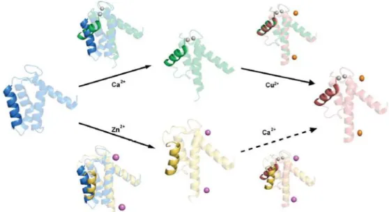
Figure 1.2.2 – Comparison of single sub-units of S100A12 to emphasize the differences in the packing of Helix III
induced by Ca2+ and transition metals. This reveals that the consequences of binding Ca2+ are much greater than those of binding transition metals. Adapted from [82].
13
Interestingly, S100 proteins bind not only Ca2+ but also transition metals, such as zinc and copper, in other sites [99]. Indeed, the binding of Zn2+ to an S100 protein was firstly reported for the S100B protein [100]. However, it is now known that Zn binding occurs in several S100 proteins (S100A1, S100A2, S100A3, S100A5, S100A6, S100A7, S100A8/A9, S100A12, S100A16 and S100B), with high affinity ranging from Kd=4 nmol L−1 (S100A3) to 100 μmol L−1 (S100A7). The Zn2+
binding S100 proteins can be classified into two categories: His-rich and Cys-rich [82].
Different studies [82] have revealed a conserved binding motif for the proteins with His-rich sites, composed by 4 His residues, or 3 His and 1 Asp residues, located at the dimer interface (Figure 1.2.3). Since the proteins are dimers, each protein binds two Zn2+ at the two symmetrically
disposed sites (Figure 1.2.4).
For the S100A7 and S100A12 it has been shown, through a detailed comparative structural analysis between all states (apo form, Ca2+- and Zn2+-loaded), that in the Zn2+-loaded structures of S100A7 and S100A12, two Zn2+ ions are bound at the symmetrically disposed sites
(Figure 1.2.4 A), coordinated by three His N2 atoms (His17, His86, His90) and an aspartate side chain (Asp24) (Figure 1.2.4 A) [99, 101]. In all cases, the primary effect of Zn2+ is to alter the orientation of Helix III (Figure 1.1.2).
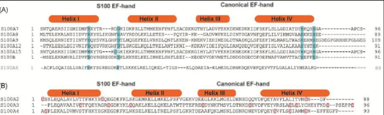
Figure 1.2.3 – Alignments of S100 proteins containing transition metal binding sites. Structure based sequence
alignment of S100 proteins from the (A) His-rich and (B) Cys-rich categories. Conserved residues in His-rich sites are highlighted with blue background, and those in Cys-rich sites in red background. Adapted from [82].
Figure 1.2.4 – Structural similarity of tetrahedral zinc and copper binding sites in S100 proteins. (A) Overlay of the
structures of (Ca2+)
4, (Zn2+)2-S100A7 (light green), (Ca2+)4, (Zn2+)2-S100B (blue) and (Ca2+)4, (Cu2+)2-S100A12 (purple)
showing that the transitions metal ions are chelated in similar manner by side chains in the same position in the
sequence. (B) Zoom in on the tetrahedral Zn2+ and Cu2+ sites showing the similar spatial disposition of the 3 His and 1
14
Regarding the Cys-rich Zn2+ binding S100 proteins (S100A2, S100A3, S100A4), these proteins are much less studied than the His-rich group [102], due to the absence of a conserved motif evident from alignment of these proteins
The possibility of different metals binding to an S100 protein, suggests scenarios where a crosstalk between metals that bind to S100 proteins is expected. In this context, it has been reported for the S100B that Zn2+ binding leads to an increase of the Ca2+ affinity by a factor of 10 and for the S10012 by ~1500 fold [102]. On the other hand, it lowers the Ca2+ affinity of S100A2 [85] and have no effect on S100A5 [103].
Due to Cu2+ and Zn2+ similar chemical properties, it was expected that Cu2+ would bind
to most zinc binding sites in proteins. Moreover, ion competition experiments demonstrated that the Cu2+ ions could be displaced by Zn2+, but not Ca2+. Thus, as expected, the S100 proteins bind Cu2+ in a similar way they bind Zn2+. There is still few information about the effects of Cu2+ binding on S100 proteins, but it seems that the overall conformational change induced by the binding of Cu2+ is very small compared with Ca2+ or Zn2+ binding [82].
1.2.3. S100A9
The work developed in this thesis is about the S100A9 homodimeric form and how it reacts to binding of metals and aggregation conditions.
S100A9, also known as Mrp14, is a Ca2+- and Zn2+-binding protein with a molecular
weight of 13.242 kDa, composed by 114 amino acids and has the longest C-terminal region in its sequence among the S100 family members (Figure 1.2.6). S100A9 is one of the most potent pro-inflammatory protein in the S100 family, being overexpressed in several inflammation scenarios, including inflammation associated with Alzheimer’s disease [104].
The S100A9 is one of the major constituents of neutrophils which plays an important role in the regulation of inflammatory and immune response [105]. This protein exists preferentially as a heterodimer or heterotetramer with S100A8 known as calprotectin [106, 107]. However, it can also exist as homodimer [108] and higher oligomeric species, including
,
fibrillary structures due to its intrinsic amyloidogenicity [91]. Dimerization is achieved by anti-parallel interactions between helices H1 and H4 of each monomer (Figure 1.2.6) [108]. Each of these forms assumes specific functions and their formation is dependent on binding of divalent cations, such as calcium and zinc [108].Current research on this protein is focused on its association with numerous human disorders, including acute and chronic inflammatory conditions, autoimmune diseases, cancer, atherosclerosis, cardiomyopathies and neurodegenerative diseases [104, 106-108], given its crucial role in normal physiological processes within cells.
Figure 1.2.5 – S100A9 homodimer crystal structure (CHAPS removed). The Ca2+ ions are colored green. Images generated in pymol using PDB entry 1IRJ.
15
Primary Structure, Ca2+- and Zn2+-Binding Sites
Like other S100 proteins, S100A9 contain two EF-hand Ca2+-binding sites: a C-terminal canonical EF-hand (site II) and a N-terminal pseudo EF-hand (site I) with a 12 and 14 amino acid residue Ca2+-binding loop positioned between two supporting α-helices, respectively (Figure
1.2.6) [108]. The alignment of these motifs with other members of the S100 family has demonstrated that in both sites S100A9 contains the conserved sequence determinants required for Ca2+-binding [108].
The Ca2+-binding site II include residues Asp67-Glu78 and interacts with Ca2+ through
seven oxygen ligands, forming a pentagonal bipyramidal coordination: the side-chain O atoms of Asp67, Asn69, Asp71 and Glu78, the main-chain carbonyl O atom and a water molecule. The Ca2+-binding site I include residues Ser23–Glu37 and interacts with Ca2+ through seven O atoms from the main-chain carbonyl groups of Ser23, Leu26, His28 and Thr31, the carboxyl group of Glu36 and a water molecule (as indicted in Figure 1.2.6) [108]]. The water molecule as a seventh ligand also forms a hydrogen bond with the side-chain of Thr31.
Although it is know that S100A9 is important for Ca2.-dependent functions during inflammatory conditions, the function behind the Ca2+-S100A9 interaction is yet to be unveiled [108]. However, it has been shown that Ca2+ is capable of inducing S100A8/A9-tetramerization [109].
Figure 1.2.6 – Amino acid sequence alignment of the human S100 family of proteins. Proteins whose three-dimensional
structures have already been analysed are marked by an asterisk. Secondary structure elements of S100A9 are given. Residues that coordinate calcium ions are marked as follows: m, main-chain carbonyl group; s, mono-dentate side-chain of Asp or Asn; b, bi-dentate side-side-chain of Glu. The residues highlighted in blue are well-conserved residues the side-chains of which coordinate calcium ions. The residues highlighted in yellow represent highly conserved hydrophobic residues forming an intra-monomer hydrophobic cluster. Residues that interact with target molecules are marked with $. The residues highlighted in pink are other conserved hydrophobic residues that form an inter-monomer hydrophobic cluster. All sequences were obtained from the SWISS-PROT protein sequence database [110]. Adapted from [108].
16
Besides Ca2+, S100A9 also bind Zn2+ ions. S100A9 is classified in the his-rich category of the Zn2+ binding S100 proteins and contain HEXXH motives in its sequences that are putative Zn2+-binding sites [111].
The role of zinc in S100A9 is still unclear, however it has been reported that S100A9 chelates zinc as a biological function [112]. Moreover, different biophysical methods have shown that Zn2+-induces S100A8/A9-tetramerization by binding to both Ca2+-specific EF-hands and Zn2+-specific binding sites [113, 114]. The researchers who carried out this study reported that twelve Zn2+ were bound to the tetramer (eight Zn2+ bound to the EF-hands and four Zn2+ to the
Zn2+-specific binding sites) [113, 114], revealing the existence two different putative Zn2+-binding
sites on the S100A8/A9 subunit interface.
Interestingly, several other S100 family members are also regulated by Zn2+ and Ca2+ [115], as mention earlier, and for some of them the Zn2+-binding sites have been well characterized (S100B, S100A2, S100A7 and S100A12) by different techniques [116, 117]. For these proteins it was observed the binding of two Zn2+-ions per homodimer and that both subunits
provide residues to form the Zn2+-binding sites. Since the Zn2+-binding residues in S100A9 and
S100A7 are fully conserved, it is likely that the S100A9 homodimer coordinates Zn2+in a similar way as S100A7 [86, 118].
1.2.4. S100A9 in Alzheimer’s disease
As aforementioned, S100A9 is involved in several human disorders, including Alzheimer’s disease (AD). AD is a neurodegenerative disorder characterized by the accumulation of amyloid-β protein (Aβ) in extracellular plaques and the deposition of hyperphosphorylated tau protein in intracellular neurofibrillary tangles [119-121], which are directly associated with prominent neuroinflammation [122]. In turn, this condition is correlated with a severe deterioration of cognitive function [123], however the molecular mechanisms underlying this observation are yet to be unveiled [124, 125]. Furthermore, aberrant metal homeostasis seems to be related with AD pathogenesis [126].
S100A9 plays an important role in regulating vascular inflammation, by recruiting leukocytes to damaged vessels, such as those injured by deposition of Aβ amyloid fibrils [127]. Moreover, an overexpression of S100A9 has been reported in microglia in the temporal cortex of both familial and sporadic AD cases [128], suggesting that S100A9 could be a neuroinflammatory marker of AD.
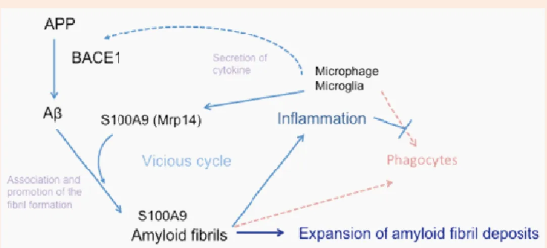
The accumulation of Aβ amyloid fibrils on cerebral vessels and in brain parenchyma leads, consequently, to local inflammation [129], which in turn results in activation and recruitment of microglia to the plaque deposition site [130], causing microgliosis. From this point, microglia acts in two ways: beneficial promotion of phagocytosis of Aβ and harmful production of neurotoxins and pro-inflammatory molecules [131-133], such as S100A9. The high levels of S100A9 on Aβ amyloid fibril deposits promotes further inflammation and enhances Aβ amyloid fibrils formation. Moreover, S100A9 drives microglia into a pro-inflammatory state thereby compromising microglial phagocytosis [131, 134].
Among this events, BACE1 activity is also elevated, resulting in increased Aβ production [135], which in turn further accelerate S100A9 expression, creating a vicious cycle that promotes and expands the deposition of Aβ amyloid fibrils including amyloid-associated proteins on cerebral vessels and in brain parenchyma (Figure 1.2.7).
17
Figure 1.2.7 – A comprehensive model of amyloid fibril formation enhancement. Local inflamation by Aβ amyloid
deposition induces S100A9 production by activated phagocytes and up-regulation of the inflamatory condition. Thereafter, S100A9 induces increased formation of Aβ amyloid fibril and futher inflamation. Moreover S100A9 drives microglia into proinflamtory state thereby compromising microglial phagocytosis. Adapted from [136].
Due to its inherent amyloidogenicity, the significantly increased levels of extracellular S100A9 leads to its amyloid aggregation [137-139] and also co-aggregation with Aβ [140], enhancing each other aggregation, suggesting a “rescue” clearance process to remove neurotoxic amyloid species from circulation. However, the consequences of this clearance process can lead to exacerbated growth of the amyloid plaques in AD brain, much more stable and protease resistant [91]. Furthermore, the plaques themselves can lead to more microglia activation, thus completing the vicious circle of amyloid-neuroinflammatory cascade.
Interestingly, the knockdown or knockout of the S100A9 gene in AD mice model (Tg2576) significantly reduced the neuropathology and the amount of Aβ, C-terminal fragments of amyloid precursor protein (APP-CT) and phosphorylated tau [141].
Therapeutic strategies for AD treatment focus on inhibiting Aβ production and/or enhancing Aβ clearance [136]. Therefore, due to its amyloidogenicity, neurotoxicity and signaling functions, S100A9 may be a promising therapeutic target. However, the specific role of S100A9 in AD as well as in aging is still far from clear.
18
1.3. Methods for structural analysis and protein
folding monitoring
1.3.1. Fluorescence spectroscopy
Fluorescence spectroscopy is an important investigation tool in different areas of analytical science, due to its high sensitivity and selectivity. Its applications include the study of protein folding, structural dynamics and protein interactions by monitoring the tertiary structure of proteins [142]. For many proteins this application is possible due to their intrinsic fluorescence granted by the presence of aromatic amino acid residues, such as the tryptophan (Trp), tyrosine (Tyr) and phenylalanine (Phe). These residues and other fluorescent molecules are called fluorophores. Besides intrinsic fluorophores there are some extrinsic probes that also give information about the protein folding, misfolding and ultimately amyloid formation. The probes used in this work were: Thioflavin-T (ThT); 8-anilino-1-naphthalenesulfonic acid (ANS); and two luminescent conjugated oligothiophenes (LCOs), p-FTAA and h-FTAA.
ThT is a benzothiazole dye that exhibits enhanced fluorescence upon binding to amyloid fibrils. Originally, it was thought that ThT only interacted with the amyloid cross-β structure [143], however, it has been shown that ThT fluorescence can bind to non-β-sheet cavities (observed for acetylcholinesterase and γ-cyclodextrin) [144]. The binding leads to an increase in fluorescence intensity and induces a shift in the maximum wavelength from 483 nm to 478 nm [145].
ANS is known to interact with hydrophobic sites, such as the ones buried within the protein. For this reason this probe has been widely used in protein folding and binding studies, where sometimes hydrophobic regions exposure occurs. Once more, this interaction leads to an increase in fluorescence intensity and induces a shift in the maximum emission wavelength, from 530 nm to 475 nm [142].
LCO’s are anionic oligothiophene based ligands capable of detecting non-thioflavinophilic aggregated species preceding amyloid fibril formation. They presumably intercalate with the amyloid fibrils by binding in grooves along the long axis of the amyloid fibril [146]. The ones used in this work where the p-FTAA and h-FTAA, each with distinct spectral signature. The binding of this probes leads to the increase of their fluorescence intensity [146].
19
1.3.2. Conformation studies by Imunodetection
The formation of amyloid deposits is a characteristic feature of many degenerative disease and its staining in human and mice AD brains has been a challenge. However, it has been overcome by the development of conformation-dependent antibodies that specifically recognize generic epitopes of a polypeptide chain with given conformation, regardless of its sequence. This antibodies include the OC and A11. The OC antibody recognizes amyloid fibrils, while the A11 specifically recognizes a generic epitope common to prefibrillar oligomers [147, 148].
Figure 1.3.2 - Dotblot resulted from detection of fibrils and oligomers, from different proteins, by the conformation-dependent antibodies A11 and OC [148].
1.3.3. Atomic Force Microscopy
Atomic Force Microscopy (AFM) is the most versatile and high-resolution scanning method for studying samples at nanoscale. An AFM uses a cantilever with a sharp tip to scan samples adsorbed on atomically flat smooth surfaces, typically mica. As it goes along the surface the cantilever deflects vertically and laterally producing a signal which is translated to generate a corresponding topography image. This technique has been used to investigate the self-assembly dynamics of several amyloidogenic proteins and other types of aggregates [149].
Figure 1.3.3 - The operating principle of AFM and AFM images of amyloid fibrils. a) Schematic representation of
AFM measurement. AFM image of b) twisted ribbons, c) helical ribbon d) multi-stranded helical fibril. Adapted from [149].
20
1.4. Objectives
Metal ions have prominent as regulators of structure and function of S100 proteins, whose functional and amyloid-type oligomerization has been implicated in pathophysiological processes related to neurodegeneration. This work aimed at investigating the metal-ion dependent oligomerization of the pro-inflammatory S100A9 protein, in an attempt to gain insights into understanding how binding of zinc and calcium influence its self-assembly. Specific objectives included:
Obtaining highly pure preparations of human homodimeric S100A9 protein through recombinant expression, chromatographic purification, and preparation and biochemical characterization of apo-forms;
Comprehensive analysis of the S100A9 self-assembly reaction in the presence of Ca2+
and Zn2+ using fluorescence based kinetic assays and extrinsic dyes;
Conformational characterization of S100A9 oligomers and aggregates combining biophysical, biochemical and immunological approaches.
The work reported in this thesis was carried out in the research laboratories of the ‘FCUL Protein Folding and misfolding laboratory’ (http://folding.fc.ul.pt/) which is affiliated to BioISI – Biosystems & Integrative Sciences Institute and is located at the FCUL Chemistry and Biochemistry Department (C8 building FCUL campus). The AFM measurements and analysis was performed by collaborators at the BioISI Magnetic Nanosystems Group / Department of Physics, also at FCUL (see acknowledgments).
21


![Figure 1.1.7 - Kinetics of amyloid formation. Adapted from [42]](https://thumb-eu.123doks.com/thumbv2/123dok_br/15571119.1048036/26.892.206.686.105.536/figure-kinetics-amyloid-formation-adapted.webp)