Universidade Católica Portuguesa
Faculdade de Engenharia
Radionuclide Therapy in Nuclear Medicine: Applying Monte
Carlo Simulation to Investigate Bremsstrahlung Imaging with
a Gamma Camera
Catarina Ramos de Mendonça
Dissertation Submitted for the Degree of Master of Science in
Clinical Engineering
Júri
Prof. Doutor Manuel José Martinho Barata Marques (Presidente)
Prof. Doutor José Pedro Miragaia Trancoso Vaz
Prof. Doutor Rui Jorge Correia Mendes Alves Pires (Co-Orientador)
Prof. Doutor Ian Cullum (Orientador)
Universidade Católica Portuguesa
Faculdade de Engenharia
Radionuclide Therapy in Nuclear Medicine: Applying Monte
Carlo Simulation to Investigate Bremsstrahlung Imaging with
a Gamma Camera
Catarina Ramos de Mendonça
Dissertation Submitted for the Degree of Master of Science in
Clinical Engineering
Orientador: Prof. Doutor Ian Cullum
Co-Orientador: Prof. Doutor Rui Jorge Correia Mendes Alves Pires
ABSTRACT
Radionuclide therapy is an innovative treatment in nuclear medicine that uses unsealed sources to treat some specific tumours in the human body. With an increasing interest in this modality, some efforts have been done to improve this field in nuclear medicine, such as the radiopharmaceuticals administered or the components of the imaging equipment.
Pure beta emitters are the radionuclides most used in radionuclide therapy. They are described to have extremely high potential in the treatment of malignant and non-malignant disorders. The energy of these radionuclides is absorbed close to the target site due to its low range (few mm in tissue). The problem is the impossibility of imaging their uptake in the interest places since there are no gamma rays emissions and the beta radiation is impossible to detect externally. As a result, the only radiation capable to be perceived by the gamma camera is the bremsstrahlung photons produced when an accelerate beta particle passes close to the atomic nucleus and is deflected towards it. The detection of these photons is relevant for dosimetric purposes, in order to detect the real uptake of the radionuclide and hence to know the absorbed dose in the patient. Thus, the current dissertation reports a study about bremsstrahlung characteristics by using a Monte Carlo simulation. The study attempts to realise in what way the bremsstrahlung photons are produced in different types of biological materials, using different beta particles energies. The simulator used was the EGSnrc (Electron Gamma Shower) system (V4 2.3.2), a package for Monte Carlo simulations. The programs were written in Mortran language and compiled to Fortran. With this study, an additional aim was to obtain a strong knowledge on EGSnrc system and to be able to modify and produce a wide range of different simulations in different physical conditions. In order to understand the bremsstrahlung photons several simulations were performed, in different media and with different energies of beta particles emissions.
Future work should be necessary in order to relate the bremsstrahlung photons understanding with the gamma camera components. The aim will be to improve the bremsstrahlung imaging and hence gathering realistic dosimetric data for the pure beta emitters.
RESUMO
A terapia com radionuclídeos é um tratamento inovador em medicina nuclear, o qual utiliza fontes não seladas para tratar tumores específicos no corpo humano. Com o crescente interesse por esta especialidade terapêutica, alguns esforços têm sido feitos para melhorar este campo da medicine nuclear, nomeadamente através dos radiofármacos administrados ou na tecnologia dos equipamentos de imagiologia.
Os emissores beta puros (electrões) são os radionuclídeos mais utlizados nesta terapia. São descritos na literatura como os que apresentam um elevado potencial no tratamento de doenças malignas e benignas, uma vez que a energia destes radionuclídeos é absorvida perto do local alvo (poucos mm de alcance nos tecidos). O problema da utilização de beta puros reside na impossibilidade de se formar uma imagem médica, uma vez que estes radionuclídeos não emitem qualquer radiação gama. Como resultado, a única radiação capaz de ser detectada pela câmara gama são os fotões bremsstrahlung (radiação de travagem), produzidos quando ocorre uma desaceleração e deflexão dos electrões acelerados quando estes passam perto dos núcleos atómicos. A detecção de bremsstrahlung é muito importante para fins dosimétricos, dado que permite conhecer a verdadeira dose absorvida pelo paciente.
A presente dissertação tem como principal objectivo estudar as características dos fotões bremsstrahlung, através da utilização de um simulador Monte Carlo - EGSnrc (Electron Gamma Shower). Pretende-se compreender de que forma os fotões bremsstrahlung são produzidos no interior de diferentes tipos de materiais biológicos, utilizando partículas beta de diferentes energias. Os códigos foram programados em linguagem Mortran e compilados para Fortran. Pretende-se também obter um forte conhecimento sobre o sistema usado, de forma a ser possível produzir qualquer tipo de simulação para diferentes condições físicas.
Torna-se necessário prosseguir este trabalho, a fim de se relacionar as características dos fotões bremsstrahlung com os componentes da câmara gama. O objectivo será melhorar as imagens criadas por fotões bremsstrahlung e, portanto, reunir dados dosimétricos realistas para os emissores beta puros.
Palavras-chave: Terapia com radionuclideos; Fotões Bremsstrahlung; Simulação de Monte Carlo; EGSnrc.
FRAMEWORK
The present dissertation encompasses a research work developed at the Institute of Nuclear Medicine (INM) of the University College London Hospital (UCLH), with a contribution of the Institute of Cancer Research (ICR) and the Department of Nuclear Medicine in Cambridge Biomedical Campus (Addenbrooke's Hospital). The work developed in UCLH was undertaken from February to November 2012, within the framework of an Erasmus exchange program.
The Institute of Nuclear Medicine (INM), actually included within the new UCL Hospital NHS Foundation Trust building, was founded in 1961 by Professor E.S. Williams (director of the institute 1963-1985) and by Professor P.J. Ell (currently consultant physician). This institute is the only academic department of nuclear medicine in the UK and its physics research group is internationally recognised. The INM is responsible for the nuclear medicine service of UCL Hospitals NHS Trust. In addition, the institute also offers the full range of nuclear medicine diagnostic, laboratory based and therapy procedures, which currently performs approximately 15,000 patient studies every year. The INM has a unique technology platform in the UK and it comprises an extensive range of state-of-the-art imaging equipment, image processing, IT systems and radiopharmacy/radiochemistry facilities. The institute is equipped with two PET-CT systems (a 16-slice system and a 64-slice system), two SPEPET-CT-PET-CT systems, two SPEPET-CT gamma camera systems, a cardiac camera (D-SPECT) and DEXA (dual-energy x-ray absorptiometry) bone densitometry. Currently, it possesses the first fully integrated 3T PET/MR in the UK, as well as a full range of supporting nuclear medicine instrumentation. Particular areas of expertise include nuclear cardiology, PET-CT and SPECT-CT imaging in oncology and radiotherapy, the sentinel lymph node technique - the application of nuclear medicine to neurology and psychiatry, and radionuclide therapy (UCLH NHS Foundation Trust & UCL, 2011).
ACKNOWLEDGMENTS
The current thesis would not have been possible without the contribute, help and support of many people who followed me in different ways, both in this phase as along to many years during which transmitted me the necessary knowledge to achieve this step.
First, I am truly thankful to my supervisor Dr. Ian Cullum, for his continuous and valuable guidance, support, understanding and help throughout this study. Thank you for all your advices, for the patience in answering all my questions and for being a good mentor.
I am also thankful to INM for the opportunity to conduct the research, as well as to Prof. Robert Speller, from University College of London (Department of Medical Physics) to have advised me to participate in this project.
I would like to take the opportunity to thank all my teachers in FEUCP for all the academic knowledge transmitted, for have guided, inspired and helped us during our academic formation and education. A special thank you to Prof. Manuel Barata Marques, Prof. Pedro Encarnação, Prof. Rui Pires, Prof. Rui Assis, and Prof. Ana Pascoal who accompanied and guided me unconditionally throughout my whole MSc course, supported and motivated me to go further on without ever giving up.
A special thanks to all my friends who supported and encouraged me. Thank you Simone, for have made my life far away from home not so lonely. Thanks very much Nanas for being there when I needed. Gonçalo, to you I am really thankful for having followed my work so closely and for have given me the strength to carry on, sharing all the tough moments, not forgetting a big thank you for providing me the huge amount of computers to undertake my simulations.
Finally, and most importantly, I would like to thanks all my family. Mother/Father/Mano thank you so much for allowing me the opportunity to fulfil my dreams, for the support, encouragement, patience and love and for having made of me what I am today. Thank you!
CONTENTS
CONTENTS ... vii
LIST OF FIGURES ... ix
LIST OF TABLES ... xii
LIST OF APPENDICES ... xiv
LIST OF ABBREVIATIONS AND ACRONYMS ... xv
CHAPTER 1. INTRODUCTION 1. INTRODUCTION ... 2
1.1. Dissertation Context ... 2
1.2. Dissertation Aim ... 4
1.3. Dissertation Structure ... 5
CHAPTER 2. GENERAL BACKGROUND 2. GENERAL BACKGROUND ... 8
2.1. General Concepts of Nuclear Physics ... 8
2.1.1. Radioactive Disintegration Processes ... 9
2.1.2. Characteristics of Radioactive Decay ... 15
2.1.3. Photons Interactions ... 18
2.1.4. Electron Interactions and Bremsstrahlung Photons Production ... 25
2.2. Gamma Camera Imaging ... 32
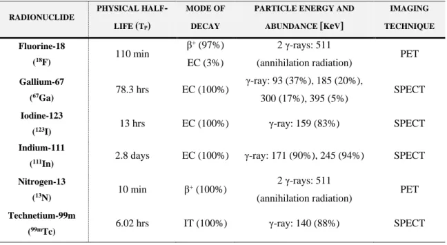
2.2.1. Radionuclides used in Diagnostic: Characteristics and Applications ... 32
2.2.2. System Components to Detect the Radiation ... 34
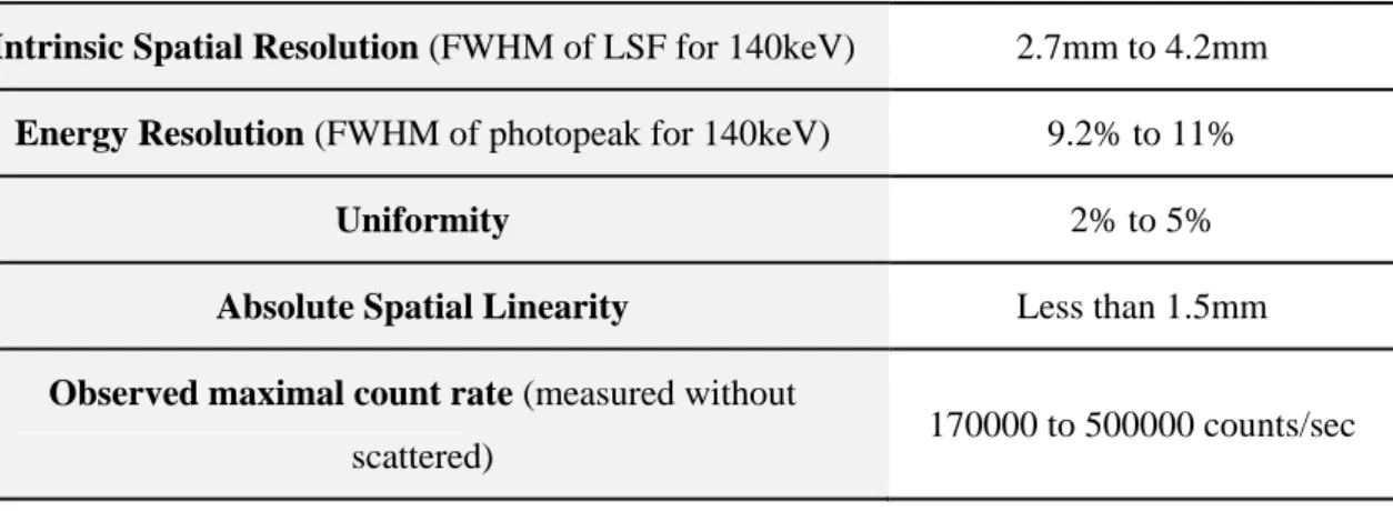
2.2.3. System Performance and Factors Affecting the Image Quality ... 43
2.2.4. Modes of Image Acquisition ... 52
2.3. Targeted Radionuclide Therapy and Imaging Radionuclide Therapy ... 57
2.3.1. Principles of Radionuclide Therapy ... 57
2.3.2. Radionuclides used in Therapy: Characteristics and Applications ... 59
2.3.3. Internal Dosimetry for Radionuclide Therapy ... 63
2.3.4. Why is Bremsstrahlung Imaging Difficult? ... 67
CHAPTER 3. DISSERTATION METHODOLOGY 3. DISSERTATION METHODOLOGY ... 70
3.1.1. Principles of Monte Carlo Simulation ... 72
3.1.2. How EGSnrc works? ... 74
3.1.3. The Models’ Code in Words ... 77
3.2. Simulations ... 83
3.2.1. Photons Simulations ... 83
3.2.2. Bremsstrahlung Simulations ... 83
3.2.3. List mode Acquisitions ... 83
CHAPTER 4. RESULTS AND DISCUSSION 4. RESULTS AND DISCUSSION ... 86
4.1. Photons Simulations ... 86
4.1.1. Verification of the Inverse Square Law ... 86
4.1.2. Analysis the Effect of the Crystal Thickness ... 90
4.1.3. Analysis the Effect of the Glass behind the Crystal ... 95
4.2. Bremsstrahlung Simulations ... 100
4.2.1. Analysis of the Bremsstrahlung Production Efficiency ... 101
4.2.2. Analysis of the Bremsstrahlung Yield (%) ... 105
4.2.3. Analysis of the Bremsstrahlung Spectrum Shapes ... 108
4.2.4. Analysis of the Angular Distribution of the Bremsstrahlung Emission ... 112
4.2.5. Analysis of the Spatial Distribution of the Bremsstrahlung Produced ... 113
4.2.6. Analysis of the 90Y and 32P Bremsstrahlung Spectra. ... 116
4.3. List-mode Acquisitions ... 125
4.3.1. List mode acquisitions with two energy windows selected (140.5±10% and 450±10%). .... 126
4.3.2. List mode acquisition with one energy window (256±100%) ... 129
CHAPTER 5. CONCLUSION 5. CONCLUSION ... 132
5.1. Future work ... 134
REFERENCES ... 138
LIST OF FIGURES
Figure 2.1. Decay scheme for 18F into a stable nuclide 18O, which may decay by both EC or β⁺, with Emax,β⁺=0.638MeV ... 12
Figure 2.2. Decay scheme diagram for 14C into a stable nuclide 14N, which decay only by β -emission, with Emax,β-=0.156MeV ... 13
Figure 2.3. Typical spectrum of beta particle energy ... 13 Figure 2.4. Simplified decay scheme of isobaric nuclides: 99Mo/99mTc/99Ru ... 14
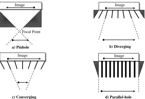
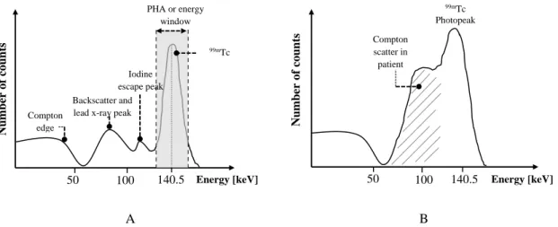
Figure 2.5. Exponential radioactivity decay law, showing the activity A of a sample as a function of time t ... 16 Figure 2.6. Representation of the photoelectric effect with characteristic x-ray emission ... 19 Figure 2.7. Schematic representation of the Compton Scatteringm where hѵ is the incident photon energy, hѵ’ the scattered photon energy, θ photon scattering angle and φ electron scattering angle ... 20 Figure 2.8. Representation of the pair production interaction with annihilation photons ... 23 Figure 2.9. Relative importance of Photoelectric effect, Compton scattering and Pair production over a wide range of energy, hѵ, of the incident photons and atomic numbers, Z, of the attenuating material ... 23 Figure 2.10. Electrons depositing energy through collisional and radiative losses when interact with the surrounding matter ... 25 Figure 2.11. Electron–nucleus interaction with bremsstrahlung production (classical description) ... 28 Figure 2.12. Representation of heavy and light charged particle tracks ... 31 Figure 2.13. Basic components of a gamma camera (Adapted from Singh & Waluch, 2000) .. 34 Figure 2.14. Types of the gamma camera collimators: a) pinhole collimator, b) diverging-hole collimator, c) converging-hole collimator and d) parallel-hole collimator ... 37 Figure 2.15. Schematic cross-section of a NaI(Tl) crystal used in a typical gamma camera ... 38 Figure 2.16. Schematic diagram of a typical photomultiplier tube ... 39 Figure 2.17. Energy spectrum of the 99mTc, when it is viewed by the gamma camera as a point source (A) and as inside a patient (B) ... 42 Figure 2.18. Representation of the LSF and the FWHM, which is the distance encompassed by the curve halfway down from its peak ... 44
Figure 2.19. Schematic representation of a parallel-hole collimator, with length or collimator
thickness t, hole diameter d, septal thickness a and source-to-collimator distance b ... 46
Figure 2.20. Effect of the septal thickness and length in parallel-hole collimators ... 47
Figure 2.21. Comparison of the intrinsic uniformity in three images as the window width is decreased ... 50
Figure 2.22. Nonlinearity of the NEMA slit phantom pattern... 51
Figure 2.23. A dual-head SPECT camera, GE NM630 model ... 54
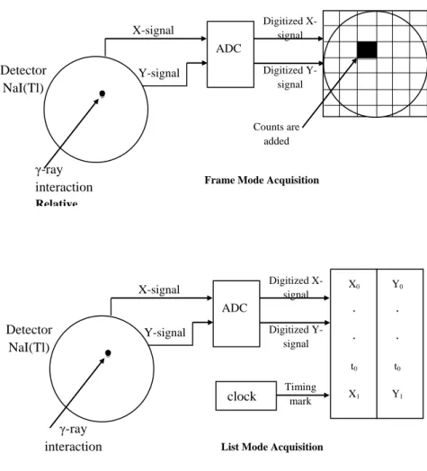
Figure 2.24. Data acquisition in frame mode and list mod ... 56
Figure 2.25. Schematic representation of the RIT, through the use of an antibody as biological vector ... 58
Figure 2.26. Application of hepatic radioembolization with 90Y glass microspheres ... 68
Figure 3.1. Structure of the EGSnrc code system ... 75
Figure 3.2. Simple geometry to the HOWFAR subroutine: two parallel planes separating three regions along z. This is a three region geometry example for HOWFAR ... 78
Figure 3.3. Scintillation detector model created in EGSnrc ... 79
Figure 3.4. Planes necessary to define the detector geometry ... 80
Figure 3.5. Regions produced by the creation of the planes... 80
Figure 3.6. Source model created in EGSnrc ... 82
Figure 4.1. Analysis of the Inverse Square Law. Number of photons detected vs distance from the crystal to the source [cm] ... 87
Figure 4.2. Comparison between simulated and theoretical of the fraction of photons reaching the crystal in order to verify the Inverse Square La ... 89
Figure 4.3. Intrinsic Efficiency of the NaI crystal. Simulation obtained for several crystal thicknesses [mm] by varying the energy of the incident photons [keV] ... 91
Figure 4.4. Absorption Efficiency of NaI(Tl) crystal ... 92
Figure 4.5. Natural Log of the fractional reduction of the intensity beam ... 93
Figure 4.6. Simulated and analytical values of the linear attenuation coefficients. ... 94
Figure 4.7. Effect of the glass thickness to analyse the photons' backscatter ... 95
Figure 4.8. Effect of the glass to analyse the photons' backscatter ... 95
Figure 4.9. Analysis of backscattering photons ... 97
Figure 4.10. Bremsstrahlung production efficiency by varying the radius of the sphere and the beta particles energy, in the four different materials. ... 102
Figure 4.12. Simulated and analytical values of bremsstrahlung yield, in percentage. Values presented for different biological material ... 106 Figure 4.13. Normalized spectra of bremsstrahlung produced, in the four different biological materials ... 109 Figure 4.14. Bremsstrahlung spectra produced and escaped in the four different materials. ... 110 Figure 4.15. Linear attenuation coefficients [cm-1] for cortical bone, inflated lung, soft tissue
and adipose tissue ... 111 Figure 4.16. Angular distribution between incident electrons and emitted bremsstrahlung photons as a function of electron kinetic energy [MeV] ... 112 Figure 4.17. Spatial distribution of bremsstrahlung photons as a fraction of the electron energy ... 114 Figure 4.18. Frequency of the number of bremsstrahlung produced within distance ranges ... 114 Figure 4.19. Total energy of the bremsstrahlung produced within distance ranges ... 114 Figure 4.20. Comparison between theoretical and simulated energy emitted spectra for A) 90Y
point source and B) 32P point source (Theoretical data from RADAR, The Decay Data, 2012) ... 118 Figure 4.21. Comparison between theoretical and simulated cumulative probabilities for A) 90Y
point source and B) 32P point source (Theoretical data from RADAR, The Decay Data, 2012) ... 118 Figure 4.22. Bremsstrahlung photons spectra created from polyenergetic point sources of 90Y
and 32P, for different biological materials ... 120 Figure 4.23. Schematic representation of the setting-up used to undertake the list mode acquisitions with two energy windows (140.5keV±10% and 450keV±10%) ... 126 Figure 4.24. 99mTc spectra acquired in a 60sec planar acquisitions, using a MEGP collimator and two energy windows 140.5±10% and 450±10% ... 127 Figure 4.25. Binary images obtained from the Matlab code ... 128 Figure 4.26. 99mTc spectrum by incident photons with energies inside the energy window
selected ... 129 Figure 4.27. 99mTc spectrum by incident photons with energies outside the energy window selected ... 129 Figure 4.28. 99mTc spectra acquired in a 60sec planar acquisitions, using a LEGP collimator and
one energy window, 256±100% ... 130 Figure 4.29. Binary image obtained from the Matlab code by using the processed list mode data ... 130
LIST OF TABLES
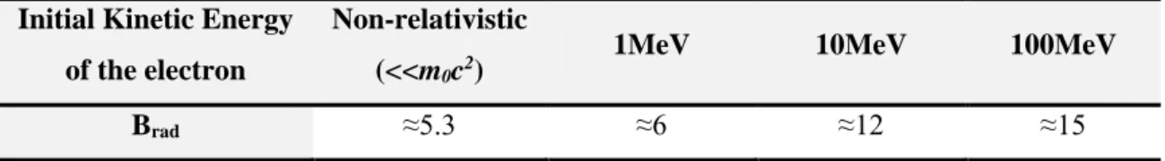
Table 2.1. Parameter Brad for some initial kinetic energies of the electrons ... 29
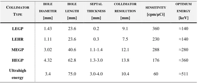
Table 2.2. Physical characteristics of the radionuclides used in diagnostic nuclear medicine ... 33 Table 2.3. Features and properties of different types of parallel-hole collimators, calculated at 10cm of collimator face ... 48 Table 2.4. Typical intrinsic gamma camera performance characteristics (Bushberg et al., 2002) ... 52 Table 2.5. Physical characteristics of the radionuclides available for common use in the therapeutic nuclear medicine ... 61 Table 2.6. Annual dose limits recommended by ICRP 1990, 2007 and IAEA. ... 64 Table 4.1. Simulated and analytical values of the fraction of photons reaching the detector by varying the source-detector distance ... 88 Table 4.2. CPU times [seconds] spent in the simulations to test the inverse square law ... 89 Table 4.3. Simulated and analytical values of the linear attenuation coefficient ... 93 Table 4.4. CPU times [seconds] spent in the simulations to test the effect of the crystal thickness ... 94 Table 4.5. Theoretical values of the energy of the scatter photons, for a point source of photons with 100, 200 and 500keV of energy ... 98 Table 4.6. CPU times [seconds] spent in the simulations to test the effect of the glass thickness. Values obtained for 106 photons emitted along the z-axis, perpendicularly to the glass ... 99 Table 4.7. CPU times [seconds] spent in the simulations to test the effect of the glass thickness. Values obtained for 106 photons emitted isotropically ... 99
Table 4.8. Effective atomic number of some biological compounds ... 101 Table 4.9. Maximum radius of the sphere, from which the number of bremsstrahlung produced inside it become constant ... 103 Table 4.10. CPU times [seconds] obtained for the maximum radius of the sphere from which the number of bremsstrahlung produced become constant ... 104 Table 4.11. Simulated and analytical values of the bremsstrahlung radiation yield ... 107 Table 4.12. CPU times [seconds] spent in the simulations to analyse the bremsstrahlung yield. ... 108 Table 4.13. CPU times [seconds] spent in the simulations to analyse the spectra shapes ... 111
Table 4.14. CPU times [seconds] spent in the simulations to analyse the angular bremsstrahlung distribution ... 113 Table 4.15. CPU times [seconds] spent in the simulations to analyse the spatial bremsstrahlung distribution ... 115 Table 4.16. Percentage of the bremsstrahlung photons produced, absorbed and escaped, for 90Y
and 32P sources ... 122 Table 4.17. Radiative yield [%] produced by 107 beta particles emitted from 90Y and 32P point sources ... 123 Table 4.18. CPU times [seconds] spent in the simulations to analyse the bremsstrahlung spectra created by polyenergetic pure beta emitters, 90Y and 32P ... 124 Table 4.19. List mode acquisition data after the use of the decoding program ... 125
LIST OF APPENDICES
APPENDIX 1. The COMMON Blocks (Kawrakow et al., 2011, pp. 110-119) ... 1500
APPENDIX 2. Specifications for AUSGAB (Kawrakow et al., 2011) ... 1533
Table 2.1. Values of IARG which are on by default and for which energy is deposited ... 1533
Table 2.2. Values of IARG which are off by default ... 1533
APPENDIX 3. Scintillation Detector Model. EGSnrc code. ... 1555
APPENDIX 4. Source Model. EGSnrc code... 1644
APPENDIX 5. Matlab code to create a binary image from the list mode data (processed list-mode data obtained from the decoding program) ... 1744
LIST OF ABBREVIATIONS AND ACRONYMS
ADC Analog-to-Digital Converters
ACHRE Advisory Committee on Human Radiation Experiments
AE Lower Electron Energy
AP Lower Photon Energy
BNMS British Nuclear Medicine Society
CT Computer Tomography
CRT Cathode Ray Tube DPK Dose Point Kernels
EANM European Association of Nuclear Medicine
EC Electron Capture
ECUT Electron Cut-off Energy EGS Electron Gamma Shower FOV Field of view
FWHM Full Width at Half Maximum HEGP High-Energy, General-Purpose HSS Health, Safety and Security IAEA Institute Atomic Energy Agency
IC Internal Conversion
ICR Institute of Cancer Research
ICRP International Commission on Radiological Protection INM Institute of Nuclear Medicine
IRS Ionizing Radiation Standards IT Internal Transition
LCD Liquid Crystal Display LEGP Low Energy General Purpose LEHR Low-Energy, High-Resolution
LET Linear Energy Transfer LSF Line Spread Function MCA Multi-Channel Analyser
MEGP Medium Energy General Purpose MR Magnetic Resonance
NaI(Tl) Thallium-doped Sodium Iodine Crystal NRC National Research Council
NEMA National Electrical Manufactures Association NIST National Institute of Standards and Technology PCUT Photon Cut-off Energy
PFD Probability Distribution Functions PHA Pulse-Height Analyser
PET Positron Emission Tomography PMT Photomultipliers Tubes
SCA Single-Channel Analyser ROI Region of Interest RIT Radioimmunotherapy RNG Random Number Generators SLAC Stanford Linear Accelerator Centre
SNMMI Society of Nuclear Medicine and Molecular Imaging SPECT Single Photon Emission Computed Tomography TRT Targeted Radionuclide Therapy
UE Upper Electron Energy
CHAPTER 1.
1. INTRODUCTION
1.1. Dissertation Context
Nuclear medicine is one of the most dynamic fields in medicine. It is defined by the British Nuclear Medicine Society (BNMS) and the Society of Nuclear Medicine and Molecular Imaging (SNMMI) as “a medical specialty that embracing all applications that leads with unsealed radioactive materials for diagnosis, therapy and research purposes”. This scientific and clinical discipline is a painless and cost-effective technique. It uses a radiopharmaceutical, a chemical or molecular agent labelled with a small amount of a radioactive material (called radionuclide, radioisotope or unsealed source), to image the patient’s body or treat diseases. Nuclear medicine is used by many medical specialties, such as paediatry, cardiology, psychiatry, angiology, and so forth, which resort to this field both to diagnostic and therapeutic purposes (Britton, 1995; SNMMI, 2004; BNMS, 2010)
The origins of nuclear medicine stem from many scientific discoveries, as the discovery of radioactivity from uranium, in 1896 by Henri Bequerel, and the finding, in 1898, of other natural radioactive compounds, as radium and polonium, by Marie Curie. But it was in 1913 that was born the real biological fundamentals to this speciality, when Georg de Hevesy developed the principles of the tracer approach, forming the first radiopharmaceutical. This event, amongst the fact of Alexander Graham Bell, in 1903, having suggested to place sources containing radium in or near tumours, which became to be the pioneering activities for the birth of radionuclide therapy (Wheat et al., 2011).
After these, the growth of the nuclear medicine history was astonishing. The “artificial radioactivity” was found with the invention of cyclotron; Lawrence made the first clinical therapeutic application to treat leukaemia with phosphorus-32 (32P) and the radioactive iodine-131 (131I) was firstly applied in the treatment of the thyroid cancer (News Medical, 2012a).
The advent of technology has enabled further the progression of this multidisciplinary medical specialty. In 1958, the first gamma camera was developed by Hal Anger - an imaging device that detects photons (γ- and x-rays) from the isotope decay, creating images reflecting the distribution of the radiopharmaceutical in the human body. The development of a generator system to produce technetium-99m (99mTc) in the 1960s was a landmark event. Today, 99mTc is the most widely used radionuclide in the nuclear medicine (Cherry et al., 2003; Graham & Metter, 2007).
The computational progress, together with the mathematics innovations to reconstruct tomographic images from a set of angular views around the patient, allowed the emergence of others image equipment, as the Single Photon Emission Computed Tomography (SPECT),
developed by David E. Kuhl, and the Positron Emission Tomography (PET), developed by Gordon (Graham & Metter, 2007).
This revolutionized the whole field of medical imaging, as it replaced the two-dimensional representation by a true three-dimensional representation of the radioactivity distribution (Cherry et al., 2003), giving to the nuclear medicine a unique property to provide information about the function of an organ (SNMMI, 2004). All these events led to the nuclear medicine as we know it today.
As previously stated, the 99mTc is the most used radionuclide in imaging diagnostic. Consequently, the actual gamma cameras are designed to image low activities of low-energy gamma emitters produced by 99mTc. Contrasting to diagnostic, therapeutic radionuclides have high activities and high energies since the objective is to destroy malignant cells. Thus, the ideal mode of decay for radioisotopes used in radionuclide therapy is the beta-minus emission (β--,
also known as electrons). Beta emitters have a high linear energy transfer (LET) and just a few millimetres of tissue range enabling the tumour’s destruction and reducing the likelihood of damaging healthy tissue (Sprawls, 1993).
The radionuclides used in diagnostic decay by emission of gamma rays. This radiation provides the ability to image the biodistribution in vivo of the radiopharmaceutical and consequently indicating the tumour localization and the non-target uptake and retention (Stanciu, 2012). There are some radionuclides used in the radionuclide therapy that emit both beta and gamma rays, as lutetium-177 (177Lu) or rhenium-188 (188Re) and therefore these ones enable
simultaneously the treatment and uptake’s visualization of the radiopharmaceutical that delivers the treatment in vivo (Flux, 2006; Stanciu, 2012), giving them a “bifunctional” property.
However, there are a few radioisotopes used in the radionuclide therapy that do not emit gamma rays. Those are the pure beta emitters, as yttrium-90 (90Y) and phosphorus-32 (32P), and their detection relies on the bremsstrahlung photons released when the electrons interact in the tissue (Heard, 2007). 90Y also does produce 511keV gamma rays by a very low probability of internal pair production followed by annihilation, so it can be imaged on a PET scanner.
Imaging pure beta emitters, used in radionuclide therapy, are useful to assess the uptake and the distribution of the radiopharmaceutical for each tumour under treatment. However, the beta emissions are completely stopped inside the tissue (a few millimetres of range) and do not emit gamma rays. For this reason, it is not possible to detect them with gamma cameras.
Nowadays, for dosimetry purposes, surrogate radionuclides with gamma rays emissions and similar chemical properties, as the radionuclides used in radionuclide therapy, are typically used for treatment planning (Minarik et al., 2009; Rong et al., 2012). In the case of 90Y, the pre-therapy dosimetry is performed by imaging the indium-111 (111In) to predict the 90Y activity
required for the treatment (Minarik et al., 2010). However, imaging the therapeutic radionuclide biodistribution is essential to confirm its uptake and estimate the absorbed dose. Therefore, imaging pure beta emitters depends on the bremsstrahlung radiation produced in the patient (Walrand et al., 2011). Problems, such as the low bremsstrahlung production efficiency, particularly in the low atomic number (low-Z) areas, such as tissue, and the continuous bremsstrahlung spectrum, prevent the easy formation of the bremsstrahlung imaging. According to Martin (2006), when beta particles are absorbed in the tissue (with a low-Z), less than 2% of the interactions produce bremsstrahlung and many of those might escape from the tissue medium, since the probability of interaction in media with low-Z is also small.
Problems related to bremsstrahlung imaging are a challenge to overcome, in order to allow a patient-specific dosimetry planning. There have been studies in this area to try to exceed the complexities and the challenges in detecting the bremsstrahlung photons, particularly in selection of the acquisition parameters of the gamma camera, such as the collimator or energy window.
1.2. Dissertation Aim
Since there is a great potential in imaging bremsstrahlung photons, created by pure beta emitters used in radionuclide therapy, and still is a challenge to do it because of all difficulties related to their emission and detection, the present investigation aims to study and understand the behaviour of the bremsstrahlung photons.
This dissertation comprises an understanding of the physical principles involved in the therapeutic process with unsealed sources when pure beta emitters are injected. The study attempts to realise the characteristics of the bremsstrahlung photons, when they are produced in different types of biological materials using different energies, i.e., aims to recognize the relationship between bremsstrahlung photons, biological materials and different energies of beta particles.
For this purpose, a Monte Carlo simulation was used to simulate realistic events. Among some coding systems that use Monte Carlo simulation, for the present work the simulation’s software called EGSnrc (Electron Gamma Shower) was chosen. EGSnrc was chosen due to its advantages with respect to electrons transport. The programs created in EGSnrc were written in Mortran language and compiled to Fortran.
A Mortran code using a point source of photons was used to validate the code model developed. The photons behaviour can be easily understood, making easier the full understanding of the EGSnrc system. The accuracy of the code may be validated by comparison of the results
obtained with the theoretical and published data. Another Mortran code was created to simulate the pure beta particles emission and hence the production of bremsstrahlung in vivo. Realistic events using beta particles source in different homogeneous media and for different energies emitted were simulated and the bremsstrahlung photons formed were recorded and studied. Also with the present study, one intends to obtain a strong knowledge of the EGSnrc system in order to be able to modify and produce a wide range of different simulations in different physical conditions.
Particular limitations in this study arise from the lack of published information in specific results, such as the bremsstrahlung spectra for different media, and the nonexistence of proved and validated evidences of imaging bremsstrahlung in patients treated with some pure beta emitters.
The results obtained may be a support tool that could be useful in future works as experimental evidence, mostly for dosimetric purposes. Due to the fact that this is an emerging area and in great development, further studies are required in order to establish and confirm the benefits of the practice, namely in the dosimetry field, to understand the real uptake of radionuclides in the patients treated by radionuclide therapy.
1.3. Dissertation Structure
The present dissertation is comprised of 5 main chapters. The second chapter presents the physics behind the nuclear medicine and all the concepts that will be drawn on throughout this study. The chapter is then divided into 3 different sections: section 2.1. summarises the general concepts of nuclear physics; section 2.2. provides an overview of the gamma camera components, imaging techniques and performance characteristics, and the last section reviews the concepts and the current practice of radionuclide therapy, the dosimetry undertaken in the treatments, as well as, it explains the difficulty in imaging bremsstrahlung photons. In chapter 3 it is clarified the methodology used in the investigation: section 3.1. summarises the general principles of Monte Carlo simulation, how EGSnrc works and likewise it described the models created in EGSnrc to achieve the proposed aim and in section 3.2. are presented the measurements undertaken. In chapter 4 all the results are described, as well as the discussion of one of them. Each simulation is properly explained in addition to the method used in each one of them. Section 4.1. refers to the validation of the Mortran code created, section 4.2. explains and discusses the bremsstrahlung results and section 4.3., mentions the validation of a program to decode the list mode data. Ultimately, the conclusions are referred in the followed chapters, along with the bibliography needed for the development of this study. The Mortran and Matlab codes developed are presented in the appendices.
CHAPTER 2.
2. GENERAL BACKGROUND
Nuclear medicine is a medical speciality used both in diagnosis and therapy of a wide range of diseases. In each one of these situations, the goals are achieved throughout the energy release and uptake. This “energy in transit” or radiation (Cherry et al., 2003) is obtained by the radionuclide decay. The radionuclide is linked to a pharmaceutical, forming the radiopharmaceutical administered to the patients. For diagnosis, the pharmaceutical is labelled with a gamma ray-emitting radionuclide or positron-emitting radionuclide, whereas in therapy the radiopharmaceutical ideally has a pure negative beta-emitting radionuclide or an alpha-emitting radionuclide.
After the radionuclide decay, the radiation interacts with the body tissues by several mechanisms, being scattered and attenuated. However, a significant fraction of the photons (gamma and x-rays) that outcome from the decay and tissues interactions can be externally detected by a sensitive gamma camera, forming an image of the distribution of the radiopharmaceutical in the body. A single photon imaging (Gamma Camera or SPECT) is used when the radionuclide decay by gamma ray emission, whilst a positron imaging (PET scanner) is used when there is a positron emission.
Due to the fact that the present study involves all the processes described above, a general physical approach will be summarised in this chapter as an introductory course of all physical mechanisms underlying in this project.
2.1. General Concepts of Nuclear Physics
All matter is composed of atoms. Each one of them contains a collection of protons and neutrons (nucleons) in the atomic nucleus and shells of electrons, with discrete energy levels, orbit the nucleus. An atom is completely identified by the formula X ZA , where A stands for the mass number, i.e., number of nucleons, Z represents the atomic number, i.e., number of electrons, (equal to the number of protons for stable atomic species), and X is the symbol for a certain chemical element. The number of neutrons is usually represented by N (A-Z).
Besides the number and organization of the orbital electrons, which defines the atomic specie or chemical element, the composition of the atomic nuclei also determines the properties of atoms (Khan, 2003). An atomic specie, characterized by a specific nuclear constitution, is called a nuclide (Turner, 2007). Nuclides with the same number of protons constitute a chemical element and each nuclide with a different number of neutrons is called an isotope of that element. A given element may have many isotopes and some of them have unstable nuclear
combinations, by excess of protons or neutrons, or even both. In the nuclei of the unstable isotopes, called radioisotopes or radionuclides, the Coulomb force (repulsive) starts to gain ground relatively to the nuclear force (attractive) and they tend to break apart, being transformed into a more stable nucleus throughout a process called radioactive disintegration or radioactive decay (Bushberg et al., 2002).
Consequently, when the neutron-proton ratio is slightly above or below the ratio of nuclear stability, the radionuclides attempt to reach stability by emitting radiation. This emission can be in the form of particles (changing the number of protons or neutrons within the nucleus) and/or photons.
The radioactive decay is a spontaneous process that continues until a stable nuclide is reached. This process results in the conversion of mass into energy and energy into mass, according to Einstein’s equation:
𝐸 = 𝑚𝑐
2 Eq. (2.1)Where m represents the nuclear rest mass (in atomic mass units, u, with an energy equivalent of 931.5 MeV) and c stands for the speed of light. Therefore, the total mass-energy conversion, or the transition energy (Q) is equal to the net decrease in the rest mass of the stable atom, from the initial (or parent, which will be represent by X) to the final (or daughter, which will be represent by Y) atomic specie. Total energy, momentum and electric charge are conserved during the process (Cherry, et al., 2003; Attix, 2004).
2.1.1. Radioactive Disintegration Processes
The three main disintegration processes in nuclear medicine are: alpha decay (α), beta decay (β), which encompasses three beta decays (β
-, β+ and electron capture, EC) and gamma (γ) decay, which incorporates two processes (isomeric transition (IT) and internal conversion(IC)).
ALPHA DECAY
(α)
Usually α-decay occurs naturally in heavy nuclides with Z≥83 (Turner, 2007). In this process the unstable nucleus emits an α-particle consisting on two neutrons and two protons. This results in a decrease of A by four, due to a reduction of both Z and N by two. This disintegration is often followed by gamma and characteristic x-ray emission, since the α-particle captures two electrons from its surroundings to become a neutral 24He atom. The general equation of α-decay can be written as:
𝑋
𝑍 𝐴→
𝛼𝑌 + 𝑄
𝛼 𝑍−2 𝐴−4 Eq. (2.2)Where the Qα is the difference in mass-energy between the parent and formed products, daughter and α-particle, and shared between the α-particle and the daughter. However, since the nuclear rest mass of the daughter is much higher than that of the α-particle, the energy released in the decay is almost all transferred to the α-particle in the form of kinetic energy.
Due to their considerable higher mass (four times heavier than the proton) and electrical charge (twice than a proton), α-particles are not used in diagnostic medical imaging. The reason is that their ranges are limited to approximately 1cm/MeV in the air and typically less than 100μm in tissue (Bushberg et al., 2002). However, they are used in radionuclide therapy or radioimmunotherapy (RIT) and the increasing of their effectiveness is being a subject of study by researchers (e.g. see Abbas et al., 2011 and Kratochwil et al., 2011).
POSITIVE BETA DECAY
(
Β⁺)
AND ELECTRON CAPTURE(
EC)
When the radionuclide has an excess of protons in the nucleus, i.e., neutron deficit (low N/Z ratio), there is a high probability to occur a beta plus emission where the radionuclide decays to a stable level. As a result, a proton is converted into a neutron, ejecting a positron (β⁺, which is the antiparticle of the electron) and a neutrino (ѵ) from the nucleus. The atomic number Z of the daughter nuclide decreases by one and N increases by one, whilst the mass number A remains constant since there is only a conversion of particles inside the nucleus. Schematically, the process of proton conversion and the positron emission equation could be represented, respectively, by:
𝑝 → 𝑛 + 𝛽
++ ѵ
Eq. (2.3)𝑋
𝑍 𝐴→
β⁺𝑌 + β
++ ѵ + 𝑄
β+ 𝑍−1𝐴 Eq. (2.4)The energy released in the decay, Qβ⁺, is shared between the positron and neutrino, which could be calculated as follow (Eq. 2.5), neglecting the electron binding energy to the nucleus, assuming that the neutrino has essentially zero rest mass and no charge and the positron mass is equal to the electron mass (Attix, 2004). It is important to refer that the recoil kinetic energy given to the daughter is extremely small because of its relatively large mass, so it may be neglected (Turner, 2007; Podgorsak, 2010).
𝑄
𝛽+= 𝑀
𝑁,𝑋− [𝑀
𝑁,𝑌+ 𝑚
𝛽+] = 𝑀
𝑋− 𝑀
𝑌− 2𝑚
𝑒Eq. (2.5)
Where MN is the nuclear mass, M the atomic mass and me the electron rest mass energy equal to 0.511MeV. According to this equation, in order for β+ decay to occur, the atomic mass of the parent must exceed the atomic mass of the daughter in at least 2me=1.022MeV.
The radionuclides that emit a positron, such as fluorine-18 (18F) or nitrogen-13 (13N), are truly
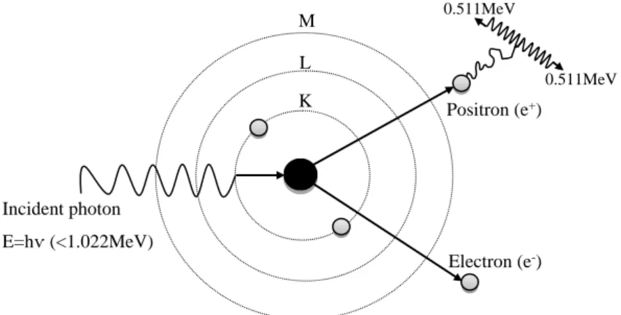
useful in diagnostic imaging. The positron, after losing its kinetic energy in collisions with the surrounding atoms, comes to rest and combines with its antiparticle, the electron, in an annihilation reaction. In this reaction, the masses of the two particles are converted into energy, which appears in form of two photons travelling in opposite directions (180° between them), each one with 0.511MeV. These two opposite photons are detected by PET scanners, enabling a spatial and functional imaging of the body (Podgorsak, 2010).
However, if the parent-daughter atomic mass difference is less than two times the electron rest mass energy, 1.022MeV, the positron emission cannot take place. Instead, an Electron Capture (EC) can occur. This is a competitive or an alternative to the positron decay for neutron-deficient radionuclides.
In the EC decay, an orbital electron, usually from the K-shell, is captured by a proton in the nucleus, converting the proton into a neutron with the ejection of a neutrino. The net effect of the EC is the same as in the positron emission: the Z is decreased by one, N is increasing by one and A remains unchanged. The general equations of EC can be written as follows:
𝑝 + 𝑒
−→ 𝑛 + ѵ
Eq. (2.6)𝑋
𝑍 𝐴 EC→
𝑌 + ѵ + 𝑄
EC 𝑍−1𝐴 Eq. (2.7)𝑄
𝐸𝐶= 𝑀
𝑁,𝑋+ 𝑚
𝑒– 𝑀
𝑁,𝑌− 𝐵
𝑒= 𝑀
𝑋– 𝑀
𝑁− 𝐵
𝑒Eq. (2.8) In this case, the neutrino carries away some of the transaction energy that equals the difference in atomic rest mass between parent and daughter, less the electron binding energy, Be, of the electron captured. The remaining energy appears in the form of characteristic x-rays and/or Auger electrons emitted by the daughter nuclide. This is due to the fact that an electron of an outer shell will fill in the K or L-shell vacancy caused by the capture electron. Characteristic x-ray energy is equal to the energy difference of the orbitals involved in the process, which can be transferred to another electron knocking it out of its shell (Cherry et al., 2003).
Positron decay and EC decay may be accompanied by gamma emission if the daughter is not in a completely stable level and the EC is always accompanied by characteristic x-rays and/or Auger electrons by the daughter nuclide. Thus, the radionuclides with parent-to-daughter transition energies greater than 1.022MeV may decay by EC or β⁺ emission, or both, as it can be seen in figure 2.1., which represents the decay scheme of the 18F into oxygen-18 (18O) (Cherry
Figure 2.1. Decay scheme for 18F into a stable nuclide 18O, which may decay by both EC or β⁺, with Emax,β⁺=0.638MeV. For the 18F, 3% of the nuclei decay by EC and 97% decay by beta plus emission (Adapted from Cherry et al., 2003; LNHB, 2006)
NEGATIVE BETA DECAY
(
Β-)
Contrary to the positron emission, negative beta decay occurs when the radionuclide nucleus has an excess of neutrons (high N/Z ratio), when compared with the number of protons. In this case, a neutron in the nucleus is transformed into a proton with a simultaneous ejection of a negative beta particle (β⁻, identical to electron, with the exception of their origin) and an antineutrino (ѵ̅). When a β⁻ particle is emitted, the N decreases by one, the Z increases by one, so that A remains unchanged. Schematically, the previous process and the negative beta emission equation may be written, respectively, as:
𝑛 → 𝑝 + 𝛽
−+ ѵ̅
Eq. (2.9)𝑋
𝑍 𝐴 β→
−𝑌 + β
−+ ѵ̅ + 𝑄
β− 𝑍+1𝐴 Eq. (2.10)The energy released in the negative beta decay, Qβ-,is the difference between the mass energies of the parent and daughter nuclides, considering the electron binding energy neglected and assuming the antineutrino, such as the neutrino, with zero rest mass and no charge (Turner, 2007; Podgorsak, 2010).
𝑄
𝛽−= 𝑀
𝑁,𝑋− [𝑀
𝑁,𝑌+ 𝑚
𝛽−] = 𝑀
𝑋− 𝑀
𝑌 Eq. (2.11)This energy is shared between the electron and the antineutrino. As explained before, the recoil nucleus (daughter), because of its relatively large mass, receives negligible energy. Therefore, if Eβ⁻ and 𝐸ѵ̅ are the initial kinetic energies of the electron and antineutrino is possible to write:
𝑄
𝛽−= 𝐸
𝛽−+ 𝐸
ѵ̅ Eq. (2.12) Decay modes, such as the beta decays (β⁻, β+and EC), in which the mass number do not change, are called isobaric transactions (Bushberg et al., 2002). A beta minus emission is shown in figure 2.2., representing the carbon-14 (14C) decay into nitrogen-14 (14N).
Relat ive M ass -En er gy [M eV] 𝐹 9 18 𝑂 8 18 2me β⁺ decay (96.86%) EC decay (3.14%) 1.660 0.638 0
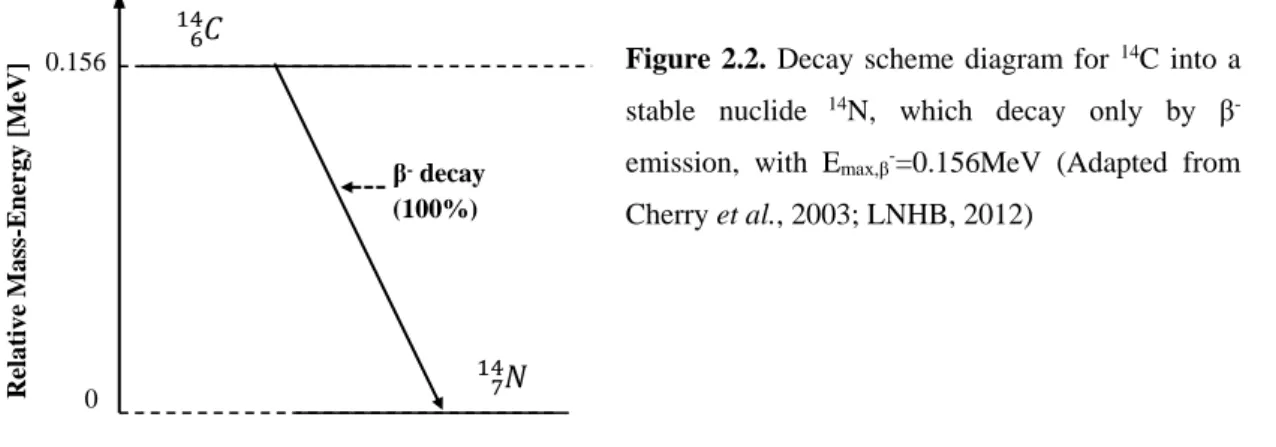
Figure 2.2. Decay scheme diagram for 14C into a stable nuclide 14N, which decay only by β -emission, with Emax,β-=0.156MeV (Adapted from Cherry et al., 2003; LNHB, 2012)
In opposition to the β⁺ emitters, β⁻ are not useful in diagnostic medical imaging because they travel a short distance inside the tissue and do not produce any gamma photons capable of being detected externally. However, according to Joseph (2006), the pure beta-minus emitters demonstrate good results in clinical therapeutic purposes, being therefore used in radionuclide therapy, as seen in section 2.3.
In contrast with α- and EC decays, where the energies of the decay products are uniquely defined by the production of a monoenergetic spectrum, β- and β+ decays emit the most of their
particles with energies lower than the maximum of the particle decay energy (Tmax). These particles exhibit a continuous spectral kinetic energy distribution, resulting in a polyenergetic spectrum ranging from zero to Tmax, calculated according to the Fermi theory of the beta decay (Podgorsak, 2010). The average energy of the β particles emitted is approximately 1/3Tmax. Typical shapes of β− and β+ spectra are presented in figure 2.3.
Figure 2.3. Typical spectrum of beta
particle energy. Energy distributions of beta particles emitted (Adapted from Podgorsak, 2010)
The differences at low kinetic energies are related to the particle charge. The electrons are attracted by the nucleus while the positrons are repelled, causing an energy shift to lower energies for electrons and to higher energies for positrons (Bushberg et al., 2002; Podgorsak, 2010). Taverage ≈ 1/3Tmax,β Relat ive nu mber of β parti cles
Kinetic energy of the beta particles Tmax,β
β -β+ Relat ive M ass -En er gy [M eV] 𝐶 6 14 𝑁 7 14 β- decay (100%) 0.156 0
ISOMERIC TRANSITION
(
IT)
AND INTERNAL CONVERSION(
IC)
After an isobaric transition, the daughter nuclide could remain with an excess of energy and it will further decay until it achieves a stable level. In order to reach stability, the daughter nucleus may emit a nonparticulate radiation in form of gamma photon, with the Z and N remaining constant. This process, in which energy is given off as a gamma photon, without change of mass and atomic number, is called isomeric transition (IT) (Mettler & Guiberteau, 2006).
The gamma rays are very useful in diagnosis imaging, since the nuclear medicine imaging equipment, as the gamma camera, are tuned to detect photons. The detected energy is determined by the difference between the intermediate and final states of the daughter nucleus and generally is in the range of 100keV to 500keV (Sprawls, 1993). It is noted that the intermediate state could emit more than one gamma ray, with different energies, until it reaches stability.
The isomeric transitions occur quickly after the isobaric ones. However, if the nuclear excited state has a long lifetime, this intermediate state is called metastable, as in the case of the 99mTc. According to Sprawls (1993), metastable states are important in nuclear medicine due to the fact that in a metastable state the only energy emitted from the nucleus and detected from the external equipment are the gamma rays. This avoids, consequently, the electron emissions in the body, which do not contribute to the medical image formation but only to the radiation dose in the patient.
Figure 2.4. represents the decay scheme of molybdenum-99 (99Mo) which undergoes an isobaric transition to technetium-99 in the metastable state, 99mTc. The 99mTc will later undergo an
isomeric transition to 99Tc.
Figure 2.4. Simplified decay scheme of isobaric nuclides: 99Mo/99mTc/99Ru. The decay starts with a β- decay of 99Mo into 99mTc, followed by a gamma ray emission of 99mTc into 99Tc, and ending with a β- emission of 99Tc into a stable 99Ru (Adapted from Podgorsak, 2010) 𝑇𝑐 43 99𝑚 𝑇𝑐 4399 𝑅𝑢 4499 𝑀𝑜 4299 Isomeric states of 99mTc Relat ive M ass -En er gy [M eV] 1.357 0.921 0.1405 0 -0.293 β- decay (17.3%) Tmax=0.436MeV β- decay (82.7%) Tmax=1.217MeV γ decay Eγ=140.5keV β- decay (100%) Tmax=0.293MeV
In some cases, the excess of energy emitted by the intermediate nucleus in the form of gamma ray (hυ) may be transferred to an orbital electron within the atom. If the energy transferred to this electron is higher than its binding energy (Be), the electron escapes from the atom with a net kinetic energy of hυ-Be (Attix, 2004).
This process is known as internal conversion (IC) and competes with the IT. However, IC just can occur when the energy transferred to the orbital electron exceeds its biding energy. After the electron being ejected, a vacancy is created. If the vacancy is replaced by an electron from a higher energy level, characteristic x-rays and/or an Auger electron are emitted (Sprawls, 1993). The ratio between the internal conversion electrons and internal transition is called by internal-conversion coefficient (α). A low internal-conversion ratio is preferable since it allows a greater number of gamma emissions used in medical imaging, avoiding the electrons production (Mettler & Guiberteau, 2006).
2.1.2. Characteristics of Radioactive Decay
According to the Advisory Committee on Human Radiation Experiments (ACHRE), the act of emitting radiation spontaneously by an unstable nuclide is called radioactivity. Radioactivity is “a physical, not a biological, phenomenon” from the radioactive nuclides (HSS, 1995). Hence, radioactive nuclei, either natural or artificially, decay by a spontaneous process in which is impossible to predict exactly the moment of its transformation into another stable nuclide. However, since nuclear medicine works, not with individual nuclei, but with a sample or collection of radioactive material, the radioactive decay could be described in terms of probabilities and average decay rates. The amount of radioactivity present in a sample is measured by its activity (A). The activity of a sample is the rate at which the nuclei, within the sample, undergo transformations. It can be measured by counting how many atoms are spontaneously decaying in each second and it is express in units of curie [Ci], or currently, in the SI unit, the Becquerel [Bq], which is defined as one disintegration per second (1Ci=3.7x10¹⁰Bq) (Sprawls, 1993; Barnes, 1996).
The activity, independently of the nature of the decay mechanism, is described mathematically as the change (dN) of the total number of radioactive atoms in a sample (N) in a given period of time (dt), or simplified, is equal to the decay constant (λ) times the total number of unstable atoms in a sample in that time. Both methods are represented in equation 2.13.
The minus sign indicates the decline of the radioactive nuclei during time. The decay constant is equal to the fraction of the number of radioactive atoms remaining in a sample, or alternatively, is the probability of any nucleus to undergo a decay per unit time. The decay constant has a characteristic value for each radionuclide and is measured in seconds-1. By integrating equation 2.13., it is possible to define the exponential radioactive decay law (Eq. 2.14), which expresses the exponential decay of every kind of radioactive nucleus over time (Bushberg et al., 2002).
𝑁 = 𝑁
0𝑒
−𝜆𝑡𝑜𝑟 𝐴 = 𝐴
0
𝑒
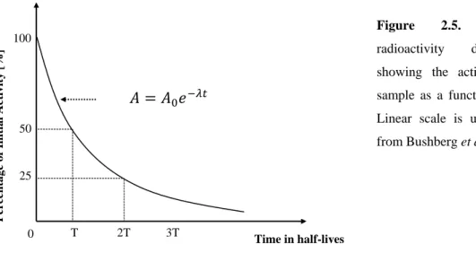
−𝜆𝑡 Eq. (2.14)This equation allows calculation of the number of unstable nuclei in the sample at any time t, knowing the initial amount of radionuclides, N0, at t=0sec. On the other hand, equation 2.14., permits to identify the remaining activity, A, in the sample after t time, assuming an initial activity equal to A0. Figure 2.5., plots equation 2.14 during successive times T, called the half-life of the radionuclide in which the activity drops by factors of one-half, as shown.
Figure 2.5. Exponential radioactivity decay law, showing the activity A of a sample as a function of time t. Linear scale is used (Adapted from Bushberg et al., 2002)
The half-life (T1/2) is a parameter related to the decay constant. It is defined as the necessary time for the radioactive atoms to decrease to one half, in a given sample, which means that the sample will be reduced to half of its existing activity. So, if in the beginning a sample has No radiative atoms, after an half-life, particular for each radionuclide, the remaining number of radioactive atoms (N) will be No/2. The decay constant and the half-life are related as follows:
𝜆 = 𝑙𝑛2 𝑇
⁄
1/2 Eq. (2.15)Both the decay constant and the half-life are unique parameters for each radionuclide (Turner, 2007). Thus, it is possible to say that the half-life, together with the nature of the decay process (α, β and γ-decay) can be a signature for the identification of the isotope. The half-lives of the radioactive isotopes range from fractions of a second to billions to years (ISNAP, 2011).
Time in half-lives Pe rce ntage of In iti al Act ivi ty [ %] 100 50 25 0 T 2T 3T
𝐴 = 𝐴
0𝑒
−𝜆𝑡In nuclear medicine, the radioactive material is administered in a living organism. Therefore, the material might be removed from the organism by normal radioactive decay of the radionuclide but also by biological transport. The half-life used to designate the normal radioactive decay, such as nominated above, is generally called physical half-life (Tp). In addition to the physical half-life, two other half-life terms are commonly used: the biological half-life (Tb), which refers to the time that an organism takes to eliminate half of the radioactive material administered by biological ways, and the effective half-life (Te) which is associated with both physical and biological half-lives (Mettler & Guiberteau, 2006). When the biological transport or elimination occurs, the lifetime of the radioactive material in the body is reduced and it is generally expressed in terms of an effective half-life, by the follow equation:
𝑇
𝑒= (𝑇
𝑝𝑥 𝑇
𝑏) (𝑇
⁄
𝑝+ 𝑇
𝑏)
Eq. (2.16)It is important to highlight that when both physical and biological half-life exist, the effective half-life is always shorter than either one or other (Sprawls, 1993).
Choosing a radiopharmaceutical with an effective half-life that agrees with the duration of the study is crucial for dosimetric purposes. If the radionuclide has a long life then the effective half-life can be reduced by choosing a chemical component with a short biological half-life (Podgorsak, 2010).
Subsequently to the radionuclide disintegration into a stable state, the particles and/or photons emitted, with certain energy according to the radionuclide, will interact with the surrounding tissues. Sometimes, photons may eventually dissipate without transferring any energy to the surrounding medium. The transferred energy to the matter is truly important for dosimetric purposes. According to Stabin (2007), it has important implications for radiation biology, radiation shielding, radiation detection, and for every practical application of radiation protection.
In the next section, the interaction mechanisms of the photons and particles with matter will be described. However, since the present work concerns, primarily, with the beta-emitting radionuclides, only the electron interactions will be discussed, as well as the photons interaction types which provide energy loss during the collision with matter, such as the photoelectric effect, Compton scattering and pair production. Photon interactions occur both in tissue and in the gamma camera.
2.1.3. Photons Interactions
Photons (x-, γ-rays or bremsstrahlung) may transfer all of their energy to matter, being totally absorbed, or they may transfer part of their energy when they are scattered or deflected from their original direction. In this last case, the photons no longer carry useful imaging information. Photons, eventually, may also penetrate the matter without interacting, and consequently, without losing any of their energy (Joseph, 2006; Turner, 2007). Most of the time, when the photons energy is low to moderate, photons interact with the whole atom or with the orbital electrons, whereas those with high energy interact with the atomic nuclei (Stabin, 2007).
One should emphasize that photons do not steadily lose energy via coulombic interactions with matter, as do charged particles (MIT NSE, 2005). This is due to the fact that photons are indirectly ionizing radiation, electromagnetic radiation with zero mass and electrically neutral. For that reason, photons travel a considerable distance before undergoing an interaction and when they do interact, the energy is transferred to the matter into two single steps (MIT OCW NSE, 2007a). The initial step involves the kinetic energy transfer to the medium, with subsequently ejection of electrons from the atoms involved in the interaction. The second phase comprises the energy transfer from these high-speed electrons (such as the beta particles) to the matter, by producing ionization and excitation of the atoms along their paths, causing biological effects (Khan, 2003).
The consequences of those facts will be analysed in section 2.3. The types of photon interactions with matter will be described below, including pair production, an uncommon interaction in gamma camera imaging, but possible to occur since in the current work the beta particles energies, and hence the bremsstrahlung photons energies, may exceed 1.022MeV.
PHOTOELECTRIC EFFECT
In the photoelectric process, a photon with energy hѵ experiences an interaction with an atom of the absorbing medium. The photon energy is completely absorbed by an orbital electron. The orbital electron is ejected from its shell with kinetic energy T=hv-Be, where Be is the binding energy of the ejected electron, and it is called of photoelectron. Thus, the incident photon completely disappears and the photoelectron carries off the resultant energy by producing ionizations and excitations, along a path until its energy is dissipated.
The photoelectron creates a vacancy in the orbital shell which is filled in by an electron from the previous shell with a lower biding energy. In turn, it creates another vacancy filled in by an electron from an even lower binding energy shell. This process keeps creating an electron cascade from outer to inner shells until the atom remains in a stable state. These electronic transitions are followed by simultaneous emission of characteristic x-rays (more frequent in
high Z-materials), with energy equal to the difference of the binding energies, or Auger electrons (more frequent in low Z-materials). Usually, the majority of photoelectric events involves interactions with the innermost orbital shell, the K-shell. The energy of the incident photon must be greater or equal than the biding energy of the K-shell, in order for the photoelectric effect occurs (Cherry et al., 2003; Khan, 2003; Attix, 2004; MIT NSE, 2005; Heard, 2007; Stabin, 2007; Tu rner, 2007). Figure 2.6., represents a photoelectric event with characteristic x-ray emission.
Figure 2.6. Representation of the photoelectric effect with characteristic x-ray emission. Note that,
although not shown on this diagram, Auger electrons can be emitted instead of the characteristic x-ray (Adapted from Alpen, 1990)
According to Attix (2004), Stabin (2007) and Turner (2007), the probability of photoelectric effect occurrence depends strongly on the photon energy, E, as well as, on the atomic number Z of the absorbing medium. The probability of producing a photoelectron per atom or the atomic cross section, symbolized by aτ, varies accordingly Z4/E3. This relationship is used for energies equal or below 0.1MeV, where the photoelectric effect becomes more important. Consequently, with base of the relation above, the photoelectric effect has a higher probability of occurring in high-Z materials and low-energy photons.
The angular distribution of the photoelectron also depends on the photon energy. The photoelectron is emitted at 90° when low-energy photons interact and in a more forward direction when high-energy photons interact with the atoms (Khan, 2003).
M
Incident photon, E=hѵ
Photoelectron, T=hѵ-Be
Characteristic radiation K