J Tissue Eng Regen Med 2011; 5: 41–49.
Published online5 July 2010 in Wiley Online Library (wileyonlinelibrary.com) DOI: 10.1002/term.287
Tissue-engineered constructs based on SPCL
scaffolds cultured with goat marrow cells:
functionality in femoral defects
M´
arcia T. Rodrigues
1,2#, Manuela E. Gomes
1,2#*, Carlos A. Viegas
1,2,3, Jorge T. Azevedo
4,
Isabel R. Dias
1,2,3, Fernando M. Guz´
on
5and Rui L. Reis
1,213Bs Research Group, University of Minho, Guimar˜aes, Portugal
2Institute for Biotechnology and Bioengineering (IBB), PT Government Associated Laboratory, Guimar˜aes, Portugal 3Department of Veterinary Sciences, University of Tr´as-os-Montes e Alto Douro, Vila Real, Portugal
4Department of Animal Science, University of Tr´as-os-Montes e Alto Douro, Vila Real, Portugal 5Veterinary Faculty, University of Santiago de Compostela, Lugo, Spain
Abstract
This study aims to assess the in vivo performance of cell–scaffold constructs composed of goat marrow stromal cells (GBMCs) and SPCL (a blend of starch with polycaprolactone) fibre mesh scaffolds at different stages of development, using an autologous model. GBMCs from iliac crests were seeded onto SPCL scaffolds and in vitro cultured for 1 and 7 days in osteogenic medium. After 1 and 7 days, the constructs were characterized for proliferation and initial osteoblastic expression by alkaline phosphatase (ALP) activity. Scanning electron microscopy analysis was performed to investigate cellular morphology and adhesion to SPCL scaffolds. Non-critical defects (diameter 6 mm, depth 3 mm) were drilled in the posterior femurs of four adult goats from which bone marrow and serum had been collected previously. Drill defects alone and defects filled with scaffolds without cells were used as controls. After implantation, intravital fluorescence markers, xylenol orange, calcein green and tetracycline, were injected subcutaneously after 2, 4 and 6 weeks, respectively, for bone formation and mineralization monitoring. Subsequently, samples were stained with L´evai–Laczk´o for bone formation and histomorphometric analysis. GBMCs adhered and proliferated on SPCL scaffolds and an initial differentiation into pre-osteoblasts was detected by an increasing level of ALP activity with the culture time. In vivo experiments indicated that bone neoformation occurred in all femoral defects. The results obtained provided important information about the performance of SPCL–GBMC constructs in an orthotopic goat model that enabled future studies to be designed to investigate in vivo the functionality of SPCL–GBMC constructs in more complex models, viz. critical sized defects, and to evaluate the influence of in vitro cultured autologous cells in the healing and bone regenerative process. Copyright 2010 John Wiley & Sons, Ltd.
Received 24 January 2010; Accepted 25 February 2010
Keywords autologous; marrow stromal cells; natural-based polymer; goat model; bone regeneration; tissue engineering
*Correspondence to: Manuela E. Gomes, 3Bs Research Group – Biomaterials, Biodegradables and Biomimetics, Department of Polymer Engineering, University of Minho, Headquarters of the European Institute of Excellence on Tissue Engineering and Regenerative Medicine, AvePark, 4806-909 Taipas, Guimar˜aes, Portugal. E-mail: megomes@dep.uminho.pt
#These authors equally contributed to this work.
1. Introduction
Skeletal regeneration is initiated by a traumatic episode involving bone damage that often includes the perios-teum, bone marrow spaces and surrounding soft tissues. Trauma such as fracture or surgical cutting and drilling causes a physical disruption of the mineralized tissue matrix, the death of many cell types and interruption of
the local blood supply. A key stage in bone regenera-tion is the differentiaregenera-tion of pluripotential mesenchymal cells from the initial granuloma containing several cellular types, towards cartilage, fibrous cartilage, fibrous tissue or bone (Dennis et al., 2001).
Bone marrow mesenchymal cells (BMCs) have been widely used in studies involving tissue-engineering (TE) strategies for bone and cartilage, as they provide a potential autologous source of cells (Mauney et al., 2005b; Muraglia et al., 2000; Pittenger et al., 1999; Tuan et al., 2003) that are able to differentiate into chondrogenic and osteogenic lineages in the presence of specific differentiation supplements, such as transforming growth factor-β and dexamethasone, respectively (Oliveira et al., 2006).
Although BMCs are abundant in skeletal tissues, damaged bone may fail to heal spontaneously and, in most cases, the use of marrow cells alone is not ideal to accomplish the necessary requirements for the repair of injured tissues. In order to overcome the limitations of current treatments, the TE field proposes the use of bioactive or inductive factors as well as a scaffold structure to support and complement the role of reparative cells (differentiated or non-differentiated) when implanted on injured or dysfunctional areas.
The selection of a scaffold material for bone TE purposes is therefore of extreme significance (Hutmacher et al., 2007). Scaffolds must be biocompatible, biodegradable and, simultaneously, promote the easy diffusion of nutrients and cellular waste products, as well as present suitable mechanical properties for cell support and new tissue ingrowth. Furthermore, the scaffold must possess adequate porosity, good interconnectivity and a degradation rate (Gomes et al., 2004) adapted to the time required for tissue regeneration.
Several biodegradable polymers have been proposed to obtain three-dimensional (3D) scaffolds for bone TE, including a new range of natural origin polymers based on starch (Gomes et al., 2004, 2008; Mendes et al., 2001). Starch-based polymers, such as starch–polycaprolactone blends (SPCL), are degradable and biocompatible poly-mers with distinct structural forms, which have been shown to be suitable for bone TE applications in sev-eral previous studies (Gomes et al., 2003, 2006a, 2006b; Mendes et al., 2003; Santos et al., 2007).
The seeding and extended in vitro culture of cells within a biodegradable scaffold before implantation is a widespread TE approach. Some studies also report the importance of a cell in vitro preculture period onto scaffolds, as it may influence the osteogenic potential of bone marrow cells (Sikavitsas et al., 2003). During the
in vitro culture, seeded cells are expected to proliferate
and secrete growth factors and matrix proteins that will possibly stimulate other cells to accelerate or recover and, in more dramatic situations, to stimulate the natural regenerative functionality when transferred to the in vivo environment.
Ultimately, and despite the relevance of results achieved from in vitro studies, the use of animal models
is an essential step in evaluating TE constructs prior to their clinical application. Different animal models, such as rat, rabbit, dog, goat, sheep or monkey, have been projected, with the purpose of fairly accurate human model requests (Buma et al., 2004) and guiding potential clinical applications.
In the last few years, goats have become increasingly popular as a valid animal subject in this research field. In fact, due to its nature of a higher-level vertebrate and non-pet status when compared to dogs, goats play a significant role in the orthopaedics field as a feasible model for orthotopic applications. In addition, goats not only have metabolic and bone remodelling rates similar to that of humans but also a comparable sequence of events in bone graft incorporation and healing capacities (Pearce
et al., 2007), which explains the fact that this model
has been frequently used in studies of bone formation and regeneration (Kruyt et al., 2004; Li et al., 2006; Zhu
et al., 2006), biocompatibility (Mendes et al., 2001) and
osteochondral regeneration (Lane et al., 2004; Niederauer
et al., 2000).
Cell-based strategies sustained by a support material have been applied to generate ectopic or orthotopic bone (Giannoni et al., 2008; Kirker-Head et al., 2007; Kruyt et al., 2007; Meinel et al., 2006). Although the latter presents a major potential for skeletal regeneration procedures, most in vivo studies are conducted using an ectopic approach and/or performed in small animal models, such as mice or rats (Kruyt et al., 2007; Livington
et al., 2002; Mauney et al., 2005a; Mastrogiacomo et al.,
2007; Mendes et al., 2003; Trojani et al., 2006). Although non-critical sized defects are usually evaluated in ectopic models, orthotopic location provides a more accurate idea of the influence or local effects of implanted cells or cell–scaffold constructs where they were initially designed to be functional.
Autologous approaches have also been considered in recent studies (Kruyt et al., 2004; Niederauer et al., 2000; Zhu et al., 2006), avoiding immune complex problems that interfere with the regenerative process as well as with the patient follow-up.
The present manuscript describes the assessment of the in vivo osteogenic ability of cell–scaffold constructs based on seeding marrow stromal cells onto SPCL fibre mesh scaffolds and in vitro cultured for different periods of time using an autologous goat model. As already mentioned, these scaffolds have demonstrated a very good in vitro functionality in several studies performed by our group. Therefore, the aim of this work was to obtain the first data concerning the
in vivo functionality of SPCL scaffolds and goat bone
marrow cells (GBMCs) constructs in new bone formation of a orthotopic non-critical defect, in order to better understand the behaviour of cell–scaffold constructs implanted in femurs and to design future in vivo studies to be performed in the critical size defect model, which will be the ultimate applications for such TE strategies.
2. Materials and methods
2.1. Production of SPCL scaffolds
The polymer scaffolds used in this study were based on a blend of corn starch and ε-polycaprolactone (SPCL; 30% : 70% wt) and were produced by a fibre-bonding method into mesh structures with a porosity of∼75% and cut into discs (diameter 6 mm, height 2 mm), as described previously (Gomes et al., 2006a, 2006b). All samples were sterilized using ethylene oxide and prior to cell seeding the scaffolds were immersed in 20 ml serum-free medium in 50 ml tubes for 30 min.
2.2. Animal study
Four skeletally adult female goats weighting 30–45 kg were used in this study. The housing care and experimental protocol were performed according to the national guidelines after approval by the National Ethical Committee for Laboratory Animals (2007-07-27; document no. 018 939) and conducted in accordance with international standards on animal welfare as defined by the European Communities Council Directive of 2 November 1986 (86/609/EEC). During the entire study, adequate measures were taken to minimize any pain and discomfort. Animals were kept in light- and temperature-controlled rooms and health parameters, such as appetite, weight maintenance or signs of infection, were monitored on a daily basis.
2.3. GBMCs harvesting and culture
In order to harvest the GBMCs, the animals were placed under general anaesthesia and iliac regions were shaved and disinfected. The animals were submitted to a pre-anaesthetic medication with acepromazine maleate (5 mg EV, Calmivet, Vet´oquinol, France) and placed under general anaesthesia by induction with thiopenthal sodium (20–25 mg/kg EV, Pentothal sodium, Abbott Laboratories, USA), maintained by inhalation of a mixture of 1.5% isoflurane (IsoFlo, Abbott Laboratories) and oxygen for a maximum of 30 min.
From each iliac crest of the goats, 10 ml samples of bone marrow aspirate were obtained, using a bone marrow aspiration needle (Inter.V, Medical Device Technologies Inc., Denmark) and a 10 ml syringe containing 1 ml heparin (5000 IU, heparin sodium, B. Braun Medical Inc., USA) to avoid marrow coagulation. The content of each syringe was then transferred into sterile 50 ml tubes and mixed with 30 ml RPMI-1640 culture medium (Sigma-Aldrich, USA), containing 1% penicillin–streptomycin (Gibco, USA) and an additional 1 ml heparin (5000 IU). Afterwards, GBMCs were centrifuged for 10 min at 1200 rpm and a dense cellular pellet was collected and cultured in 75 cm2 flasks (Corning, USA) using
basic culture medium (Dulbecco’s modified Eagle’s
medium – DMEM; Sigma-Aldrich, USA) supplemented with 10% autologous serum isolated from goat peripheral blood and 1% antibiotic/antimicotic solution (A/B, Invitrogen, Spain). Four days after the harvesting procedure, the medium containing non-adherent cells was removed and the adherent cells were rinsed with a sterile phosphate-buffered saline (PBS; Sigma) solution and fresh medium was added. The cells were expanded in basic culture medium until about 80% of confluence before being seeded onto SPCL scaffolds at passage 3.
During this study, every 3 weeks, goat peripheral blood samples were collected from the jugular vein of each animal in order to obtain autologous serum, and collected into serum tubes without anticoagulant (Sarstedt, Monovette, Serum Gel S, Germany); 30 min after collection the blood samples were centrifuged at 3000 rpm for 10 min. Harvested sera were immediately stored in appropriate tubes and preserved at −20◦C until use.
2.4. In vitro cell seeding and culture
In order to cell-seed the scaffolds, GBMCs were thawed and expanded until 90% confluence. Afterwards, the cells were enzymatically lifted with 0.05% trypsin–EDTA (Invitrogen, Spain) and at passage 2 a cell suspension was prepared (2.5 million cells/ml) and seeded onto the SPCL porous scaffolds in a dropwise manner, at a cellular density of 0.5 million cells/scaffold and using seeding chambers in order to improve cell seeding efficiency by avoiding cellular dispersion. After in vitro seeding, cell–scaffold constructs were cultured in non-adherent 12-well plates (Costar, Becton Dickinson, USA) to avoid cellular adhesion to the bottom of the plates, and using minimal essential medium Eagle’s α-modification (α-MEM; Sigma-Aldrich), autologous sera (10%), A/B (1%) and osteogenic supplements, viz. dexamethasone (10−8M;Sigma-Aldrich), ascorbic acid (50µg/ml; Sigma) and β-glycerophosphate (10 mM;Sigma) for 1 and 7 days prior to implantation. An in vitro control of the experiment was kept, consisting of cell–scaffold constructs seeded and cultured under the same conditions and for the same periods of time. Autologous culture medium was changed twice a week in all cell cultures.
2.5. In vitro characterization of cell–scaffold
constructs
Samples were collected on days 1 and 7 after seeding for the assessment of proliferation by DNA quantification and initial osteogenic differentiation studies by ALP activity analysis. For these purposes, samples removed from culture were rinsed twice in a PBS solution and transferred into 1.5 ml microtubes containing 1 ml ultra-pure water. Then, GBMC–SPCL constructs were incubated for 1 h at 37◦C in a water-bath and stored in a −80◦C freezer, promoting a thermal shock variation and thus inducing
cell lysis. Before assessing DNA and ALP levels, constructs were thawed and sonicated for 15 min.
A fluorimetric dsDNA quantification kit (PicoGreen, Molecular Probes, USA) was used to determine the proliferation of cells in GBMC–SPCL constructs. Samples and standards (0–2µg/ml) were prepared. Triplicates were made for samples and standards. Afterwards, the 96-well white plate (Costar; Becton-Dickinson) was incubated for 10 min in the dark and the fluorescence was read using a microplate ELISA reader (BioTek, USA) at an excitation of 485/20 nm and an emission of 528/20 nm. A standard curve was developed in order to read DNA values of samples from the standard graph.
ALP activity was measured to detect initial osteogenic differentiation on days 1 and 7. For this purpose, to each well of a 96-well plate (Costar; Becton-Dickinson) were added 20µl sample plus 60 µl substrate solution consist-ing of 0.2% (wt/v) p-nytrophenyl phosphate (Sigma) in a substrate buffer with 1Mdiethanolamine HCl (Merck, Germany), pH 9.8. The plate was then incubated in the dark for 45 min at 37◦C. After the incubation period, 80µl stop solution [2M NaOH (Panreac, Spain) plus 0.2 mM EDTA (Sigma)] was added to each well. Stan-dards were prepared with p-nytrophenol (10µM/ml; Sigma) in order to achieve final concentrations in the range 0–0.3µM/ml. Samples and standards were pre-pared in triplicates. Absorbance was read at 405 nm and sample concentrations were read from the standard graph.
GBMCs adhesion and morphology was also investigated using scanning electron microscopy (SEM) by previously fixing cell–scaffold constructs in a 2.5% glutaraldehyde solution (Sigma), rinsing and dehydrating through a series of ethanol concentrations before sputter-coating them in gold.
2.6. Implantation procedures
Surgical procedures were performed under standard conditions and the drill-hole technique selected was based on the one described by Hallfeldt et al. (1995). Animals were submitted to the anaesthetics protocol described in the previous section. After general anaesthesia, each goat was positioned in lateral recumbency, prepared and draped in a sterile manner to perform a surgical access to the lateral diaphysis of the femur. A skin incision was then performed from the greater trochanter and continued distally to the lateral femoral condyle. The subcutaneous tissue, tensor fascia lata and lateral fascia of the vastus muscle were incised. The biceps femoris muscles were retracted posteriorly, and the vastus muscle was retracted anteriorly, after being detached from the linea aspera and femora shaft, like the periosteum. Non-critical size defects (diameter 6 mm, depth 3 mm) were drilled in the lateral diaphysis of both posterior femurs of the four adult goats with a bone drill (Synthes, Switzerland). Eight drill holes were made in each posterior femur,
with a separation distance between drill holes of 3 cm, in two non-parallel sections in order to avoid fracture tension in the bone and also to avoid new bone formation among the drill holes. Two of these were left empty and two were filled with scaffolds without cells, which were the controls for this experiment. The remaining drill holes were filled in with cell–scaffold constructs cultured for 1 day (two defects) and 7 days (two defects). The implants were carefully press-fitted and placed into the bilateral defects. The muscle was replaced over the bone, and the fascia lata and skin were closed with resorbable and non-resorbable sutures, respectively.
After implantation, intravital fluorescence markers, viz. xylenol orange (90 mg/kg; Sigma Aldrich), calcein green (10 mg/kg; Sigma) and tetracycline (25 mg/kg; Sigma) were injected subcutaneously (after 2, 4 and 6 weeks, respectively) for bone formation and mineralization monitoring, along with the implantation period.
The animals were observed in ambulation on a daily basis and signs of infection or pain were monitored. During the first postoperative week, the animals were subjected to analgesic medication with flunixin meglumin [1 mg/kg intramuscular (i.m.) each 24 h; FinadyneP.A., Schering-Plough II, USA] for 2 days and to antibiotic therapy with amoxicillin (15 mg/kg i.m. each 24 h; ClamoxylL.A., Pfizer, USA) for 7 days.
After implantation, the animals were kept in a 25 m2 room, with freedom of movement and full weight-bearing of the posterior limbs during the complete post-operative period.
2.7. Harvesting samples after implantation
Six weeks after the implantation procedure and 24 h after tetracycline injection, animal euthanasia was performed, using an overdose of pentobarbital sodium (Eutasil, Sanofi, France). The femurs were then removed and cut into single defect-sections. The sections were fixed in 4% formaldehyde solution, pH 7.2 (Sigma), and embed-ded in glycol methacrylate blocks (Technovit 7200, VLC–Heraus Kulzer GmbH, Germany). Thin sections of about 30µm were prepared, using a special micro-tome according to the Donath et al. (1995) technique, using an Exakt-Cuttingsystem (Aparatebau GMBH, Ger-many) in order to slice the calcified bone. Only the mid-section of each block was used for observation using a fluorescence microscope (Olympus BX51, Ger-many) and for histomorphometric analysis. Additional sections were also stained with L´evai–Laczk´o (Jeno and Geza, 1975) to observe new bone formation, using a stereo-microscope (Olympus SZX9, Germany). Quantita-tive measurement for bone neoformation was carried out after selecting relevant drill surrounding areas, where neobone was marked and quantified using Microimage 4.0 software.2.8. Statistical analysis
Statistical analysis was carried out by mean ± standard error of mean (SEM), using t-Test and two-way ANOVA for in vitro and for in vivo measurements, respectively. At least four samples were considered in the in vitro assays (DNA, ALP, SEM), while 16 samples were considered
in vivo for each condition; (a) empty drill hole; (b) drill
hole with SPCL scaffold; drill holes with SPCL seeded with GBMCs for either (c) 1 day or (d) 7 days in osteogenic medium.
In the present study, controls of the experiment were considered indirectly by in vitro assessments of DNA and ALP activity studies and directly by the histometric analysis of induced drill holes made in the posterior femurs of each animal.
3. Results and discussion
3.1. In vitro characterization of autologous
GBMC–SPCL constructs
SEM micrographs (Figure 1) indicated that GBMCs attached to the SPCL fibre meshes presented a typical elon-gated morphology and were homogeneously distributed throughout the surface of the SPCL scaffold. These pic-tures also showed an increase in GBMCs proliferation with the culture time, as observed by the cell layer formed on top of the fibres when compared to cells cultured for 1 day under the same conditions.
Regarding cell proliferation results, data obtained from the DNA content test (Figure 2) indicated that proliferation of GBMCs seeded onto SPCL scaffolds seemed to decrease slightly after 7 days in culture, although no statistically differences were found (p < 0.05,
t-test) to support these results. Nevertheless, the tendency
to cell proliferation decrease could be directly associated to the increment of ALP activity of GBMCs.
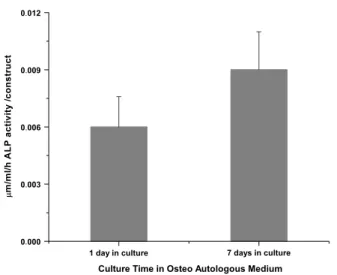
Results obtained from the ALP assay revealed that after 7 days in culture with osteogenic medium there was a significant increment in ALP activity levels (Figure 3;
Figure 2. In vitro double-stranded DNA concentration in SPCL scaffolds seeded with GBMCs cultured in osteogenic culture for 1 and 7 days
p < 0.05, t-test) when compared to levels obtained for
1 day of culture, as expected, since these cells were biochemically stimulated towards the osteogenic pathway (Oliveira et al., 2006; Rodrigues et al., 2006, 2007).
3.2. In vivo studies
All animals completed the study. No weight differences were observed and no signs of infection or inflammation were found nearby the implantation areas after 6 weeks.
In this pilot study, orthotopic defects were drilled to determine the influence of SPCL scaffolds alone and osteogenic differentiation stage of GBMCs seeded onto SPCL scaffolds in bone neoformation. In this way, cells were cultured on SPCL scaffolds, after 1 day in osteogenic culture, practically undifferentiated, or after 7 days in culture, when the cells had already initiated the osteogenic process, as indicated by in vitro ALP activity levels and by previous studies performed by our group.
Prior to construct characterizations, the femurs were cleaned of muscle, as the induced drill holes were not
Figure 1. (A) SEM micrographs of SPCL scaffolds seeded with GBMCs and in vitro cultured in osteogenic culture for 1 day. (B) SEM micrographs of SPCL scaffolds seeded with GBMCs and in vitro cultured in osteogenic culture for 7 days
Figure 3. In vitro ALP activity in SPCL scaffolds seeded with GBMCs cultured in osteogenic culture for 1 or 7 days
easily detected 6 weeks after implantation (both controls and drills containing the cell–SPCL constructs) due to an excellent regeneration process, which occurred in all cases. In order to expose the drill holes and access the inner region, the bone was longitudinally cut. Under macroscopic observation of femoral defects, defects filled with cell–SPCL constructs seemed to have higher bone
growth than empty drill defects or defects filled with SPCL material alone.
3.3. Histological and fluorescence analysis
The observation of sections stained with L´evai–Laczk´o, a specific neobone marker (Figure 4), shows that there was bone formation in all drill defects, as expected in non-critical defects. Nevertheless, there was an enhanced neobone formation in defects containing SPCL scaffolds seeded with GBMCs, which suggests the importance of the presence of these cells in the constructs to stimulate bone formation.Giant cells were present in one of the studied samples, which indicated that after 6 weeks scaffold materials are being absorbed by the body, according to some research works (Pego et al., 2003).
The sequential administration of fluorescent dyes allowed bone ingrowth to be monitored during the overall period of implantation.
Again, the presence of GBMCs seems to positively influ-ence bone ingrowth with time, especially 2 weeks after implantation, when calcein green was subcutaneously injected (Figure 5).
However, the degradation rate of each staining was faster than expected, probably because of the rapid
Figure 4. Drill sections marked with L´evai–Laczk´o staining: (A) control 1, empty drill defects; (B) control 2, defects filled with SPCL (no cells); (C) defects filled with cells–SPCL constructs after (C1) 1 day of culture and (C2) 7 days of culture in osteogenic medium
Figure 5. Drill sections marked with xylenol orange (red), calcein green (green) and tetracycline (not observed) fluorescence stainings: (A) control 1, empty drill defects; (B) control 2, defects filled with SPCL (no cells); (C) defects filled with cell–SPCL constructs after (C1) 1 day of culture and (C2) 7 days of culture in osteogenic medium
metabolism of these animals. Due to this, in some section regions, bone formation was dark just before the subsequent fluorescent stain was injected. In some pictures is still possible to observe dark neobone areas, as the new bone was formed. As tetracycline dye was injected just 24 h before the retrieval of the samples, the fluorescence mark was too light to be easily observed in the drill defect images. Even though the stain had reached the bone drill holes, 24 h was probably an insufficient period of time for the fluorescence to be imprinted in the fresh bone.
Besides a qualitative analysis, histomorphological parameters of bone neoformation were measured to obtain quantitative data regarding the percentage of new bone formation and new bone roundness. The percentage of neobone present in each drill hole was compared with the remaining drill holes and statistically analysed (Figure 6).
The amount of new bone formation tended to increase in the presence of cell-seeded scaffolds, although the quantitative analysis performed did not reveal significant differences between the values (%) measured for new bone formation between defects with and without GBMCs. Other parameters, viz. roundness of new bone formation, were also considered in order to evaluate the spreading of neobone tissue into the induced defects. As can be observed in Figure 7, there was a tendency to an increment in new bone roundness obtained in
Figure 6. New bone formation percentage in the different induced drills: (A) empty drill defects; (B) defects filled with SPCL (no cells); defects filled with cell–SPCL constructs after (C) 1 day of culture and (D) 7 days of culture in osteogenic medium
defects filled with cell-seeded scaffolds (with increasing culture times) when compared to empty defects. This fact may be associated with cellular stimulation provided by autologous cells implanted in the animals.
Neither inflammatory response after 6 weeks of implantation nor animal behaviour modifications in terms
Figure 7. New bone roundness measured for the different induced drills: (A) empty drill defects; (B) defects filled with SPCL (no cells); defects filled with cell–SPCL constructs after (C) 1 day of culture and (D) 7 days of culture in osteogenic medium
of food intake, movement or general health were detected. The presence of fibre mesh SPCL scaffolds allowed for neobone ingrowth in the induced defects. Furthermore, traces of cell-mediated absorption were observed in one of the retrieved samples by the presence of fibrous tissue containing an occasional multinucleated giant cell on the implant surface. This may indicate that locally, material absorption can initiate at an early stage of bone regeneration.
4. Conclusions
In the present study it was possible to observe the neoformation of bone in all orthotopic induced drill
holes in the goat femurs, as expected for non-critical defects. Nevertheless, increased neobone formation was found, as well as cellular distribution into the defect where the SPCL–GBMCs constructs were implanted. This increment was enhanced with the in vitro culture time, which indicates that the in vitro culture time of GBMCs onto the SPCL constructs may also play an important role in new bone growth. The data obtained concerning in vitro proliferation and differentiation of these constructs suggest that the in vitro culture time of GBMCs in osteogenic medium, although still at a very early stage, is likely to play an important role in bone growth onto these defects. However, further studies should provide better understanding of the importance of the number of cells and their differentiation stage, as well as the time needed for osteogenic supplementation, to induce significant changes in bone formation in vivo. Nevertheless, SPCL fibre mesh scaffolds showed a great potential for the development of adequate tissue 3D support for the regeneration of bone in non-critical defects. Thus, the results obtained provided important information about the performance of SPCL–GBMC constructs in an orthotopic goat model that enabled future studies to be designed to investigate the in vivo functionality of GBMC–SPCL constructs in more complex models, viz. in induced critical sized defects, and to evaluate the influence of in vitro cultured autologous cells in the healing and bone regenerative process.
Acknowledgements
M´arcia T. Rodrigues acknowledges the Portuguese Foundation for Science and Technology (FCT) for her PhD scholarship (Grant No. SFRH/BD/30745/2006). This work was partially supported by the European Union-funded STREP Project HIPPOCRATES (Grant No. NMP3-CT-2003-505758) and was carried out under the scope of the European NoE EXPERTISSUES (Grant No. NMP3-CT-2004-500283).
References
Buma P, Schreurs W, Verdonschot N. 2004; Skeletal tissue engineering-from in vitro studies to large animal models. Biomaterials 25(9): 1487–1495. Donath K. 1995; Preparation of histologic
sections by the cutting-grinding technique for hard tissue and other material not suitable to be sectioned by routine methods. In Equipment and Methodical Performance. Exakt-Kulzer-Publications: Norderstedt, Germany.
Dennis R, Carter GSB. 2001; Skeletal tissue regeneration. In Skeletal Function and Form – Mechanobiology of Skeletal Development, Aging and Regeneration. Cambridge University Press: Cambridge, UK; 161–200.
Giannoni P, Mastrogiacomo M, Alini M, et al. 2008; Regeneration of large bone defects in sheep using bone marrow stromal cells. J Tissue Eng Regen Med 2(5): 253–262.
Gomes ME, Azevedo HS, Moreira AR, et al. 2008; Starch–poly(ε-caprolactone) and
starch–poly(lactic acid) fibre-mesh scaf-folds for bone tissue engineering appli-cations: structure, mechanical properties and degradation behaviour. J Tissue Eng Regen Med 2(5): 243–252.
Gomes ME, Bossano CM, Johnston CM, et al. 2006a; In vitro localization of bone growth factors in constructs of biodegradable scaffolds seeded with marrow stromal cells and cultured in a flow perfusion bioreactor. Tissue Eng 12(1): 177–188. Gomes ME, Holtorf HL, Reis RL, et al.
2006b; Influence of the porosity of starch-based fiber mesh scaffolds on the proliferation and osteogenic differentiation of bone marrow stromal cells cultured in a flow perfusion bioreactor. Tissue Eng 12(4): 801–809. Gomes ME, Reis RL. 2004; Biodegradable
polymers and composites in biomedical applications: from catgut to tissue engineering. Part 1. Available systems and their properties. Int Mater Rev 49(5): 261–273.
Gomes ME, Sikavitsas VI, Behravesh E, et al. 2003; Effect of flow perfusion on the osteogenic differentiation of bone marrow stromal cells cultured on starch-based three-dimensional scaffolds. J Biomed Mater Res A 67(1): 87–95.
Hallfeldt KK, Stutzle H, Puhlmann M, et al. 1995; Sterilization of partially dem-ineralized bone matrix: the effects of different sterilization techniques on osteo-genetic properties. J Surg Res 59(5): 614–620.
Hutmacher DW, Schantz J, Lam CXF, et al. 2007; State of the art and future directions of scaffold-based bone engineering from a biomaterials perspective. J Tissue Eng Regen Med 1(4): 245–260.
Jeno L, Geza L. 1975; A simple differential staining method for semi-thin sections of ossifying cartilage and bone tissues embedded in epoxy resin. Mikroskopie 31(1–2): 1–4.
Kirker-Head C, Karageorgiou V, Hofmann S, et al. 2007; BMP–silk composite matrices
heal critically sized femoral defects. Bone 41(2): 247–255.
Livingston T, Ducheyne P, Garino J. 2002; In vivo evaluation of a bioactive scaffold for bone tissue engineering. J Biomed Mater Res 62(1): 1–13.
Kruyt MC, Dhert WJ, Oner FC, et al. 2007; Analysis of ectopic and orthotopic bone formation in cell-based tissue-engineered constructs in goats. Biomaterials 28(10): 1798–1805.
Kruyt MC, Dhert WJ, Yuan H, et al. 2004; Bone tissue engineering in a critical size defect compared to ectopic implantations in the goat. J Orthop Res 22(3): 544–551. Lane JG, Massie JB, Ball ST, et al. 2004; Follow-up of osteochondral plug transfers in a goat model: a 6-month study. Am J Sports Med 32(6): 1440–1450.
Li X, Feng Q, Liu X, et al. 2006; Collagen-based implants reinforced by chitin fibres in a goat shank bone defect model. Biomaterials 27(9): 1917–1923. Mastrogiacomo M, Papadimitropoulos A,
Cedola A, et al. 2007; Engineering of bone using bone marrow stromal cells and a silicon-stabilized tricalcium phos-phate bioceramic: evidence for a cou-pling between bone formation and scaffold resorption. Biomaterials 28(7): 1376–1384.
Mauney JR, Jaquiery C, Volloch V, et al. 2005a; In vitro and in vivo evaluation of differentially demineralized cancellous bone scaffolds combined with human bone marrow stromal cells for tissue engineering. Biomaterials 26(16): 3173–3185.
Mauney JR, Volloch V, Kaplan DL. 2005b; Role of adult mesenchymal stem cells in bone tissue engineering applications: current status and future prospects. Tissue Eng 11(5–6): 787–802.
Meinel L, Betz O, Fajardo R, et al. 2006; Silk-based biomaterials to heal critical sized femur defects. Bone 39(4): 922–931.
Mendes SC, Bezemer J, Claase MB, et al. 2003; Evaluation of two biodegradable polymeric systems as substrates for bone tissue engineering. Tissue Eng 9(suppl 1): S91–101.
Mendes SC, Reis RL, Bovell YP, et al. 2001; Biocompatibility testing of novel starch-based materials with potential application in orthopaedic surgery: a preliminary study. Biomaterials 22(14): 2057–2064. Muraglia A, Cancedda R, Quarto R. 2000;
Clonal mesenchymal progenitors from human bone marrow differentiate in vitro according to a hierarchical model. J Cell Sci 113(7): 1161–1166.
Niederauer GG, Slivka MA, Leatherbury NC, et al. 2000; Evaluation of multiphase implants for repair of focal osteochondral defects in goats. Biomaterials 21(24): 2561–2574.
Oliveira JM, Rodrigues MT, Silva SS, et al. 2006; Novel hydroxyapatite/ chitosan bilayered scaffold for osteochon-dral tissue-engineering applications: scaf-fold design and its performance when seeded with goat bone marrow stromal cells. Biomaterials 27(36): 6123–6137. Pearce AI, Richards RG, Milz S, et al. 2007;
Animal models for implant biomaterial research in bone: a review. Eur Cell Mater 13: 1–10.
Pego AP, Van Luyn MJ, Brouwer LA, et al. 2003; In vivo behavior of poly(1,3-trimethylene carbonate) and copolymers of 1,3-trimethylene carbonate with D,L -lactide or ε-caprolactone: degradation and tissue response. J Biomed Mater Res A 67(3): 1044–1054.
Pittenger MF, Mackay AM, Beck SC, et al. 1999; Multilineage potential of adult human mesenchymal stem cells. Science 284(5411): 143–147.
Rodrigues MT, Oliveira JM, Gomes ME, et al. 2006; Novel hydroxyapatite/ chitosan bilayer scaffolds for the regeneration of osteochondral defects using a tissue engineering approach
based on culturing and differentiation of goat marrow cells into osteoblasts or chondrocytes. In World Congress on Tissue Engineering and Regenerative Medicine, Pittsburg, PA, USA.
Rodrigues MT, Leonor I, Tuzlakoglu K, et al. 2007; Novel in situ approach for the design of osteoconductive and osteoinductive 3D wet-spun fibre mesh scaffolds for bone tissue engineering. In Tissue Engineering International and Regenerative Medicine Society – Asia Pacific Chapter Meeting, Tokyo, Japan.
Santos MI, Fuchs S, Gomes ME, et al. 2007; Response of micro- and macrovascular endothelial cells to starch-based fiber meshes for bone tissue engineering. Biomaterials 28(2): 240–248.
Sikavitsas VI, van den Dolder J, Ban-croft GN, et al. 2003; Influence of the in vitro culture period on the in vivo per-formance of cell/titanium bone tissue-engineered constructs using a rat cranial critical size defect model. J Biomed Mater Res A 67(3): 944–951.
Trojani C, Boukhechba F, Scimeca JC, et al. 2006; Ectopic bone formation using an injectable biphasic calcium phos-phate–SiHPMC hydrogel composite loaded with undifferentiated bone mar-row stromal cells. Biomaterials 27(17): 3256–3264.
Tuan RS, Boland G, Tuli R. 2003; Adult mesenchymal stem cells and cell-based tissue engineering. Arthritis Res Ther 5(1): 32–45.
Zhu L, Liu W, Cui L, et al. 2006; Tissue-engineered bone repair of goat-femur defects with osteogenically induced bone marrow stromal cells. Tissue Eng 12(3): 423–433.