Received: 09 July 2015 Revised: 10 March 2016 Accepted: 18 April 2016
Cite this article as:
Lopes Dias J, Borges A, Lima Rego R. Primary intraosseous squamous cell carcinoma of the mandible: a case with atypical imaging features. BJR Case Rep 2016; 2: 20150276.
CASE REPORT
Primary intraosseous squamous cell carcinoma of the
mandible: a case with atypical imaging features
1JOÃO LOPES DIAS,MD,2ALEXANDRA BORGES,MDand3RAFAELA LIMA REGO,MD 1Department of Radiology, Hospital de São José, Centro Hospitalar de Lisboa Central, Lisbon, Portugal 2Department of Radiology, Instituto Português de Oncologia de Lisboa Francisco Gentil, Lisbon, Portugal 3Department of Pathology, Instituto Português de Oncologia de Lisboa Francisco Gentil, Lisbon, Portugal Address correspondence to: João Lopes Dias
E-mail: [email protected]
ABSTRACT
Primary intraosseous squamous cell carcinoma is a rare malignant tumour that exclusively arises within the jaws. Its diagnosis requires an appropriate clinical, imaging and histological correlation. The exclusion of primary oral mucosa lesions and metastatic disease is mandatory. We report an atypical imaging appearance of this uncommon entity, characterized by new bone formation and periosteal reaction that resemble sarcomatous or malignant odontogenic tumours. A comprehensive discussion on the embryological principles of primary intraosseous squamous cell carcinoma is also provided.
BACKGROUND
Primary intraosseous squamous cell carcinoma (PIOSCC) is a rare malignant tumour that arises within the jaws and derives from odontogenic epithelial remnants.1–4 Owing to its rarity, clinical and radiological diagnostic criteria are not well established. Varied nomenclature has been used for this tumour since Loos first described it in 1913.2 Terminologies such as intra-alveolar epidermoid carcinoma or primary intra-alveolar epidermoid carci-noma have fallen in disuse and the most recent World Health Organization Classification of Tumours in 2005 used the term PIOSCC for this tumour.2 According to this classification, three subcategories of PIOSCC may be found: (1) solid tumours that invade the marrow spaces and induce osseous resorption; (2) squamous cancer aris-ing from the linaris-ing of an odontogenic cyst; (3) squamous cell carcinoma (SCC) in association with other benign epithelial odontogenic tumours.4
Clinical and imaging features are non-specific and the first impression of both clinicians and radiologists usually favours the diagnosis of alveolar or gingival SCC with bone invasion, or metastatic SCC.5–7We report a PIOSCC with atypical imaging findings on both CT and MRI. A discus-sion on the relevant embryological and histological features is also provided.
CASE REPORT
A 42-year-old male presented to the Department of Head and Neck Surgery of a tertiary oncological centre because
of right mandibular swelling and trismus. The patient had already been admitted to a secondary care hospital 3 months earlier with complaints of right mandibular dis-comfort and slight tumefaction. A biopsy was then per-formed and the diagnosis of ameloblastic carcinoma was made histologically. The patient was otherwise healthy with no significant past medical history, including alcohol, smoking or tobacco abuse.
A complete head and neck examination revealed a painless, firm and fixed right mandibular mass with no cutaneous inflammatory signs. No ulcers or mucosal lesions were found in the oral cavity. Laboratory evaluation, chest radiograph and respiratory function tests were unremark-able. The patient underwent bronchofibroscopy, which revealed only mild laryngeal hyperaemia.
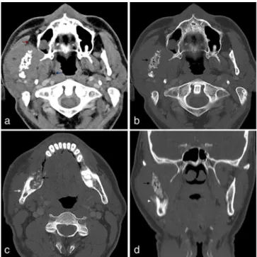
Both neck CT (Figure 1) and MRI (Figure 2) were per-formed, showing a large, solid tumour arising from the ramus and posterior body of the right mandible. The lesion extended to the surrounding soft tissues, with invasion of the masseter and medial pterygoid muscles and caused bulging of the buccal mucosa. The soft tissue component was hypointense on T1 weighted and hyperintense on
T2 weighted MR images and showed avid enhancement
after gadolinium administration on MR examination. CT scan disclosed striking sclerosis and irregularity of the mandibular ramus with some gas bubbles inside the med-ullary cavity and an expansive lytic component in the pos-terior body and angle with some bone-forming matrix
inside. Prominent periosteal reaction was also identified, partic-ularly in the outer cortical surface of the mandibular ramus with the typical pattern of a ruptured Codman triangle. No associated cystic lesion was found in the mandible. No enlarged lymph nodes were detected and the evaluation of the remaining cervical
After surgery, the patient underwent adjuvant chemotherapy (cisplatin-based regimen) and radiotherapy.
DISCUSSION
PIOSCC is a rare tumour with only a few cases reported in the liter-ature. It is exclusively found within the jaws, the only bones that may give origin to both connective and epithelial tumours. The mandible is much more often involved than the maxilla.8The
asso-ciation of the PIOSCC with odontogenic cysts has already been reported, particularly with residual and radicular cysts, and less
fre-Figure 1. (a) Axial enhanced CT image in soft tissue window settings reveals a large right mandibular tumour consisting of a soft tissue component surrounding a markedly irregular and sclerotic mandibular ramus with some gas bubbles within the medullary cavity. The soft tissue component invades the mas-seter muscle (m) laterally and the pterygoid muscles (p) medi-ally. The ipsilateral parapharyngeal space (blue arrow) and the Bichat’s fat pad (red arrow) are medially and anteriorly dis-placed, respectively. (b–d) Axial and coronal CT images in bone window settings better depict the heterogeneous bone involvement with a more irregular and sclerotic pattern in the mandibular ramus occupying the central portion of the tumour (black arrows) and a more expansive, remodelling pattern in the angle and posterior body with a ground-glass pattern of bone-forming matrix. Different patterns of periosteal reaction are depicted lengthwise in the mandibular ramus, including a thick regular deposition resembling an “onion skin” (white arrow) and a ruptured Codman triangle (white arrowhead).
Figure 2. (a, b) Synchronized axial T1 weighted and
T2weighted MR images show multiple coalescent hypointense
foci in the region of the mandibular ramus (white arrow) and a surrounding soft tissue component isointense and hyperin-tense in relation to the remaining masseter (blue arrows) on
T1 weighted image and T2 weighted image, respectively.
(c) Axial enhanced fat-suppressed T1weighted MR image
dem-onstrates avid tumour enhancement and better depicts the involvement of the pterygoid space (asterisk). (d) Axial
T1weighted MR image at a lower level shows the replacement
of the normal fatty marrow by an expansive soft tissue mass expanding and remodelling the cortical bone (red arrow).
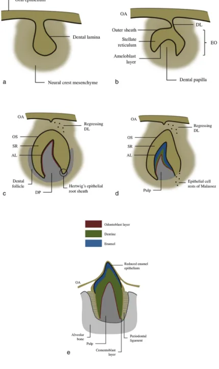
Figure 4. Diagram of tooth development during (a) the bud stage (8th week), (b) cap stage (10th week), (c) bell stage (12th week), (d) apposition stage (variable) and (e) maturation stage (variable). The primitive oral ectoderm thickens and invaginates to form a C-shaped primary dental lamina. It progressively grows into the subjacent neural crest mesenchyme, giving rise to a bell-shaped tooth primordium. The epithelium of the tooth primordium, known as enamel organ, remains connected to the oral epithelium by a stalk of dental lamina that will soon regress. An outer epithelial sheath, a mesenchyme stellate reticulum and an inner epithelial ameloblast layer constitute the enamel organ. After the ameloblast layer secretes the definitive enamel of the tooth, the reduced enamel epithelium appears superficially, resulting from the fusion of the three primordial layers of the enamel organ. It plays an essential role in the tooth eruption process by secreting connective-tissue-breaking proteases. Deep under the concave surface of the enamel organ, two neural-crest-derived ectomesenchymal structures are separated by the Hertwig’s epithelial root sheath, a double-layered covering of epithelial cells that plays an important role during the tooth root development. While the dental papilla gives rise to the dentin, the dental follicle has the capacity to differentiate into cementoblasts, fibroblasts and osteoblasts, thus giving rise to the cementum. The Hertwig’s epithe-lial root sheath starts to disintegrate after the deposition of the first dentine layer, leading to the formation of residual epitheepithe-lial filaments known as epithelial cell rests of Malassez. These are the only odontogenic epithelial cells that may be found in the adult periodontal space.13–16The three recognized sources of epithelial remnants within the mandible are illustrated. AL, ameloblast layer; DL, dental lam-ina; DP, dental papilla; EO, enamel organ; OA, oral epithelium; OS, outer sheath; SR, stellate reticulum.
periodontal cysts.7,9,10In rare cases, PIOSCC results from dediffer-entiation of a benign ameloblastoma.4
Risk factors and causes are not well established, but since PIOSCC develops without initial communication with the oral mucosa, exogenous carcinogens such as tobacco and alcohol are unlikely to be involved.7 Other factors such as long-standing chronic inflammation and keratinization have been postulated to increase the risk of malignant transformation within an odontogenic cyst.8
A predilection for males (2:1) is described, as well as an increased incidence in the fifth decade.11 Symptoms are non-specific and depend on the location, size and aggressiveness of the tumour. Pain, swelling, sensory disturbance (due to inferior alveolar nerve involvement) and odontogenic disorders are commonly found.11
Bodner et al9analysed the clinical and pathological features of 116 PIOSCCs arising in odontogenic cysts, and reported a large majority of well-to-moderately differentiated tumours (85%). In the same study, the overall survival rate was 62% at 2 years and 38% at 5 years.12
Several hypotheses have been postulated to explain the origin of PIOSCC. Epithelial cells have to be present within the bone to give rise to a squamous cell tumour, and the presence of epithe-lial remnants constitutes the most consensual theory.8The jaws
arise from the first pharyngeal arch, which appears on day 22 of embryonic development. It is posteriorly remodelled to form cranial maxillary and caudal mandibular prominences, which give rise to the upper and lower jaws, respectively. Each swelling contains a central cartilage that is produced by neural crest cells. In the mandibular swellings, the central cartilage is known as Meckel’s cartilage. The mandible results from the enlargement and fusion of the bilateral mandibular prominences.13 Since Meckel’s cartilage has no epithelium or stem cells that might undergo epithelial differentiation, it cannot be stated as the source of epithelial remnants. Actually, some authors argue in favour of a direct transformation of remnants of the odonto-genic epithelium, which may include the remnants of dental lamina in the hard and soft tissue spaces, the epithelial cell rests of Malassez in the periodontal ligament space and the reduced enamel epithelium.4,5,7,13–16 Figure 4summarizes the stages of tooth development and illustrates the three recognized sources of epithelial remnants within the mandible.
radiation and usually suffice in the diagnosis of tooth-related tumours. The imaging findings of the majority of benign cysts are typical and usually offer no doubt, unless invasive features or multiple lesions are found. In the setting of aggressive lesions, CT is more accurate than orthopantomography by allowing vol-umetric acquisitions and multiplanar reconstruction. CT is then able to detect small cortical erosions, periosteal reaction and soft tissue calcified components. MRI remains a second-line choice in the evaluation of tooth-related diseases. However, despite being less accurate in the detection of calcified matrices, it better depicts soft tissue involvement and perineural invasion owing to its higher contrast resolution.13
The imaging appearance of PIOSCC is variable, ranging from well-defined benign-like masses to ill-defined and often bone-destructing lytic lesions.9 Although more typical of tumours such as metastatic SCC or sarcomatous tumours, bone-forming PIOSCCs have also been described. Some authors have reported small radiopaque foci within these tumours that were due to cal-cification or dentinoid structures. Ground-glass opacity has also been reported, potentially mimicking fibrous dysplasia or ossify-ing fibroma.11 In this case we report a prominent interrupted type of periosteal reaction apart from a medullary new bone for-mation. This kind of periosteal reaction suggests an aggressive malignancy such as sarcoma, and is not commonly found in metastases or odontogenic tumours. A Codman’s triangle was also found, corresponding to a segment of the periosteum that was elevated from the cortex by the underlying rapidly growing tumour.17
As previously stated, PIOSCC is often found in association with odontogenic cysts, so particular attention should be provided to patients with a previous diagnosis of probably benign jaw lesion who present with new symptoms and volumetric changes upon physical examination. Malignant transformation within an odontogenic cyst should be suspected on imaging modalities if the typical unilocular, homogeneous cyst increases in size or develops hazy borders or bone erosions.8
This article reports a peculiar imaging presentation of a rare tumour. The imaging findings of an aggressive bone-centred tumour with irregular periosteal reaction are more often seen in a sarcoma or a primary malignant odontogenic tumour. The above described ossification pattern is uncommon in the setting of an SCC, either primary oral or metastatic. With regard to the
3. The exclusion of primary oral mucosa lesions and metastatic disease is mandatory.
4. The imaging appearance of PIOSCC ranges from well-defined masses to ill-defined and bone-destructing lytic lesions.
5. Ossification and irregular periosteal reaction are uncommon in the setting of an SCC.
CONSENT
Informed consent was obtained.
REFERENCES
1. Nomura T, Monobe H, Tamaruya N, Kishishita S, Saito K, Miyamoto R, et al. Primary intraosseous squamous cell carcinoma of the jaw: two new cases and review of the literature. Eur Arch Otorhinolaryngol 2013; 270: 375–9. doi:http://dx.doi.org/10.1007/ s00405-012-2235-9
2. Huang JW, Luo HY, Li Q, Li TJ. Primary intraosseous squamous cell carcinoma of the jaws. Clinicopathologic presentation and prognostic factors. Arch Pathol Lab Med 2009; 133: 1834–40. doi:http://dx.doi.org/ 10.1043/1543-2165-133.11.1834
3. Zwetyenga N, Pinsolle J, Rivel J, Majoufre-Lefebvre C, Faucher A,
Pinsolle V. Primary intraosseous carcinoma of the jaws. Arch Pathol Lab Med 2001; 127: 794–7.
4. Eversole L, Siar C, van der Waal I. Primary intraosseous squamous cell carcinomas. In: Barnes L, Eveson JW, Reichart P,
Sidransky D eds. World Health Organization classification of tumours (Pathology and Genetics of Head and Neck Tumours). 2005. pp. 290–1.
5. Adachi M, Inagaki T, Ehara Y, Azuma M, Kurenuma A, Motohashi M, et al. Primary intraosseous carcinoma arising from an odontogenic cyst: a case report. Oncol Lett 2014; 8: 1265–8. doi:http://dx.doi.org/10. 3892/ol.2014.2248
6. Thomas G, Sreelatha KT, Balan A, Ambika K. Primary intraosseous carcinoma of the mandible: a case report and review of the literature. Eur J Surg Oncol 2000;
26: 82–6. doi:http://dx.doi.org/10.1053/ejso.
1999.0746
7. Jain M, Mittal S, Gupta DK. Primary intraosseous squamous cell carcinoma arising in odontogenic cysts: an insight in pathogenesis. J Oral Maxillofac Surg 2013; 71: e7–14. doi:http://dx.doi.org/10.1016/j.
joms.2012.08.031
8. Bereket C, Bekçioğlu B, Koyuncu M, Şener İ, Kandemir B, Türer A. Intraosseous carcinoma arising from an odontogenic cyst: a case report. Oral Surg Oral Med Oral Pathol Oral Radiol 2013; 116: e445–9. doi:http://dx.doi.org/10.1016/j. oooo.2012.02.029
9. Bodner L, Manor E, Shear M, van der Waal I. Primary intraosseous squamous cell carcinoma arising in an odontogenic cyst: a clinicopathologic analysis of 116 reported cases. J Oral Pathol Med 2011; 40: 733–8. doi:
http://dx.doi.org/10.1111/j.1600-0714.2011. 01058.x
10. Prasad H, Anuthama K, Chandramohan M, Sri Chinthu KK, Ilayaraja V, Rajmohan M. Squamous cell carcinoma arising from a dentigerous cyst——report of a case and review of literature. J Oral Maxillofac Surg Med Pathol 2015; 27: 121–5. doi:http://dx. doi.org/10.1016/j.ajoms.2013.10.002
11. Matsuzaki H, Katase N, Matsumura T, Hara M, Yanagi Y, Nagatsuka H, et al. Solid-type primary intraosseous squamous cell carcinoma of the mandible: a case report with histopathological and imaging features. Oral Surg Oral Med Oral Pathol Oral Radiol
2012; 114: e71–7. doi:http://dx.doi.org/10. 1016/j.oooo.2012.05.019
12. Tan B, Yan TS, Shermin L, Teck KC, Yoke PC, Goh C, et al. Malignant transformation of keratocystic odontogenic tumor: two case reports. Am J Otolaryngol 2013; 34: 357–61. doi:http://dx.doi.org/10. 1016/j.amjoto.2013.01.002
13. Dunfee BL, Sakai O, Pistey R, Gohel A. Radiologic and pathologic characteristics of benign and malignant lesions of the mandible. Radiographics 2006; 26: 1751–68. doi:http://dx.doi.org/10.1148/rg. 266055189
14. McClatchey KD. Tumors of the dental lamina: a selective review. Semin Diagn Pathol 1987; 4: 200–4.
15. Park SJ, Bae HS, Cho YS, Lim SR, Kang SA, Park JC. Apoptosis of the reduced enamel epithelium and its implications for bone resorption during tooth eruption. J Mol Histol 2013; 44: 65–73. doi:http://dx.doi.org/ 10.1007/s10735-012-9465-4
16. Xiong J, Gronthos S, Bartold PM. Role of the epithelial cell rests of Malassez in the development, maintenance and regeneration of periodontal ligament tissues. Periodontol 2000 2013; 63: 217–33. doi:http://dx.doi.org/ 10.1111/prd.12023
17. Bisseret D, Kaci R, Lafage-Proust MH, Alison M, Parlier-Cuau C, Laredo JD, et al. Periosteum: characteristic imaging findings with emphasis on radiologic-pathologic comparisons. Skeletal Radiol 2015; 44: 321–38. doi:http://dx.doi.org/10.1007/ s00256-014-1976-5