Review
The role of ophthalmic imaging in central nervous system degeneration
in systemic lupus erythematosus
Arnaldo Dias-Santos
a,b,c,⁎
, Rita Pinto Proença
a, Joana Tavares Ferreira
a,b,c, So
fia Pinheiro
d, João Paulo Cunha
a,c,
Rui Proença
e,f, Maria Francisca Moraes-Fontes
c,g,ha
Department of Ophthalmology, Centro Hospitalar de Lisboa Central, Lisbon, Portugal
b
Department of Ophthalmology, Hospital CUF Descobertas, Lisbon, Portugal
c
NOVA Medical School, Universidade NOVA de Lisboa, Lisbon, Portugal
d
Autoimmune Disease Unit, Unidade de Doenças Auto-imunes/Serviço Medicina 3, Hospital de Santo António dos Capuchos, Centro Hospitalar de Lisboa Central, Lisbon, Portugal
eDepartment of Ophthalmology, Centro Hospitalar e Universitário de Coimbra, Coimbra, Portugal f
Faculty of Medicine, University of Coimbra, Coimbra, Portugal
g
Autoimmune Disease Unit, Unidade de Doenças Auto-imunes/Serviço de Medicina 7.2, Hospital Curry Cabral, Centro Hospitalar de Lisboa Central, Lisbon, Portugal
h
Instituto Gulbenkian de Ciência, Oeiras, Portugal
a b s t r a c t
a r t i c l e i n f o
Article history: Received 9 January 2018 Accepted 14 January 2018 Available online 7 April 2018
Systemic lupus erythematosus (SLE) is an autoimmune connective tissue disorder that can involve any organ sys-tem. Central nervous system involvement can be a severe life threatening complication, ultimately resulting in severe neurodegenerative changes. Magnetic resonance imaging suggests that neurodegeneration, which may have deleterious effects on brain function, may occur early in SLE and experimental models suggest that neuro-protection may be feasible and beneficial.
The retina is an extension of the brain. Recent ophthalmic imaging technologies are capable of identifying early changes in retinal and choroidal morphology and circulation that may reflect CNS degeneration. However, their utility in monitoring CNS involvement in SLE has been poorly studied as these have only been performed in small cohorts, in a cross-sectional design, non-quantitatively and without correlation to disease activity.
The authors aim to review the current understanding of neurodegeneration associated with SLE, with particular focus on the visual pathway. We describe the neuropathology of the visual system in SLE and the evidence for retinal and choroidal neurodegenerative and microvascular changes using optical coherence tomography tech-nology. We aim to describe the potential role of optical imaging modalities in NPSLE diagnosis and their likely im-pact on the study of neuronal function.
© 2018 Elsevier B.V. All rights reserved. Keywords:
SLE
Neuropsychiatric lupus Neurodegeneration Visual pathway
Optical coherence tomography
Contents
1. Introduction . . . 618
1.1. Systemic lupus erythematosus . . . 618
1.2. Neuropsychiatric SLE (NPSLE)– clinical features . . . 618
1.3. Neuropsychiatric SLE (NPSLE)– pathophysiology . . . 618
1.4. Central nervous system degeneration in SLE . . . 619
2. The eye in SLE . . . 619
2.1. The optic nerve . . . 619
2.2. The choroid . . . 619
2.3. The retina . . . 619
2.4. The retrochiasmal visual pathway . . . 620
3. Imaging the retina, optic nerve and choroid in SLE . . . 620
3.1. Fundusfluorescein angiography and indocyanine angiography . . . 620
3.2. Spectral domain optical coherence tomography . . . 620 ⁎ Corresponding author at: Hospital de Santo António dos Capuchos, Alameda de Santo António dos Capuchos, 1169-050 Lisboa, Portugal.
E-mail address:[email protected]. (A. Dias-Santos).
https://doi.org/10.1016/j.autrev.2018.01.011
1568-9972/© 2018 Elsevier B.V. All rights reserved.
Contents lists available atScienceDirect
Autoimmunity Reviews
4. Conclusion . . . 621 Take-home messages . . . 622 Funding . . . . References. . . 622
1. Introduction
1.1. Systemic lupus erythematosus
Systemic lupus erythematosus (SLE) is a systemic, autoimmune dis-order that can involve multiple organ systems. It has a global prevalence of 20–150 cases per 100,000 people [1,2], preferentially affecting women of childbearing age (female-to-male ratio is close to 9:1). Over the last four decades, there has been an increase in incidence as well as survival, reflecting the better diagnostic acuity, a better understand-ing of the pathogenesis of the disease and advances in the therapeutic approach [3]. The main cause of death is also changing. While in the past decade most patients died from infection and complications of ac-tive SLE, nowadays thrombotic events are becoming the most important cause of mortality [4–6].
1.2. Neuropsychiatric SLE (NPSLE)– clinical features
Central nervous system (CNS) involvement has been reported to occur in 12% to 95% of SLE patients [7]. This wide range in prevalence re-sults from the multitude of manifestations recognized as neuropsychiat-ric (NP) systemic lupus erythematosus. In 1999, the Ameneuropsychiat-rican College of Rheumatology has defined 19 NPSLE syndromes [8], including 12 CNS and 7 peripheral nervous system manifestations (Table 1). Acquisition of valuable treatment strategies poses the need for early recognition of nervous system involvement in SLE and responses to the medication. However, there is also the issue of attribution of NP events to SLE as these conditions may arise from multiple causes, making it difficult to distinguish between NPSLE and other neurologic conditions [9]. More recently, in addition, SLE has been associated to an increased risk of de-mentia [10].
NPSLE remains a diagnostic challenge as there are no widely ac-cepted biomarkers for patients who have subclinical involvement. Moreover, for patients with neuropsychiatric events, their lack of spec-ificity for SLE makes attribution difficult despite advances in neuroimag-ing and other diagnostic strategies. It has been demonstrated that SLE patients have higher rates of post-steroid NP symptoms which might have erroneously been diagnosed as NPSLE, accounting for the wide range in prevalence [11]. Magnetic resonance imaging (MRI) is the im-aging method of choice, where atrophy involving mainly the frontal and
temporal grey matter and white matter is the hallmark of NPSLE [12]. Additionally, different NP syndromes and immunological patterns have been associated to specific imagiologic findings [13,14]. However, up to 50% of NPSLE patients have a normal exam [15]. On the other hand, non-NPSLE patients also have high rates of abnormal brain scans, indicating that MRI is not sufficient to diagnose CNS involvement [16]. Studies with functional MRI revealed an altered pattern of cortical activation in sensorimotor areas, as well as in some regions of the frontal and parietal lobes and in the visual pathway [17]. A study with
18fluorodeoxyglucose (18FDG) PET imaging to measure bloodflow and
glucose uptake in the brains of newly diagnosed SLE patients without neurologic symptoms revealed increased18
FDG uptake (hypermetabo-lism) in the white matter, which correlated with higher scores of dis-ease activity index [18]. Taken together, thesefindings suggest that overall lupus inflammatory activity is associated with inflammation in the white matter of patients with SLE, irrespective of NP manifestations.
1.3. Neuropsychiatric SLE (NPSLE)– pathophysiology
NPSLE is a complex and incompletely understood medical condition. Its pathophysiology is multifactorial and involves auto-antibody medi-ated neuronal cell damage, immune complex depositions, inflammatory and/or thrombotic microangiopathy, damage to the blood-brain barrier and intrathecal production of proinflammatory cytokines [19,20]. An in-flammatory state may begin early in the course of the disease, accom-pany disease relapses and eventually result in neuronal death [18].
More specifically, increased susceptibility to NPSLE has been de-scribed in patients with apolipoprotein E polymorphism [21] which is itself associated to an increased risk of Alzheimer's disease [22]. More recently, an increased risk of NPSLE has been described to occur in pa-tients with TREX 1 gene variants, involved in the regulation of apoptosis and oxidative stress [23]. Anti-phospholipid antibodies are the autoan-tibodies with the highest potential to cause brain damage in LES pa-tients. A significantly greater proportion of NPSLE patients have positive titers as compared to non-NPSLE [24]. The exact pathogenic mechanism of these antibodies is unknown but increasing evidence supports the idea that, besides having a direct prothrombotic effect, anti-phospholipid antibodies increase the expression of cell-adhesion molecules and proinflammatory cytokines in the endothelium, thereby increasing local inflammatory response [25,26]. Some studies also sug-gest that anti-phospholipid antibodies directly bind to the neural tissue, deregulating their functions and having an immediate pathogenic effect [27]. Anti-neuronal antibodies with direct cytotoxic effects also have an important role in the physiopathology of NPSLE [24]. Anti-ribosomal P were related to hippocampal atrophy and memory impairment in these patients [28] [29]. Elevated anti-ribossomal P, both in serum and cerebrospinalfluid, was also reported to have a strong association with lupus related psychosis [30,31]. Antibodies against NMDA recep-tor, which is responsible for activity-dependent synaptic plasticity and long-term potentiation that underlie memory and learning [32,33], are significantly augmented in the serum of NPSLE patients [34]. The pres-ence of anti-microtubule-associated protein 2 antibodies also correlated with neuropsychiatric manifestations in SLE, namely psychosis, sei-zures, neuropathy, and cerebritis [35].Increased cerebrospinalfluid levels of several cytokines, namely interleukin-6, interleukin-8 [36], in-terleukin-1ß, interleukin-10 and tumor necrosis factorα (TNFα) were demonstrated in several studies [37]. These proinflammatory cytokines promote the synthesis of proteolytic enzymes, metalloproteinases, Table 1
Neuropsychiatric syndromes in systemic lupus erythematosus as defined using the American College of Rheumatology nomenclature [8].
Central nervous system Peripheral nervous system Aseptic meningitis Guillain Barré syndrome Cerebrovascular disease Autonomic neuropathy Demyelinating syndrome Mononeuropathy
Headache Myastenia gravis
Movement disorder Cranial neuropathy
Myelopathy Plexopathy
Seizure disorder Polyneuropathy Acute confusional state
Anxiety disorder Cognitive disfunction Mood disorder Psychosis
which in turn induce damage to the brain parenchyma. Intrathecal levels of matrix metalloproteinase-9 are elevated in SLE with CNS in-volvement and its levels correlate with those of interleukin 6 and 8 [25]. Corroborating this data, a significant increase in soluble bio-markers of neuronal and astrocytic cell death, such as neurofilament, Tau and astroglialfibrillary acidic protein in the cerebrospinal fluid of NPSLE patients has been demonstrated [38]. Recent research revealed the importance of type I interferonɑ signalling as cytokine abrogation was sufficient to prevent synapse loss and some behavioural pheno-types in lupus-prone mice [39]. In addition, mitochondrial dysfunction and disruption of neuron-glia metabolic coupling occurring as a result of the increased oxidative stress are observed in the brain of patients with systemic inflammation. This leads to decreased expression of genes associated with synaptic plasticity and increased expression of stress-response genes [40,41]. Additionally, reactive oxygen species, to-gether with activated immune cells and proinflammatory cytokins, like TNF-α and interleukin-1ß, can directly induce neuronal apoptosis [7,42]. Taken together thesefindings indicate that systemic inflamma-tion leads to an energy crisis of the brain that reduces its synaptic activ-ity, thus resembling the hallmarks of the aged brain [7].
1.4. Central nervous system degeneration in SLE
The definition of neurodegeneration is neither simple nor consen-sual, although universally accepted. In the strict sense of the word, neu-rodegeneration corresponds to any pathological condition in which there is primary loss of structure and/or function of neurons. In practice, neurodegenerative diseases represent a large group of neurological dis-orders with heterogeneous clinical and pathological expressions affect-ing specific subsets of neurons in specific functional anatomic systems. Furthermore, they are characterized by having a chronic relentless course and an essentially unknown cause. Even when its cause is known, for example in the case of Huntington's disease (defective huntingtin caused by a mutation in HTT gene), the mechanism by which it initiates the disease remains speculative [43]. Currently the number of neurodegenerative syndromes reaches a few hundred, the vast majority of which exhibits diffuse or focal loss of neurons with re-active gliosis [43]. At least four main types of distinct neuronal death have been defined: apoptotic, necrotic, autophagic, and cytoplasmic [44]. A common misconcept is the belief that only necrosis elicits in-flammation. The higher grade of inflammatory reaction in regions of ne-crosis may simply reflect the higher number of dead cells. In the brain microenvironment, especially in the context of neurodegeneration, the inflammatory response is mainly local, even for necrosis. In other words, the main cellular effectors are resident microglia and astrocytes, instead of blood-borne neutrophils and monocytes [45].
The immune and nervous systems have coevolved from early inver-tebrates to higher mammals, creating intricate cross-talk mechanisms. This is not surprising since glial cells constitute no less than half the cells in a mammalian brain [7]. Data from animal studies suggest that both acute high bursts of systemic inflammation [46] or transient low dose bouts can induce permanent brain dysfunction [47]. On the other hand, intracerebroventricular minocycline injection, a tetracycline de-rivative which reduces blood-brain barrier permeability and inhibits ac-tivation and proliferation of microglia, protects septic mice against long-term memory impairment [48]. Animal studies also demonstrate that systemic inflammation induces brain NADPH oxidative activity and ni-tric oxide synthase (iNOS), with a significant increase in oxidative stress. Moreover, experimental administration of the anti-oxidants N-acetylcysteine and deferoxamine shortly after murine sepsis resulted in long-term neuroprotective effects [49].
2. The eye in SLE
SLE may affect almost any ocular structure, namely the eyelid, con-junctiva, episclera, sclera, cornea, retina, retinal vasculature, uveal
tract, optic nerve and orbit (1). Up to one-third of patients present ocu-lar manifestations, which may precede extra-ocuocu-lar systemic disease [50]. Among these, posterior segment involvement may correlate with SLE activity and/or CNS lupus (Table 2) [50].
Next we shall describe in, more detail, manifestations in specific oc-ular compartments that may be associated to clinical or subclinical neu-ronal involvement.
2.1. The optic nerve
Neuro-ophthalmic manifestations are rare, optic neuropathy being the most common complication [52]. It most frequently manifests as optic neuritis, with a moderate to poor visual prognosis [51] and a favourable response to prompt steroid therapy [57,58]. Neuromyelitis optica in patients testing positive for aquaporin-4 autoantibody has also been described in SLE patients [59]. In this case the prognosis for functional recovery is worse, even with high-dose corticotherapy or plasmapheresis. Ischemic optic neuropathy [60,61] or chiasmopathy [57] may also occur.
2.2. The choroid
Histopathology studies of the choroid have shown inflammatory cell infiltrates within the choroid, as well as immunoglobulin and comple-ment deposition in the choroidal vasculature and damage to the retinal pigment epithelium [62]. There is a wide spectrum of manifestations. Lupus choroidopathy can occur isolated or associated with retinopathy. Subtle and subclinical changes in choroidal circulation have also been demonstrated with indocyanine angiography in SLE patients with ne-phropathy and no other signs of ophthalmic involvement [54]. On the other hand it can present with serous retinal detachment, retinal pig-ment epithelium detachpig-ment, retinal pigpig-ment epitheliopathy, choroidal ischemia or effusion [50]. Lupus choroidopathy is usually a marker of high disease activity, often correlated with CNS and renal disease [63]. 2.3. The retina
Retinal involvement depends on the systemic control of disease ac-tivity [64] and is strongly correlated to CNS involvement [55]. Damage to the retina can be induced directly as a result of three main mecha-nisms: microangiopathy, retinal vasculitis and/or severe vaso-occlusion. Microangiopathy results from immune complexes deposition in vessel walls and microemboli, which translates in intraretinal haemorrhages, hard exudates, microaneurysms and cotton wool spots. Histopathology reveals immunoglobulin and complement deposits, perivascular monocellular infiltrate and rarely fibrinoid necrosis [63,65]. Retinal vas-culitis, affecting both veins and arterioles, is a much rarer presentation with a significantly worse prognosis than microangiopathy. The finding of retinal vasculitis correlates with anti-phospholipid syndrome as well as with the presence of CNS vasculitis [56,66]. Histopathologic speci-mens showfibrinoid change with thrombus formation without a true arteritis [67]. Severe vaso-occlusion has a spectrum of clinical presenta-tions ranging from occlusion of major retinal veins or arteries to microembolisation of multiple small vessels presenting as Purtscher-like retinopathy [50]. Indirect retinal damage can also occur secondary to systemic hypertension from renal involvement or prolonged cortico-steroid therapy.
Besides the above-mentioned clinicalfindings, retinal involvement may be slowly progressive and subclinical. The retina is a neuronal tis-sue with the same embryologic origin as the brain. It also contains neu-rons– the ganglion cells – and unmyelinated axons – the retinal nerve fiber layer (RNFL) – making it an ideal tissue to study CNS alterations. In fact, early retinal changes that occur in neurodegenerative diseases such as multiple sclerosis [68], Alzheimer's disease [69] and Parkinson's disease [70] can be tracked with optical coherence tomography (OCT). These changes consist of macular thinning, mainly involving retinal
nervefiber layer, ganglion cell layer and ganglion cell complex (GCC), comprising the ganglion cell layer and inner plexiform layer. Neuronal retinal loss can actually be detected even in patients without afferent vi-sual defects.
The rational for retinal thinning in SLE involves both direct retinal damage and trans-synaptic retrograde degeneration (TRD). IgG im-mune complexes in the walls of retinal vessels are reportedly associated to RNFL microinfarcts and ganglion cell atrophy [71]. Anti-NMDA anti-bodies directed against NMDA receptors present on the surface of gan-glion cells may also be pathogenic [72]. Moreover, TRD of neurons is a process occurring after central nervous system lesions, in which degen-eration of the axon proceeds towards the cell body, as opposed to anter-ograde (Wallerian) degeneration. This results in a neurotoxic microenvironment that leads to secondary degeneration of adjacent neurons [73]. Optic disc pallor and band atrophy in individuals with congenital occipital lesions have been described [74] and TRD of retinal ganglion cells has been further described in acquired occipital lobe le-sions [75–77]. Overall, both inflammation and neurodegeneration of the posterior visual pathway may be accompanied by early retinal changes.
2.4. The retrochiasmal visual pathway
Eye movement disorders are common in SLE. They have been re-ported in up to 29% patients, often as a result of brainstem infarction [78]. Visual cortex involvement can cause visual hallucinations, visual field defects, nystagmus, and cortical blindness [63]. Idiopathic intracra-nial hypertension has also been reported both in children and adults and may rarely be the presenting manifestation of SLE [79,80]. 3. Imaging the retina, optic nerve and choroid in SLE 3.1. Fundusfluorescein angiography and indocyanine angiography
Fundusfluorescein angiography (FFA) is an invasive method that may be helpful in identifying subclinical signs of retinopathy, manifest-ing as leakage, retinal capillary dilatation and microaneurysms in pa-tients with mild-to-moderate disease activity. It can also reveal optic disc edema in cases of optic neuropathy. In severe vaso-occlusive reti-nopathy, FFA may present major artery or vein occlusions or multiple microembolisation in small vessels presenting as Purtscher-like reti-nopathy [81,82]. The choroid can also be evaluated with FFA, which may present delayed choroidal filling or areas of choroidal nonperfusion. In more severe choroidopathy, FFA may reveal multifocal areas of subretinal leakage with pooling, corresponding to the areas of exudative retinal detachment [53]. However, indocyanine green angi-ography (IGA) is a more valuable exam to evaluate choroidal pathology. Typically, it presents with focal, transient early-phase hypofluorescence secondary to perfusion delay followed by late-phase diffuse hyperfluorescence due to vascular hyperpermeability. More subtle find-ings include distortion of the large choroidal vessels and also pinpoint
clusters of choroidal hyperfluorescence in the intermediate phase that may represent immune deposition in deeper layers of choroidal stroma or Bruch membrane [83].
3.2. Spectral domain optical coherence tomography
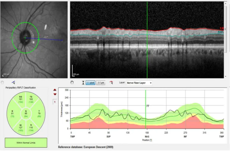
Spectral domain optical coherence tomography (SD-OCT) is a re-liable, non-invasive, trans-pupillary technique that provides high-resolution cross-sectional images of the retina and the optic nerve head, using a principle analogous to B-scan ultrasound. A beam of in-frared light strikes the retina and the delayed light reflected from the various layers of the retina and choroid is compared to a reference beam projected against a mirror. The elaboration of the three-di-mensional image is made based on the principles of low-coherence interferometry. This method provides detailed data on the morphol-ogy and reproducibly measures the thickness and volume of optic nerve, peripapillary area and the various retinal layers at the macular level (Figs. 1 and 2).
Classical OCT devices like time-domain OCT were unhelpful in choroidal imaging, as the retinal pigment epithelium blocked the signal from the choroid. New software for SD-OCT– enhanced depth imaging (EDI)– maximizes sensitivity and detail by moving the peak of the sensitivity curve to the sclera (Fig. 3). Thus it provides a better view of the choroidal cross-sectional structure, thickness and choroidal-scleral interface [84]. However, the few studies on choroidal thickness in SLE patients revealed contradictory results. According to Altinkaynak et al. SLE patients had thinner choroids than age and gender-matched controls [85]. On the other hand, Ferreira et al. reported thicker choroids in these patients [86]. This difference may be due to the systemic disease activity state as in thefirst study all patients were “inactive” whereas in the latter study the disease activity state was not accessed. An increase in cho-roidal thickness with systemic inflammatory activity has also been described in Behçet disease [87,88].
More recently, a new OCT system– optical coherence tomography angiography (OCTA)– has the ability to show both structural and bloodflow information without requiring intravenous contrast. This in-novative technology, called split-spectrum amplitude decorrelation an-giography, is based on the comparison of multiple B scans acquired consecutively in the same spot. In the case of stationary tissues or cells, there is a high correlation between consecutive images. For mov-ing cells, such as blood cells inside the vessels, there is a low correlation (or high decorrelation) between consecutive images, thereby revealing the microvascular architecture [89].
Studies regarding retinal thickness measurements have yielded con-tradictory results (Table 3) likely explained on the basis of small sample size and heterogeneity of clinical NPSLE manifestations and SLE sys-temic disease activity [90,91].
Longitudinal studies as well as studies with larger samples are needed to evaluate the real potential of OCT to detect early neurodegen-erative changes and to eliminate the effect of potential confounders Table 2
Posterior segment involvement in SLE.
Optic nerve Choroid Retina
Prevalence 1% b1% 3% to 29%
Presentation Optic neuritis, ischemic optic neuropathy
Serous retinal detachment, retinal pigment epithelium detachment, retinal pigment epitheliopathy, choroidal ischemia, choroidal effusion
Microangiopathy, retinal vasculitis and severe vaso-occlusion Visual prognosis Moderate to poor Variable Depends on the type of presentation Association to systemic
lupus erythematosus
Yes Yes Yes
Association to CNS lupus No Yes Yes
References Frigui et al. [51] Man et al. [52]
Nguyen et al. [53] Baglio et al. [54]
Stafford-Brady et al. [55] Jabs et al. [56]
such as the effect of ocular axial length, intraocular pressure, ophthalmic pathologies, systemic medications like hydroxychloroquine and other systemic comorbidities, like diabetes mellitus, that potentially affect ret-inal thickness [92]. Standardised questionnaires may also offer valuable help for screening practices.
4. Conclusion
The retina is an extension of the central nervous system; retinal gan-glion cells and their axons forming the optic nerve are similar to CNS neurons and the retinal and cerebral vasculature share anatomic,
Fig. 2. Macular segmentation. Macular scan obtained with spectral domain optical coherence tomography, showing the segmentation of the various retinal layers. BM– Bruch membrane; ELM– external limiting membrane; GCL – ganglion cell layer; ILM – internal limiting membrane; INL – inner nuclear layer; IPL – inner plexiform layer; ONL – outer nuclear layer; OPL – outer plexiform layer; PR1– photoreceptors inner segments; PR2 – photoreceptors outer segments; RPE – retinal pigment epithelium.
Fig. 1. Peripapillary retinal nervefiber layer. Peripapillary retinal nerve fiber layer thickness measurement with spectral domain optical coherence tomography (Spectralis Heidelberg®) and comparison to a sex and age-matched database. ILM– internal limiting membrane; RNFL – retinal nerve fiber layer.
physiological, and embryological similarities. Therefore, in some way the retina can be considered a“window to the brain”. The utility of OCT in tracking early signs of neurodegeneration has been demon-strated in other pathologies. The idea of monitoring the neurodegener-ative process associated with SLE would undoubtedly be an appealing one. It would enable a better control of CNS involvement since the early stages and open the way to studies in neuroprotection. However, so far, the studies conducted in SLE have given inconsistent results. Ro-bust studies are further needed to access the real potential of ophthal-mic imaging in studying neurodegenerative changes associated with SLE with respect to disease activity and cognition.
NPSLE is a frequent and severe complication of SLE, with a significant impact in quality of life and life expectancy. However, its diagnosis is an ongoing challenge given the multitude of clinical syndromes and the ab-sence of reliable diagnostic tools. Imagiologic signs of brain in flamma-tion or atrophy are a frequentfinding, not only in NPSLE but also in SLE patients without neuropsychiatric manifestations. This suggests that neurodegeneration in LES is a relentless continuous process, starting long before the appearance of clinical signs of CNS involvement. On the other hand, one can discuss the importance of this relent-less neurodegenerative process in the algorithms of NPSLE: is it a pathophysiologic process behind several NP syndromes like move-ment disorders, anxiety, mood disorder, psychosis or cognitive dis-function? Or should we consider lupic neurodegeneration the 20th NP syndrome?
Take-home messages
• NPSLE is associated with a significant impact in morbidity and life ex-pectancy.
• Early diagnosis of NPSLE is difficult as there are no widely accepted biomarkers.
• CNS inflammation and atrophy is present in lupus patients without NPSLE.
• Retinal thinning on SD-OCT is an established biomarker of neurode-generation.
• Experimental models suggest that neuroprotection may be feasible and beneficial in systemic inflammation.
Funding
This research did not receive any specific grant from funding agen-cies in the public, commercial, or not-for-profit sectors.
References
[1] Pons-Estel GJ, Alarcón GS, Scofield L, Reinlib L, Cooper GS. Understanding the epide-miology and progression of systemic lupus erythematosus. Semin Arthritis Rheum 2010;39:257–68.https://doi.org/10.1016/j.semarthrit.2008.10.007.
[2] Danchenko N, Satia JA, Anthony MS. Epidemiology of systemic lupus erythematosus: a comparison of worldwide disease burden. Lupus 2006;15:308–18.https://doi.org/ 10.1191/0961203306lu2305xx.
[3] Alonso MD, Llorca J, Martinez-Vazquez F, Miranda-Filloy JA, Diaz de Teran T, Dierssen T, et al. Systemic lupus erythematosus in northwestern Spain: a 20-year epidemiologic study. Medicine (Baltimore) 2011;90:350–8.https://doi.org/10. 1097/MD.0b013e31822edf7f.
[4] Cervera R, Khamashta MA, Hughes GRV. The Euro-lupus project: epidemiology of systemic lupus erythematosus in Europe. Lupus 2009;18:869–74.https://doi.org/ 10.1177/0961203309106831.
[5] Cervera R, Khamashta MA, Font J, Sebastiani GD, Gil A, Lavilla P, et al. Morbidity and mortality in systemic lupus erythematosus during a 10-year period a comparison of early and late manifestations in a cohort of. Mortality 2003;82:299–308.https://doi. org/10.1097/01.md.0000091181.93122.55.
Table 3
Published studies comparing retinal thickness and volume between NPSLE, non-NPSLE and healthy controls. N Peripapillary retinal nerve
fiber layer Central macular thickness Central macular volume Macular inner retinal complexa Macular ganglion cell complexb Macular inner nuclear layer Liu et al. NPSLE patients
versus non-NPSLE [90]
15 NPSLE vs 16 non-NPSLE
No difference No difference No difference
No difference No difference No difference Liu et al. NPSLE patients
versus healthy controls [90]
15 NPSLE vs 16 healthy controls
↓ thickness
Global, temporal superior and nasal (pb 0.05) No difference ↓ thickness (pb 0.05) ↓ thickness (pb 0.05) ↓ thickness (pb 0.05) No difference Liu et al.
SLE versus healthy controls [90]
31 SLE vs 16 healthy controls
↓ thickness
Global, temporal superior and nasal (pb 0.05) ↓ thickness (pb 0.05) ↓ thickness (pb 0.05) ↓ thickness (pb 0.05) ↓ thickness (pb 0.05) ↓ thickness (pb 0.05)
Shulman et al. [91] 14 NPSLE, 7 non-NPSLE, 11 healthy controls
No difference. Trend towards lower absolute values in NPSLE
N.A. N.A. N.A. N.A. N.A.
a
Macular inner retinal complex: between the internal limiting membrane and the inner edge of the inner nuclear layer.
b
Ganglion cell complex: between the outer edge of the RNFL and the inner edge of the inner nuclear layer.
[6]Abu-Shakra M, Urowitz MB, Gladman DD, Gough J. Mortality studies in systemic lupus erythematosus. Results from a single center. II. Predictor variables for mortal-ity. J Rheumatol 1995;22:1265–70.
[7] Sankowski R, Mader S, Valdés-Ferrer SI. Systemic inflammation and the brain: novel roles of genetic, molecular, and environmental cues as drivers of neurodegeneration. Front Cell Neurosci 2015;9:28.https://doi.org/10.3389/fncel.2015.00028. [8] Liang MH, Corzillius M, Bae SC, Lew RA, Fortin PR, Gordon C, et al. The American
Col-lege of Rheumatology nomenclature and case definitions for neuropsychiatric lupus syndromes. Arthritis Rheum 1999;42:599–608.https://doi.org/10.1002/1529-0131 (199904)42:4b599::AID-ANR2N3.0.CO;2-F.
[9] Preble JM, Silpa-archa S, Foster CS. Ocular involvement in systemic lupus erythema-tosus. Curr Opin Ophthalmol 2015;26:540–5. https://doi.org/10.1097/ICU. 0000000000000209.
[10] Goldacre MJ, Wotton CJ. Associations between specific autoimmune diseases and subsequent dementia: retrospective record-linkage cohort study, UK. J Epidemiol Community Health 2017;71:576–83.https://doi.org/10.1136/jech-2016-207809. [11] Shimizu Y, Yasuda S, Kako Y, Nakagawa S, Kanda M, Hisada R, et al. Post-steroid
neu-ropsychiatric manifestations are significantly more frequent in SLE compared with other systemic autoimmune diseases and predict better prognosis compared with de novo neuropsychiatric SLE. Autoimmun Rev 2016;15:786–94.https://doi.org/ 10.1016/j.autrev.2016.03.017.
[12] Sarbu N, Toledano P, Calvo A, Roura E, Sarbu MI, Espinosa G, et al. Advanced MRI techniques: biomarkers in neuropsychiatric lupus. Lupus 2017;26:510–6.https:// doi.org/10.1177/0961203316674820.
[13] Sarbu N, Alobeidi F, Toledano P, Espinosa G, Giles I, Rahman A, et al. Brain abnormal-ities in newly diagnosed neuropsychiatric lupus: systematic MRI approach and cor-relation with clinical and laboratory data in a large multicenter cohort. Autoimmun Rev 2015;14:153–9.https://doi.org/10.1016/j.autrev.2014.11.001.
[14] Toledano P, Sarbu N, Espinosa G, Bargalló N, Cervera R. Neuropsychiatric systemic lupus erythematosus: magnetic resonance imagingfindings and correlation with clinical and immunological features. Autoimmun Rev 2013;12:1166–70.https:// doi.org/10.1016/j.autrev.2013.07.004.
[15] Bertsias GK, Ioannidis JPA, Aringer M, Bollen E, Bombardieri S, Bruce IN, et al. EULAR recommendations for the management of systemic lupus erythematosus with neu-ropsychiatric manifestations: report of a task force of the EULAR standing committee for clinical affairs. Ann Rheum Dis 2010;69:2074–82.https://doi.org/10.1136/ard. 2010.130476.
[16] Sabbadini MG, Manfredi AA, Bozzolo E, Ferrario L, Rugarli C, Scorza R, et al. Central nervous system involvement in systemic lupus erythematosus patients without overt neuropsychiatric manifestations. Lupus 1999;8:11–9.https://doi.org/10. 1191/096120399678847344.
[17]Postal M, Lapa AT, Reis F, Rittner LAS. Magnetic resonance imaging in neuropsychi-atric systemic lupus erythematosus: current state of the art and novel approaches. Lupus 2017;26:517–21.
[18] Ramage AE, Fox PT, Brey RL, Narayana S, Cykowski MD, Naqibuddin M, et al. Neuro-imaging evidence of white matter inflammation in newly diagnosed systemic lupus erythematosus. Arthritis Rheum 2011;63:3048–57.https://doi.org/10.1002/art. 30458.
[19] Brey RL. Neuropsychiatric lupus: clinical and imaging aspects. Bull NYU Hosp Jt Dis 2007;65:194–9.https://doi.org/10.1016/j.rdc.2005.01.007.
[20] Stock AD, Gelb S, Pasternak O, Ben-Zvi A, Putterman C. The blood brain barrier and neuropsychiatric lupus: new perspectives in light of advances in understanding the neuroimmune interface. Autoimmun Rev 2017;16:612–9.https://doi.org/10. 1016/j.autrev.2017.04.008.
[21] Pullmann Jr R, Skerenova M, Hybenova J, Lukac J, Rovensky J, Pullmann R. Apolipo-protein E polymorphism in patients with neuropsychiatric SLE. Clin Rheumatol 2004;23:97–101.https://doi.org/10.1007/s10067-003-0796-0.
[22] van der Weide J, Steijns LS, Teepen JL, Noback WJ, Klaverwijden G. Apolipoprotein E polymorphism and Alzheimer disease. Tijdschr Gerontol Geriatr 1996;27:73–7.
https://doi.org/10.1001/archneur.57.6.824.
[23] de Vries B, Steup-Beekman GM, Haan J, Bollen EL, Luyendijk J, Frants RR, et al. TREX1 gene variant in neuropsychiatric systemic lupus erythematosus. Ann Rheum Dis 2010;69:1886–7.https://doi.org/10.1136/ard.2009.114157.
[24] Ho RC, Thiaghu C, Ong H, Lu Y, Ho CS, Tam WW, et al. A meta-analysis of serum and cerebrospinalfluid autoantibodies in neuropsychiatric systemic lupus erythemato-sus. Autoimmun Rev 2016;15:124–38.https://doi.org/10.1016/j.autrev.2015.10.003. [25] Trysberg E, Tarkowski A. Cerebral inflammation and degeneration in systemic lupus erythematosus. Curr Opin Rheumatol 2004;16:527–33.https://doi.org/10.1097/01. bor.0000135451.85671.14.
[26] Negrini S, Pappalardo F, Murdaca G, Indiveri F, Puppo F. The antiphospholipid syn-drome: from pathophysiology to treatment. Clin Exp Med 2016:1–11.https://doi. org/10.1007/s10238-016-0430-5.
[27] Rodrigues CEM, Carvalho JF, Shoenfeld Y. Neurological manifestations of antiphospholipid syndrome. Eur J Clin Invest 2010;40:350–9.https://doi.org/10. 1111/j.1365-2362.2010.02263.x.
[28] Appenzeller S, Carnevalle A, Li L, Costallat L, Cendes F. Hippocampal atrophy in sys-temic lupus erythematosus. Ann Rheum Dis 2006;65:1585–9.https://doi.org/10. 1136/ard.2005.049486.
[29] Bravo-Zehnder M, Toledo EM, Segovia-Miranda F, Serrano FG, Benito MJ, Metz C, et al. Anti-ribosomal p protein autoantibodies from patients with neuropsychiatric lupus impair memory in mice. Arthritis Rheumatol 2015;67:204–14.https://doi. org/10.1002/art.38900.
[30] Hirohata S, Arinuma Y, Takayama M, Yoshio T. Association of cerebrospinalfluid anti-ribosomal p protein antibodies with diffuse psychiatric/neuropsychological syndromes in systemic lupus erythematosus. Arthritis Res Ther 2007;9:R44.
https://doi.org/10.1186/ar2184.
[31] Briani C, Lucchetta M, Ghirardello A, Toffanin E, Zampieri S, Ruggero S, et al. Neurolupus is associated with anti-ribosomal P protein antibodies: an inception co-hort study. J Autoimmun 2009;32:79–84.https://doi.org/10.1016/j.jaut.2008.12. 002.
[32] Lauvsnes MB, Omdal R. Systemic lupus erythematosus, the brain, and anti-NR2 an-tibodies. J Neurol 2012;259:622–9.https://doi.org/10.1007/s00415-011-6232-5. [33] Lapteva L, Nowak M, Yarboro CH, Takada K, Roebuck-Spencer T, Weickert T, et al.
Anti-N-methyl-D-aspartate receptor antibodies, cognitive dysfunction, and depres-sion in systemic lupus erythematosus. Arthritis Rheum 2006;54:2505–14.https:// doi.org/10.1002/art.22031.
[34] Tay SH, Fairhurst AM, Mak A. Clinical utility of circulating anti-N-methyl-D-aspartate receptor subunits NR2A/B antibody for the diagnosis of neuropsychiatric syndromes in systemic lupus erythematosus and Sjögren's syndrome: an updated meta-analy-sis. Autoimmun Rev 2017;16:114–22.https://doi.org/10.1016/j.autrev.2016.12.002. [35] Williams RC, Sugiura K, Tan EM. Antibodies to microtubule-associated protein 2 in patients with neuropsychiatric systemic lupus erythematosus. Arthritis Rheum 2004;50:1239–47.https://doi.org/10.1002/art.20156.
[36] Trysberg E, Carlsten H. Tarkowski a. Intrathecal cytokines in systemic lupus erythe-matosus with central nervous system involvement. Lupus 2000;9:498–503.https:// doi.org/10.1177/096120330000900704.
[37]Dellalibera-Joviliano R, Dos Reis ML, Queiroz Cunha F, De Donadi EA. Kinins and cy-tokines in plasma and cerebrospinalfluid of patients with neuropsychiatric lupus. J Rheumatol 2003;30:485–92 [doi:0315162X-30-485 [pii].
[38]Trysberg E, Nylen K, Rosengren LE, Tarkowski A. Neuronal and astrocytic damage in systemic lupus erythematosus patients with central nervous system involvement. Arthritis Rheum 2003;48:2881–7.
[39] Bialas AR, Presumey J, Das A, van der Poel CE, Lapchak PH, Mesin L, et al. Microglia-dependent synapse loss in type I interferon-mediated lupus. Nature 2017.https:// doi.org/10.1038/nature22821.
[40] Lu T, Pan Y, Kao S-Y, Li C, Kohane I, Chan J, et al. Gene regulation and DNA damage in the ageing human brain. Nature 2004;429:883–91. https://doi.org/10.1038/ nature02661.
[41] Lin MT, Beal MF. Mitochondrial dysfunction and oxidative stress in neurodegenera-tive diseases. Nature 2006;443:787–95.https://doi.org/10.1038/nature05292. [42] Kaur C, Sivakumar V, Zou Z, Ling EA. Microglia-derived proinflammatory cytokines
tumor necrosis factor-alpha and interleukin-1beta induce Purkinje neuronal apopto-sis via their receptors in hypoxic neonatal rat brain. Brain Struct Funct 2014;219: 151–70.https://doi.org/10.1007/s00429-012-0491-5.
[43] Przedborski S, Vila M, Jackson-Lewis V. Neurodegeneration: what is it and where are we? J Clin Invest 2003;111:3–10.https://doi.org/10.1172/JCI200317522. [44] Clarke PGH. Developmental cell death: morphological diversity and multiple
mech-anisms. Anat Embryol (Berl) 1990;181:195–213. https://doi.org/10.1007/ BF00174615.
[45] Clarke PGH. Apoptosis versus necrosis. Cell death. Dis Nerv Syst 1999:3–28.https:// doi.org/10.1007/978-1-4612-1602-5_1.
[46] d'Avila J da CP, Santiago APSA, Amâncio RT, Galina A, Oliveira MF, Bozza FA. Sepsis induces brain mitochondrial dysfunction. Crit Care Med 2008;36:1925–32.https:// doi.org/10.1097/CCM.0b013e3181760c4b.
[47] Anderson ST, Commins S, Moynagh PN, Coogan AN. Lipopolysaccharide-induced sepsis induces long-lasting affective changes in the mouse. Brain Behav Immun 2015;43:98–109.https://doi.org/10.1016/j.bbi.2014.07.007.
[48]Michels M, Vieira AS, Vuolo F, Zapelini HG, Mendonça B, Mina F, et al. The role of mi-croglia activation in the development of sepsisinduced long-term cognitive impair-ment. Brain Behav Immun 2015;43:54–9.
[49] Barichello T, Machado RA, Constantino L, Valvassori SS, Réus GZ, Martins MR, et al. Antioxidant treatment prevented late memory impairment in an animal model of sepsis*. Crit Care Med 2007;35:2186–90. https://doi.org/10.1097/01.CCM. 0000281452.60683.96.
[50] Silpa-Archa S, Lee JJ, Foster CS. Ocular manifestations in systemic lupus erythemato-sus. Br J Ophthalmol 2016;100:135–41. https://doi.org/10.1136/bjophthalmol-2015-306629.
[51] Frigui M, Frikha F, Sellemi D, Chouayakh F, Feki J, Bahloul Z. Optic neuropathy as a presenting feature of systemic lupus erythematosus: two case reports and literature review. Lupus 2011;20:1214–8.https://doi.org/10.1177/0961203311403344. [52] Man BL, Mok CC, Fu YP. Neuro-ophthalmologic manifestations of systemic lupus
er-ythematosus: a systematic review. Int J Rheum Dis 2014;17:494–501.https://doi. org/10.1111/1756-185X.12337.
[53] Nguyen QD, Uy HS, Akpek EK, Harper SL, Zacks DN, Foster CS. Choroidopathy of sys-temic lupus erythematosus. Lupus 2000;9:288–98. https://doi.org/10.1191/ 096120300680199024.
[54] Baglio V, Gharbiya M, Balacco-Gabrieli C, Mascaro T, Gangemi C, di Franco M, et al. Choroidopathy in patients with systemic lupus erythematosus with or without ne-phropathy. J Nephrol 2011;24:522–9.https://doi.org/10.5301/JN.2011.6244. [55]Stafford-Brady FJ, Urowitz MB, Gladman DD, Easterbrook M. Lupus retinopathy.
Pat-terns, associations, and prognosis. Arthritis Rheum 1988;31:1105–10.
[56] Jabs DA, Fine SL, Hochberg MC, Newman SA, Heiner GG, Stevens MB. Severe retinal vaso-occlusive disease in systemic lupus erythematous. Arch Ophthalmol 1986;104: 558–63.https://doi.org/10.1001/archopht.1986.01050160114025.
[57]Siatkowski RM, Scott IU, Verm AM, Warn AA, Farris BK, Strominger MB, et al. Optic neuropathy and chiasmopathy in the diagnosis of systemic lupus erythematosus. J Neuroophthalmol 2001;21:193–8.
[58] Lin Y-C, Wang A-G, Yen M-Y. Systemic lupus erythematosus-associated optic neuri-tis: clinical experience and literature review. Acta Ophthalmol 2009;87:204–10.
https://doi.org/10.1111/j.1755-3768.2008.01193.x.
[59] Závada J, Nytrová P, Wandinger KP, Jarius S, Svobodová R, Probst C, et al. Seroprev-alence and specificity of NMO-IgG (anti-aquaporin 4 antibodies) in patients with
neuropsychiatric systemic lupus erythematosus. Rheumatol Int 2013;33:259–63.
https://doi.org/10.1007/s00296-011-2176-4.
[60]Cordeiro MF, Lloyd ME, Spalton DJ, et al. Ischaemic optic neuropathy, transverse my-elitis, and epilepsy in an anti-phospholipid positive patient with systemic lupus er-ythematosus. J Neurol Neurosurg Psychiatry 1994;57:1142–3.
[61]Massin M, Berche C, Ullern M, et al. Acute anterior ischemic optic neuropathy dis-closing disseminated lupus erythematosus. Ophtalmologie 1987;1:61–3.
[62] Hannouche D, Korobelnik JF, Cochereau I, Hayem G, Beaudreuil J, Meyer O, et al. Sys-temic lupus erythematosus with choroidopathy and serous retinal detachment. Int Ophthalmol 1995;19:125–7.https://doi.org/10.1007/BF00133184.
[63] Palejwala NV, Walia HS, Yeh S. Ocular manifestations of systemic lupus erythemato-sus: a review of the literature. Autoimmune Dis 2012:1.https://doi.org/10.1155/ 2012/290898.
[64] Davies JB, Rao PK. Ocular manifestations of systemic lupus erythematosus. Curr Opin Ophthalmol 2008;19:512–8.https://doi.org/10.1097/ICU.0b013e3283126d34. [65] Aronson AJ, Ordoñez NG, Diddie KR, Ernest JT. Immune-complex deposition in the
eye in systemic lupus erythematosus. 1979;139.https://doi.org/10.1001/archinte. 1979.03630480084026.
[66] Montehermoso A, Cervera R, Font J, Ramos-Casals M, Garcia-Carrasco M, Formiga F, et al. Association of antiphospholipid antibodies with retinal vascular disease in sys-temic lupus erythematosus. Semin Arthritis Rheum 1999;28:326–32.https://doi. org/10.1016/S0049-0172(99)80017-1.
[67] Au A, O'Day J. Review of severe vaso-occlusive retinopathy in systemic lupus erythe-matosus and the antiphospholipid syndrome: associations, visual outcomes, compli-cations and treatment. Clin Experiment Ophthalmol 2004;32:87–100.https://doi. org/10.1046/j.1442-9071.2004.00766.x.
[68] Gordon-Lipkin E, Chodkowski B, Reich DS, Smith SA, Pulicken M, Balcer LJ, et al. Ret-inal nervefiber layer is associated with brain atrophy in multiple sclerosis. Neurol-ogy 2007;69:1603–9.https://doi.org/10.1212/01.wnl.0000295995.46586.ae. [69] Cunha JP, Proença R, Dias-Santos A, Almeida R, Águas H, Alves M, et al. OCT in
Alzheimer's disease: thinning of the RNFL and superior hemiretina. Graefes Arch Clin Exp Ophthalmol 2017.https://doi.org/10.1007/s00417-017-3715-9. [70] Moschos MM, Chatziralli IP. Evaluation of choroidal and retinal thickness changes in
Parkinson's disease using spectral domain optical coherence tomography. Semin Ophthalmol 2017:1–4.https://doi.org/10.1080/08820538.2017.1307423. [71] Karpik AG, Schwartz MM, Dickey LE, Streeten BW, Roberts JL. Ocular immune
reac-tants in patients dying with systemic lupus erythematosus. Clin Immunol Immunopathol 1985;35:295–312.https://doi.org/10.1016/0090-1229(85)90091-1. [72] Diamond B. Antibodies and the brain: lessons from lupus. J Immunol 2010;185:
2637–40.https://doi.org/10.4049/jimmunol.1090080.
[73]Vanburen JM. Trans-synaptic retrograde degeneration in the visual system of pri-mates. J Neurol Neurosurg Psychiatry 1963;26:402–9.
[74]Fletcher WA, Hoyt WF, Narahara MH. Congenital quadrantanopia with occipital lobe ganglioglioma. Neurology 1988;38:1892–4.
[75] Jindahra P, Petrie A, Plant GT. The time course of retrograde trans-synaptic degener-ation following occipital lobe damage in humans. Brain 2012;135:534–41.https:// doi.org/10.1093/brain/awr324.
[76] Goto K, Miki A, Yamashita T, Araki S, Takizawa G, Nakagawa M, et al. Sectoral anal-ysis of the retinal nervefiber layer thinning and its association with visual field loss in homonymous hemianopia caused by post-geniculate lesions using spectral-do-main optical coherence tomography. Graefes Arch Clin Exp Ophthalmol 2016;254: 745–56.https://doi.org/10.1007/s00417-015-3181-1.
[77] Anjos R, Vieira L, Costa L, Vicente A, Santos A, Alves N, et al. Macular ganglion cell layer and peripapillary retinal nervefibre layer thickness in patients with unilateral posterior cerebral artery ischaemic lesion: an optical coherence tomography study. Neuro-Ophthalmol 2016;40.https://doi.org/10.3109/01658107.2015.1122814. [78]Keane JR. Eye movement abnormalities in systemic lupus erythematosus. Arch
Neurol 1995;52:1145–9.
[79]DelGiudice GC, Scher CA, Athreya BH, Diamond GR. Pseudotumor cerebri and child-hood systemic lupus erythematosus. J Rheumatol 1986;13:748–52.
[80]Kuyucu S, Argin A, Kuyucu N, Ozen S. Systemic lupus erythematosus presenting with pseudotumor cerebri: a rare association. Turk J Pediatr 2007;49:98–101.
[81] Kunavisarut P, Pathanapitoon K, Rothova A. Purtscher-like retinopathy associated with systemic lupus erythematosus. Ocul Immunol Inflamm 2016;24:60–8.
https://doi.org/10.3109/09273948.2014.932816.
[82] Wu C, Dai R, Dong F, Wang Q. Purtscher-like retinopathy in systemic lupus erythe-matosus. Am J Ophthalmol 2014;158:1335–1341.e1.https://doi.org/10.1016/j.ajo. 2014.09.001.
[83]Gharbiya M, Pecci G, Baglio V, Gargiulo A, Allievi F, Balacco-Gabrieli C. Indocyanine green angiographicfindings for patients with systemic lupus erythematosus ne-phropathy. Retina 2006;26:159–64.
[84] Spaide RF, Koizumi H, Pozonni MC. Enhanced depth imaging spectral-domain opti-cal coherence tomography. Am J Ophthalmol 2008;146:496–500.https://doi.org/ 10.1016/j.ajo.2008.05.032.
[85] Altinkaynak H, Duru N, Uysal BS, ErtenŞ, Kürkcüoğlu PZ, Yüksel N, et al. Choroidal thickness in patients with systemic lupus erythematosus analyzed by spectral-do-main optical coherence tomography. Ocul Immunol Inflamm 2015:1–7.https:// doi.org/10.3109/09273948.2015.1006790.
[86]Ferreira CS, Beato J, Falcão MS, Brandão E, Falcão-Reis FCÂ. Choroidal thickness in multisystemic autoimmune diseases without ophthalmologic manifestations. Retina 2017;37:529–35.
[87] Kim M, Kim H, Kwon HJ, Kim SS, Koh HJ, Lee SC. Choroidal thickness in Behcet's uve-itis: an enhanced depth imaging-optical coherence tomography and its association with angiographic changes. Invest Ophthalmol Vis Sci 2013;54(9):6033.https:// doi.org/10.1167/iovs.13-12231.
[88] Yesilirmak N, Lee W-H, Gur Gungor S, Yaman Pinarci E, Akkoyun I, Yilmaz G. En-hanced depth imaging optical coherence tomography in patients with different phases of Behcet's panuveitis. Can J Ophthalmol 2017;52:48–53.https://doi.org/10. 1016/j.jcjo.2016.07.020.
[89] Chalam K, Sambhav K. Optical coherence tomography angiography in retinal dis-eases. J Ophthalmic Vis Res 2016;11:84–92.https://doi.org/10.4103/2008-322X. 180709.
[90] Liu GY, Utset TO, Bernard JT. Retinal nervefiber layer and macular thinning in sys-temic lupus erythematosus: an optical coherence tomography study comparing SLE and neuropsychiatric SLE. Lupus 2015;24:1169–76.https://doi.org/10.1177/ 0961203315582285.
[91] Shulman S, Shorer R, Wollman J, Dotan G, Paran D. Retinal nervefiber layer thick-ness and neuropsychiatric manifestations in systemic lupus erythematosus. Lupus 2017:961203317703496.https://doi.org/10.1177/0961203317703496.
[92] Tavares Ferreira J, Alves M, Dias-Santos A, Costa L, Santos BO, Cunha JP, et al. Retinal neurodegeneration in diabetic patients without diabetic retinopathy. Invest Ophthalmol Vis Sci 2016;57.https://doi.org/10.1167/iovs.16-20215.