D
EVELOPMENT OF QUERCETIN BRAIN
DELIVERY SYSTEMS FOR THE TREATMENT
OF
A
LZHEIMER
’
S DISEASE
Rúben Gonçalo Rodrigues Pinheiro
Master Thesis for the degree in Master in Bioengineering
Specialization in Molecular Biotechnology
Supervisor: Doutora Salette Reis
Co-supervisor: Doutora Ana Rute Neves
i
ii
A
BSTRACT
Quercetin is a flavonol present in many vegetables and fruits with many beneficial effects such as anti-cancer, anti-inflammatory, anti-oxidant and neuroprotection action. However, due to its lipophilicity, low water solubility, and extensive metabolism, quercetin presents poor bioavailability and cannot be used efficiently for diseases prevention or treatment. In this context, nanotechnology can help to overcome this problem by encapsulating quercetin inside nanoparticles to increase its bioavailability and potentiate its efficacy by directing to a specific target of interest.
The main goal of this work was the development of quercetin-loaded lipid nanoparticles (SLN and NLC) functionalized with transferrin and RVG29 peptide for enhancing the bioavailability of quercetin, promoting its site-specific delivery through the blood-brain barrier (BBB) into the brain with the utmost purpose of treating or preventing Alzheimer’s disease. In fact, transferrin and nicotinic acetylcholine receptors are highly expressed on BBB, potentiating receptor-mediated transcytosis of transferrin- and RVG29-coated nanoparticles, respectively, to the brain.
Functionalization has been confirmed using nuclear magnetic resonance and Fourier transform infrared spectroscopies. All nanoparticles appeared to have a spherical and uniform morphology with smooth surfaces, analyzed by transmission electron microscopy. Transferrin and RVG29 nanoparticles were capable of encapsulating around 80-90% of quercetin and this value remained almost unchanged over 3 months of stability study. The size of both modified nanoparticles was between 150 and 250 nm and zeta potential was around -30 mV, which is a good indicator of formulation stability. Photostability study showed that free quercetin was degraded (55%) after 6 hours of UV light exposure, but lipid nanoparticles protected the compound against photodegradation.
The cytotoxicity of nanoparticles was tested by lactate dehydrogenase assay in hCMEC/D3 cell line, a model of human BBB. This assay showed almost no cytotoxicity even for the highest concentration tested. The BBB permeability of nanoparticles across hCMEC/D3 cells monolayer was tested using Transwell devices. The results showed that RVG29 nanoparticles can pass through the monolayer more efficiently than transferrin-functionalized or non-functionalized nanoparticles, probably due to the active expression of nicotinic acetylcholine receptors on brain endothelial cells. In order to test the efficacy of the developed nanoparticles for preventing Aβ(1-42) peptide fibrillation in Alzheimer’s disease, a ThT binding assay was performed. Almost all
iii
quercetin-loaded nanoparticles were able to inhibit fibrils formation, but especially transferrin- and RVG29-functionalized NLC may have a greater potential to be used in Alzheimer’s prevention or therapy.
iv
R
ESUMO
A quercetina é um flavonóide presente em muitas frutas e vegetais, com efeitos benéficos para a saúde, nomeadamente, propriedades anti-cancerígenas, antioxidantes e anti-inflamatórias, e ainda um efeito neuroprotetor. No entanto, devido à sua lipofilicidade, baixa solubilidade em água e extenso metabolismo, a quercetina apresenta baixa biodisponibilidade, não podendo ser utilizada de forma eficiente para a prevenção ou o tratamento de doenças. Neste contexto, a nanotecnologia pode ajudar a ultrapassar esse problema através da encapsulação da quercetina dentro de nanopartículas para aumentar a sua biodisponibilidade e potenciar a sua eficácia, direcionando-a para um alvo específico de interesse.
Neste trabalho, pensando numa abordagem para o cérebro, foram usadas nanopartículas lipídicas (SLN e NLC) funcionalizadas com Transferrina e RVG29, com intuito de as direcionar especificamente para o cérebro e, desta forma, a quercetina poder exercer o seu efeito neuroprotetor na prevenção ou tratamento da doença de Alzheimer. De facto, o receptor de transferrina e o receptor nicotínico da acetilcolina estão altamente expressos na barreira hematoencefálica (BHE), potenciando a transcitose mediada por receptores de nanopartículas revestidas com transferrina e RVG29, respectivamente, para o cérebro.
As funcionalizações com estes ligandos foram confirmadas utilizando as técnicas de espectroscopia por ressonância magnética nuclear e espectroscopia de infravermelho. Para além disso, a microscopia electrónica de transmissão permitiu ver que as partículas apresentavam uma morfologia esférica e uniforme. O tamanho medido por dispersão dinâmica da luz ficou compreendido entre os 150 e 250 nanómetros o que é adequado para atravessar a BHE. Por sua vez, os valores de potencial zeta rondaram os -30 mV o que garante a estabilidade das formulações. A taxa de encapsulação obtida foi alta, por volta dos 80-90%, mantendo-se estável ao longo de três meses de estudo. Quanto ao estudo da fotoestabilidade, foi possível observar que a quercetina encapsulada era protegida da degradação exercida pela radiação UV, contrariamente ao que se passava com a quercetina na forma livre que sofreu uma fotodegradação na ordem dos 55% ao fim de 6 horas de exposição.
Após esta etapa de caracterização, foram feitos estudos de citotoxicidade usando o ensaio da quantificação da enzima lactato desidrogenase, num modelo in vitro da barreira hematoencefálica composto pela linha celular hCMEC/D3. Os resultados mostraram que as partículas não induzem toxicidade, mesmo para as concentrações
v
mais altas estudadas. Foram ainda feitos estudos de permeabilidade usando uma monocamada de células hCMEC/D3 num ensaio feito com dispositivos Transwell, para testar a capacidade de permeação das nanopartículas através da BHE. As nanopartículas funcionalizadas com RVG29 foram as mais eficientes no aumento da permeabilidade através da barreira de células, tirando partido da presença dos receptores nicotínicos da acetilcolina presentes nas células endoteliais do cérebro. Por fim, para aferir o potencial terapêutico/preventivo destes nanosistemas, foi usado um modelo in vitro da doença de Alzheimer com o péptido Aβ(1-42). Os resultados mostraram que quase todas as nanopartículas carregadas com quercetina foram capazes de inibir a agregação do péptido, especialmente as partículas NLC funcionalizadas com transferrina ou RVG29, o que abre excelentes perspectivas para esta abordagem poder ser futuramente pensada como uma estratégia preventiva ou mesmo terapêutica da doença de Alzheimer.
vi
A
CKNOWLEDGMENTS
I am profoundly grateful to professor Salette Reis for giving me the opportunity to work in her research team and for all her kindness and comprehension in more difficult times.
I would like to thank to Dr. Ana Rute Neves for her guidance during these two semesters, for all the opportunities she has giving me, all her patience to listen to my concerns and all the encouragement.
I would like to thank to Andreia Granja for helping me with some of my experimental work, particularly for teaching me all about cells. Thanks for the support, good vibrations and all the hours that you spent with me in the lab.
I am deeply grateful to Alexandre Vieira for his kindness and support in some difficult stages of this work.
I would also want to thank Claudia, Joana and Pris for helping me to solve some pratical problems.
vii
viii
TABLE OF CONTENTS
ABSTRACT ... II ACKNOWLEDGMENTS ... VII CHAPTER 1. INTRODUCTION ... 1 1.1.QUERCETIN...1 1.2.NANOTECHNOLOGY ...81.3.DISSERTATION WORK PLAN... 16
CHAPTER 2. MATERIALS AND METHODS... 20
2.1. PREPARATION OF NANOPARTICLES ... 20
2.2.FUNCTIONALIZATION OF NANOPARTICLES WITH TRANSFERRIN ... 21
2.3.FUNCTIONALIZATION OF NANOPARTICLES WITH RVG29 ... 22
2.4.NUCLEAR MAGNETIC RESONANCE SPECTROSCOPY ... 23
2.5.FOURIER TRANSFORM INFRARED SPECTROSCOPY ... 24
2.6.TRANSMISSION ELECTRON MICROSCOPY ... 24
2.7.DYNAMIC LIGHT SCATTERING ... 24
2.8.ZETA POTENTIAL ANALYZER ... 25
2.9.ENTRAPMENT EFFICIENCY DETERMINATION ... 25
2.10.PHOTOSTABILITY STUDY ... 25
2.11. HCMEC/D3 CELL CULTURE ... 25
2.12.LDH CYTOTOXICITY ASSAY ... 26
2.13.TRANSWELL PERMEABILITY ASSAY ... 26
2.14.AMYLOID-BETA PEPTIDE PREPARATION ... 27
2.15.THIOFLAVIN T BINDING ASSAY AND FLUORESCENCE MEASUREMENTS ... 27
2.16.STATISTICAL ANALYSIS ... 27
CHAPTER 3. RESULTS AND DISCUSSION ... 28
3.1.1H-NMR CHARACTERIZATION OF TRANSFERRIN-FUNCTIONALIZED CONJUGATE ... 28
3.2.1H-NMR CHARACTERIZATION OF RVG29-FUNCTIONALIZED CONJUGATE ... 28
ix
3.4.MORPHOLOGY DETERMINATION... 32
3.5.PHYSICOCHEMICAL CHARACTERIZATION AND STABILITY STUDY OF NANOPARTICLES ... 33
3.6.QUERCETIN PHOTOSTABILITY STUDY ... 39
3.7.CYTOTOXICITY STUDY ... 40
3.8.BBB PERMEABILITY STUDY ... 42
3.9.AMYLOID-BETA PEPTIDE STUDY ... 43
CONCLUDING REMARKS ... 48
REFERENCES ... 50
x
A
BBREVIATIONS AND SYMBOLS
DLS Dynamic light scattering
EE Encapsulation Efficiency
LDH Lactate Dehydrogenase
ROS Reactive Oxygen Species
SLN Solid Lipid Nanoparticles
TEM Transmission Electron Microscopy CoA Coenzyme A
IUPAC International Union of Pure and Applied Chemistry LUMO Lowest unoccupied molecular orbital
HOMO Highest unoccupied molecular orbital UVA Ultraviolet A
UVB Ultraviolet B
COMT Catechol-O-methyltransferase AhR Aryl hydrocarbon receptor PAH Polycyclic aromatic hydrocarbon HAH Halogenated aromatic hydrocarbon
CYP1A1 Cytochromes P450, family 1, subfamily A, polypeptide 1 CYP1A2 Cytochromes P450, family 1, subfamily A, polypeptide 2 CYP1B1 Cytochromes P450, family 1, subfamily B, polypeptide 1 DR5 Death receptor 5
TRAIL NF-related apoptosis-inducing ligand NF-kB Factor nuclear kappa B
RVG-29 Rabie virus glycoprotein DLS Dynamic light scattering BBB Blood brain barrier MNPs Magnetic nanoparticles PEG Polyethylene glycol
PLGA Poly(lactic- co-glycolic acid PLA Polylactic acid
xi
MTT Methylthiazolyldiphenyl-tetratozium Bromide NO Nitric Oxide
PCL Polycaprolactone
MWCO Molecular Weight Cut Off
NADH Nicotinamide Adenine Dinucleotide – Hydrogen CHD Coronary heart diseases
u-PA Urokinase-type plasminogen activator
t-PA Tissue-type plasminogen activator
PAI-1 Plasminogen activator inhibitor
u-PAR
Urokinase-type plasminogen activator receptor
MCP-1 Monocyte chemoattractant protein 1 IL-1β Interleukin 1 beta
PMA Phorbol 12-myristate 13-acetate IL-6 Interleukin 6
IL-8 Interleukin 8
eNOS Endothelial nitric oxide synthase PC Phosphatidylcholine
EPC Egg phosphatidylcholine Chol Cholesterol
ESM Egg sphingomyelin GMS Glycerol monostearate SA Stearic acid
MCT Medium chain triglycerides
GT Glyceryltridecanoate TG Glyceryl tripalmitate
AEMA 2-aminoethyl methacrylamide FA Folic acid
TPGS d-α-tocopheryl polyethylene glycol 1000 succinate E137S18E137 Block copolymer of ethylene oxide and styrene oxide
APTS 3-aminopropyl triethoxysilane TEOS Tetraethyl orthosilicate βCD β-cyclodextrin
HP-βCD Hydroxypropyl-β-cyclodextrin SBE-βCD Sulfobutyl ether-β-cyclodextrin SBE-7βCD Sulfobutyl ether-7β-cyclodextrin MLV Multi-lamellar vesicles
xii
Boc-AEMA Hydrophilic part and N-2-[(tert-butoxycarbonyl)amino] ethyl
methacrylamide
mPLGA Methacrylated poly(lactic-co-glycolic acid) SPION Superparamagnetic iron oxide
NLC Nanostructured lipid carriers
LNCaP Lymph Node Carcinoma of the Prostate
hCMEC Human cerebral microvascular endothelial cells iNOS Inducible nitric oxide synthase
F3O4 Magnetite
xiii
1
C
HAPTER
1.
I
NTRODUCTION
1.1.
Q
UERCETIN
1.1.1
H
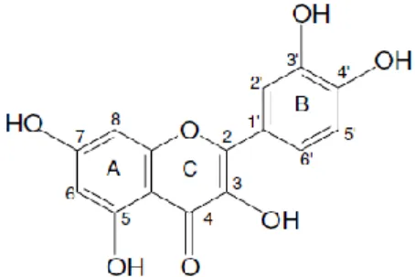
ISTORY OF QUERCETINQuercetin (3,3´,4´,5,7-pentahydroxyflavone) is a flavonol isolated and identified for the first time by Szent-Gyorgyi in 1936 (Figure 1) [1]. In the beginning, and similarly to what happened with other flavonoids, quercetin was identified as a vitamin showing to be important in the maintenance of capillary wall integrity and capillary resistance antihypertensive and antiarrhythmic activity. In posterior studies it was proven to have anti-inflammatory and antiallergic properties, hypocholesterolaemic activity, platelet and mast cell stabilization, antihepatotoxic activity and antitumor activity [2, 3]. Later on, more profound works explained the reasons for this beneficial effects of quercetin based on antioxidant (free-radical scavenging) and anti-inflammatory activity [4].
Figure 1. Chemical structure of quercetin.
1.1.2.
N
ATURAL SOURCES OF QUERCETINQuercetin is one of the most abundant flavonoids in vegetables and fruits, mainly in onions, chilli, berries and apples (Table 1) [5-7]. It is present mainly in leaves as aglycones or glycosides (3-position or/and 4´-position). The most common sugar group found is glucose, however lactose and rhamnose can also be bound to phenolic groups of quercetin [5]. It is important to refer that in onion quercetin appears in the form of 4´-O-β-glucoside and quercetin-3,4´-O-β-diglucoside, representing 80% of the total content of quercetin flavonoid types, being present in high quantities in 28 vegetables and 9 fruits studied [8]. In a different study, quercetin was found in all 25 eatable berries studied and the highest concentration was found
2
in bog whortleberry (158 mg/kg, fresh weight) [9]. In a different work, quercetin-3-O-β-glucoside was found in a considerable amount in apple, pear peels, Hypericum perforatum leaves or flowers [5, 10]. Moreover, other studies have shown that black tea, red wine and various fruit juices can be valid choices as sources for quercetin [11].
Table 1. Quercetin natural sources [5, 9, 11].
Sources Total quercetin
(mg/Kg)
White onion bulbs 2604 Onion dry outer skin 960 Spring onion leaves 450 Chilli powder 400 Bog whortleberry 158 Lingonberry 146 Cranberry 121 Kale 110 Chokeberry 89 Sweet rowan 85 Rowanberry 63 Sea buckthorn berry 62 Apples red delicious 58 Crowberry 56 Broccoli 30 Green beans 25 Apple peel 21 Tomato 13
1.1.3
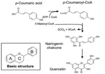
B
IOSYNTHESIS OF QUERCETINThe biosynthesis of quercetin shares almost the same steps in terms of metabolic pathway of the other flavonoids. The three-ring structure (A, B and C) with a diphenyl propane skeleton (C-6-C-3-C-6) is a fingerprint of quercetin (Figure 1) [3]. The A and B are benzene rings linked by oxygen containing pyrene ring (C) [12]. The A ring is biosynthesized by the condensation of three moles of malonyl-coenzyme A (CoA) originated from the metabolism of glucose [3]. The C and B rings are also derived from glucose metabolism by way of the shikimic acid pathway to produce cinnamic acid and its reduced product, coumaric acid [3]. As CoA derivatives, this C-9 p-coumaroyl-CoA
3
condenses with three moles of C-3 malonyl-CoA to form a C-15 chalcone. After that, the ring closes and the hydration gives rise to quercetin (Figure 2) [13].
Figure 2. Schematic representation of quercetin biosynthesis.
1.1.4.
C
HEMICAL STRUCTURE AND PROPERTIES OF QUERCETINThe IUPAC name of quercetin is 2-(3,4-dihydroxyphenyl)-3,5,7-trihydroxy-4H-chromen-4-one. This is composed by two benzene rings and one oxygen containing pyrene ring (Figure 1) [3]. Quercetin has amphipathic behavior due to the phenyl rings (hydrophobic part) and the hydroxyl groups (hydrophilic part) [14, 15]. Nevertheless, quercetin presents low water solubility. There is some controversy in the literature concerning its solubility value, however most studies indicate approximately 0.01 mg/mL (25°C) [16-18]. Besides that, photodegradation of quercetin is also an important issue to concern while working with this flavonoid. A photostability study has been conducted with quercetin alcoholic solutions and the results revealed the appearance of some products of degradation measured by spectrophotometry under the exposition to UVB and UVA radiation, indicating the degradation of this polyphenol compound [19]. Another important property relates with quercetin stability at different pH values. One study has demonstrated that quercetin is degraded at weak basic pH 8, while at pH 5 almost 75% of quercetin remains in solution, indicating that quercetin is more stable in a protic medium [20]. Furthermore, a magnetic study of quercetin indicates this compound displays diamagnetic properties [14]. This feature shows that valence electrons are paired and consequently the compound seems to be stable. However the
4
study of LUMO (lowest unoccupied molecular orbital) and HOMO (highest unoccupied molecular orbital) showed a little energy gap between those orbitals indicating that an electron can transit from LUMO to HOMO orbitals and by this way can easily react [14]. Additionally, the ionization potential study showed that it is easy to take away an electron from the valence orbital [14]. These two properties may explain the antioxidant capacity of quercetin.
1.1.5.
A
BSORPTION,
DISTRIBUTION,
METABOLISM AND ELIMINATIONQuercetin is a highly hydrophobic compound, so when it arrives to the small intestine, it can be absorbed by the epithelial cells passing through cellular membranes (phospholipid bilayers) by simple diffusion pathway [21]. Chen et al. performed some absorption experiments in Sprague–Dawley rats and found that almost 60% of quercetin orally administered was absorbed [22]. This result was comparable to the work developed by Walle et al. where 53% of quercetin administered was absorbed [23]. Inside the epithelial cells (enterocytes), the compound then suffers glucuronidation and sulfatation at one of the hydroxyl groups by glucuronosyltransferases and sulfotransferases (phase II enzymes) in order to confer hydrophilicity [24-27]. Quercetin can also be O-methylated, primarily resulting in the formation of 3´-O-methylquercetin (isorhamnetin) and, to a smaller extent, 4´-O-methylquercetin (tamaraxetin) [26, 27]. When quercetin is conjugated with sugars, the first step is to remove this group by β-glucosidase present in enterocytes and intestinal flora and after that the quercetin can be conjugated as previously mentioned [24]. However, some molecules of quercetin can enter the circulation without suffering any conjugation. These molecules will reach the liver through the portal vein and there will occur metabolization by glucuronosyltransferases and sulfotransferases, which are enzymes largely expressed [28]. Moreover the catechol-O-methyltransferase (COMT) enzymes present in liver and kidney can methylate quercetin [27, 29, 30]. If quercetin conjugates are excreted for bile they flow in the small intestine and they reach the hindgut where they can be hydrolyzed by the β-glucuronidase and sulfatase activities of the microflora, which allows the enterohepatic cycling, increasing the circulation time [28]. Although liver is the central organ of metabolization, a study shows that 90% of quercetin absorbed was metabolized in the gut [22]. All these enzymatic changes transform quercetin in a compound more soluble and allow blood circulation freely or bound to blood proteins, such as albumin [31, 32]. The entrance of quercetin in blood circulation will allow tissue distribution. It is also important to refer that quercetin can be absorbed from the gastrointestinal tract to the lymphatic system [26]. The regular ingestion of the
5
compound appears to accumulate in many organs (i.e., lung, kidney, thymus, heart, liver) with the highest concentrations of quercetin and its methylated derivatives, particularly isorhamnetin, found in the pulmonary tissue [33]. Quercetin remains in the organism for a long period of time (20-72 h) and probably this may be due to enterohepatic recycling [23]. However, quercetin can be degraded by microflora in the colon in phenolic acids and carbon dioxide, which is expelled in breath [34]. Consequently, the phenolic acids can be excreted in feces. Some quercetin may be also eliminated in urine [35-37]. All this routes are valid for quercetin elimination. In a study conducted by Ueno et al., quercetin was excreted as expired CO2 (35%), or via the feces (45%) and urine (10%) as glucuronide or sulfate conjugates following oral administration [38]. However, in a more recent study only a small amount of absorbed quercetin was eliminated in urine (3.3–5.7%) and feces (0.21–4.6%) [23]. The majority of quercetin was eliminated under the form of carbon dioxide (41.8–63.9%) [23].
1.1.6.
T
OXICITY OF QUERCETINIn terms of toxicity, there are still some contradictory results. The in vitro studies revealed that quercetin induces SOS activity, reverse mutations and DNA single strand breaks in bacteria (Salmonella typhimurium strains and Escherichia coli) and in eukaryotic cells, including yeast cells, at relatively high concentrations (up to 10 mg/incubation mixture) in the last case [39-42]. In hamster and mouse cells and human lymphocytes were verified special effects as chromosomal aberrations, DNA single strand breaks and micronucleus formation [43, 44]. However, this toxicity was not proven to occur in in vivo studies. Mice and rats oral administered with quercetin consistently did not induce any significant changes in several mutagenicity/genotoxicity endpoints, i.e., micronuclei, chromosomal aberrations, sister chromatid exchange, unscheduled DNA synthesis, and alkali-labile DNA damage [44-46]. Based on the toxicity founded by in vitro assays, the carcinogenity of quercetin was questioned using animal models. F344 rats receiving quercetin as 0.1% of the diet (50 mg/kg body weight/day) for a period of 540 days, did not differ from the control rats in terms of tumor incidence, with the exception of lung adenoma and one jejunal adenocarcinoma [47]. In order to comprove the connection between quercetin and lung cancer, quercetin was administered in the diet of A/JJms mice at 5% (7500 mg/kg body weight/day) for a period of 23 weeks, and this compound did not induce a significant difference in the incidence of lung tumors, discarding the possibility of quercetin to induce lung cancer [48]. Therefore, all studies conducted in vivo with animal models seem to indicate that quercetin is a safe compound.
6
1.1.7.
H
EALTH BENEFITS OF QUERCETIN1.1.7.1.
C
ANCER CHEMOPREVENTIONQuercetin has been reported as a cancer chemopreventive compound. This feature is associated to its ability to inhibit carcinogenesis via antimutagenic activity, antioxidant activity, anti-inflammatory mechanisms, modulation of signal transduction pathways, and apoptosis-inducing and anti-proliferative activity [49]. Aryl hydrocarbon receptor (AhR) which binds to polycyclic aromatic hydrocarbons (PAHs) and halogenated aromatic hydrocarbons (HAHs) can activate the expression of CYP1A1, CYP1A2, and CYP1B1 and consequently these enzymes have the capacity of activating procarcinogens resulting in lung cancer [50]. Quercetin seems to naturally bind to AhR, being capable of preventing this signalization cascade and consequently cancer [50, 51]. Quercetin have also the capacity of inducing the expression of death receptor 5 (DR5) in lung cancer cells which binds to TNF-related apoptosis-inducing ligand (TRAIL) and promotes apoptosis [52]. Additionally, quercetin can interfere with ErbB signalization pathway reducing the expression of ErbB2 and ErbB3 in HT-29 colon cancer cells resulting in the inhibition of cell growth and the induction of apoptosis [53]. Another important example relates with prostate cancer prevention. Quercetin demonstrates capacity to reduce the expression of androgen receptor (AR) in human prostate cancer cell lines, LNCaP and/or LAPC-4 slowing down the progression of cancer [54]. All these studies clearly show the capacity of quercetin to prevent and slowdown cancer and impair its progression.
1.1.7.2.
C
ARDIOVASCULAR PROTECTIONQuercetin can protect the cardiovascular system by multiple pathways. Galindo et al. conducted a study with rats and showed that regularly intake of quercetin reduces the systolic blood pressure, normalizes the heart rate, reduces heart hypertrophy and allows aortic relaxation by increasing nitric oxide and reducing some subunits expression of NADPH oxidase [55, 56]. Furthermore, quercetin has the ability to activate fibrinolytic proteins (t-PA, u-PA, PAI-1, u-PAR and Annexin-II) in mice, which disrupt fibrin clots in blood vessels contributing to eliminate the thrombi and consequently lowering the risk of coronary heart diseases (CHD) [57]. Despite several studies showed the decrease of blood pressure in rats, it is necessary to transfer these studies into humans. Therefore, Edwards et al. proved that quercetin can lower the blood pressure in stage 1 hypertensive patients [58].
7
1.1.7.3.
A
NTI-
INFLAMMATORY ACTIONNowadays, many studies illustrate the anti-inflammatory capacity of this polyphenol compound. Mamani-Matsuda and colleagues have worked with rat models of arthritis, which correlates well to what happens in humans in terms of macrophage markers, and demonstrated that quercetin reduced the production of nitric oxide (NO), tumor necrosis factor (TNF-α), monocyte chemoattractant protein 1 (MCP-1) and interleukin 1 beta (IL-1β) which are the major inflammatory and pro-arthritic mediators of macrophages [59]. Additionally, Rogerio et al. used murine models of asthma to prove the capacity of quercetin to lower the number of white blood cells and eosinophil in the bronchoalveolar lavage fluid, blood and lung parenchyma [60]. Besides these studies in animal models, the anti-inflammatory ability of quercetin was also tested in human cells. Human mast cell lines were stimulated with phorbol 12-myristate 13-acetate (PMA) and calcium ionophore. Quercetin decreased the gene expression and production of TNF-α, interleukin-1β, IL-6, and IL-8 by reducing the activation of NF-kB and p38 mitogen-activated protein kinase [61]. Also with human mast cells, Kimata et al. demonstrated the capacity of quercetin to impair the release of histamine, leukotrienes, prostaglandin D2, and granulocyte macrophage-colony stimulating factor [62]. All these studies are strong evidences of the anti-inflammatory capacity of quercetin.
1.1.7.4.
A
NTIOXIDANT ACTIVITYIt is well known the antioxidant activity of quercetin. This natural compound participates in many protective mechanisms, such as, scavenging reactive oxygen species (ROS) and preventing ROS formation by chelating transition metal ions, such as, iron and copper [63, 64]. The radical scavenging ability of this flavonol is due to its chemical structure, particularly the hydroxyl (-OH) substitutions and the catechol-type B-ring [65]. There is many studies proving this capacity, namely the works developed by Kim and Jang who showed that quercetin had the ability to protect against the oxidative stress provoked by hydrogen peroxide in HepG2 cell line [66]. Moreover, Sánchez et al. proved that quercetin downregulates NADPH oxidase, increases endothelial nitric oxide synthase (eNOS) activity and prevents endothelial dysfunction in spontaneously hypertensive rats, highlighting also its cardiovascular protection through the oxidative stress reduction [56]. Besides that, Chen et al. demonstrated the inhibition of iNOS gene expression by quercetin in mouse BV-2 microglia [67].
8
1.1.7.5.
N
EUROPROTECTION EFFECTSSeveral studies bring strong and solid evidences of quercetin neuroprotection activity. In fact, it can protect nerve cells from oxidative stress increasing the survival of neurons [68-70]. Simultaneously, it can induce neuron differentiation contributing to maintain the balance of neuron number [71]. Not only in preventing but also in the treatment of neurodegenerative disease, quercetin can be an interesting natural compound to attenuate the progressive degradation and loss of cell neurons. In this context, the accumulation of aggregates can cause a massive oxidative stress and, here, quercetin may play an important role. For example, in the case of Alzheimer’s disease, Ansari et al. showed that quercetin was capable of reducing protein oxidation, lipid peroxidation and apoptosis caused by amyloid beta-protein, Aβ(1-42), in primary hippocampal cultures [72]. Moreover, this flavonol has also shown ability to inhibit the fibril formation of Aβ in a study conducted by Kim et al. [73]. Besides that, quercetin shows a varied spectrum of action in brain protection, such as increased mitochondrial biogenesis [74]. In fact, this is very important because impaired mitochondrial activity seems to be correlated with neurodegenerative diseases [49]. At same time, this natural compound is a potent anti-inflammatory and can reduce the expression of proinflammatory molecules contributing to reduce the damage associated to this destructive inflammation process [75].
1.2.
N
ANOTECHNOLOGY
1.2.1
N
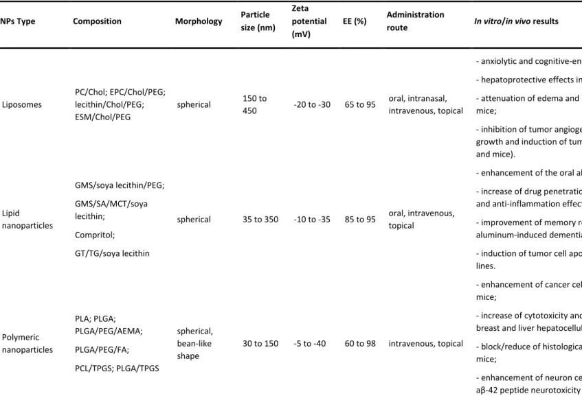
ANOPARTICLES TO DELIVER QUERCETINNowadays, the challenge consists in trying to deliver drugs to their target places, allowing to increase compounds bioavailability, thereby lowering the amount of administered substances and, consequently, minimizing their side-effects while enhancing their therapeutic efficacy. This question is particularly important while dealing with hydrophobic molecules, being crucial to find the best vehicles to increase the solubility and bioavailability of these compounds, in order to reach their target areas. Quercetin is one of these examples and several studies reported on the literature devote great attention to the development of several nanotechnological approaches which try to establish the best strategy to encapsulate and deliver quercetin for different applications. Table 2 summarizes some parameters that characterize different quercetin delivery systems, namely their composition, particle size, zeta potential, entrapment efficiency, administration route and in vitro/in vivo results.
10
Table 2. Properties of different quercetin-loaded carriers. NPs Type Composition Morphology Particle
size (nm) Zeta potential (mV)
EE (%) Administration
route In vitro/in vivo results Refs
Liposomes PC/Chol; EPC/Chol/PEG; lecithin/Chol/PEG; ESM/Chol/PEG spherical 150 to 450 -20 to -30 65 to 95 oral, intranasal, intravenous, topical
- anxiolytic and cognitive-enhancing effects in rats; - hepatoprotective effects in rodents;
- attenuation of edema and inflammation in irradiated mice;
- inhibition of tumor angiogenesis, inhibition of tumor cell growth and induction of tumor cell apoptosis (cell lines and mice). [76-81] Lipid nanoparticles GMS/soya lecithin/PEG; GMS/SA/MCT/soya lecithin; Compritol; GT/TG/soya lecithin
spherical 35 to 350 -10 to -35 85 to 95 oral, intravenous, topical
- enhancement of the oral absorption in rats;
- increase of drug penetration in skin and anti-oxidation and anti-inflammation effect;
- improvement of memory retention in rats with aluminum-induced dementia;
- induction of tumor cell apoptosis in breast cancer cell lines. [82-86] Polymeric nanoparticles PLA; PLGA; PLGA/PEG/AEMA; PLGA/PEG/FA; PCL/TPGS; PLGA/TPGS spherical, bean-like shape 30 to 150 -5 to -40 60 to 98 intravenous, topical
- enhancement of cancer cell uptake in tumor-bearing mice;
- increase of cytotoxicity and induction of apoptosis in breast and liver hepatocellular carcinoma cell lines; - block/reduce of histological alterations in irradiated mice;
- enhancement of neuron cells viability due to inhibition of aβ-42 peptide neurotoxicity in cell lines;
- improvements of cognition and memory impairments in
11
APP/PS1 mice model of Alzheimer's disease.
Magnetic nanoparticles Fe3O4; Fe3O4/E137S18E137; Fe3O4/APTS/PEG/FA; Fe3O4/PLGA
spherical 10 to 300 around +6 around
80 Intranasal
- induction of cytotoxicity in human lung carcinoma, breast
cancer and glioblastoma cell lines. [95-98]
Mesoporous silica nanoparticles TEOS/APTS; TEOS/FA spherical 200 to 250 -25 to +13 around 99 n.i.
- inhibition of tumor cell proliferation and induction of
tumor cell apoptosis (cell lines). [99, 100]
Cyclodextrins βCD; HP-βCD; SBE-βCD; SBE-7βCD
truncated cone; fibers
around
270 n.i. n.i. Oral
- increase of quercetin solubility and photostability; - enhancement of quercetin antioxidant capacity; - inhibition of tumor cell proliferation in cell lines; - impairment of tumor growth in B16F10 mouse melanoma model.
[101-104]
n.i. – not indicated; PC – phosphatidylcholine; EPC – egg phosphatidylcholine; Chol – cholesterol; PEG – poly(ethyleneglycol); ESM – egg sphingomyelin; GMS – glycerol monostearate; SA – stearic acid; MCT – medium chain triglycerides; GT – glyceryltridecanoate; TG – glyceryl tripalmitate; PLA – poly(lactic acid); PLGA – poly(lactic-co-glycolic) acid; PCL – poly(ε-caprolactone); AEMA – 2-aminoethyl methacrylamide; FA – folic acid; TPGS – d-α-tocopheryl polyethylene glycol 1000 succinate; E137S18E137 – block copolymer of ethylene oxide and styrene oxide; APTS –
3-aminopropyl triethoxysilane; TEOS – tetraethyl orthosilicate; βCD – β-cyclodextrin; HP-βCD – hydroxypropyl-β-cyclodextrin; SBE-βCD – sulfobutyl ether-β-cyclodextrin; SBE-7βCD – sulfobutyl ether-7β-cyclodextrin.
12
1.2.2.
N
ANOTECHNOLOGY STRATEGIES FOR THE DELIVERY OF QUERCETIN1.2.2.1.
L
IPOSOMESLiposomes can be developed as nanocarriers which mimetize the cellular phospholipid bilayers. The phospholipids used to produce liposomes have hydrophilic and hydrophobic portions allowing the spontaneous formation of spherical lipid bilayers in water formulations. The type of lipids chosen influences the properties of liposomes, such as charge, size, rigidity, etc. [105]. Liposomes composed by natural phospholipids are biologically inert and weakly immunogenic which is crucial to biological applications [106]. Liposomes have been used to encapsulate quercetin for many purposes [76-81, 107]. Liu et al. have developed liposomes composed by phosphatidylcholine, cholesterol and tween 80 to encapsulate quercetin in order to protect the hairless skin of Kun Ming mice against photodamage provoked by UVB [79]. Shaji et al. used multi-lamellar vesicles (MLV) with phosphatidylcholine and cholesterol in ratio of 9:1 to encapsulate quercetin and showed hepatoprotective activity in rats [77]. Thinking in tumoral therapy, Yuan et al. made a work with tumor-bearing mice model using lecithin/cholesterol/PEG4000/quercetin in 13:4:1:6 ratio and showed tumors growth inhibition [78]. Particularly, Long et al. used PEGylated liposomes composed by lecithin and cholesterol to encapsulate quercetin showing anti-tumor and anti-angiogenesis properties in ovarian cancer mouse model [79]. Many times, it is advantageous to use the synergistic effect of drugs to treat cancer. With this in mind, Wong et al. used liposomes to encapsulate vincristine and quercetin for treatment trastuzumab-insensitive breast tumor xenograft model [80]. This formulation was capable of increasing the circulation time of both drugs in plasma and inhibiting tumor growth [80]. For brain application area, Priprem et al. used quercetin liposomes in rats, with a mixture of egg phosphatidylcholine, cholesterol, and quercetin (2:1:1) and dispersed in 50% polyethylene glycol in water and they managed to reduce the anxiety and verified cognitive-enhancing in rats [76], while Phachonpai et al. have also developed egg phosphatidylcholine/cholesterol liposomes to encapsulate quercetin and showed promising results via intracerebroventricular route administration, reducing degeneration of cholinergic neurons in hippocampus [107]. Finally, in human cell lines, Goniotaki et al. demonstrated that quercetin encapsulated in egg phosphatidylcholine liposomes can inhibit the growth of many human cancer cells [81].
13
1.2.2.2.
L
IPID NANOPARTICLESLipid nanoparticles can be classified as nanostructured lipid carriers (NLC) and solid lipid nanoparticles (SLN) [108]. The last one is composed by one or more solid lipids which form a solid matrix, being excellent vehicles for drug delivery because of its physical stability, protection of the incorporated drug from degradation, controlled release and low cytotoxicity [109]. Despite these advantages, their lipid matrix may suffer recrystallization while stored and form more perfect matrices (β-modifications) that can prematurely release the encapsulated compound [110]. In this context, NLC have been developed to overcome this problem, because they blend liquid lipids with solid ones, creating an imperfect structure with more cavities and capacity to encapsulate drugs and avoiding premature release of the encapsulated compounds [110]. Several approaches using lipid nanoparticles have been developed in order to increase quercetin bioavailability and target specific places [82-86]. Li et al. produced SLN with glyceryl monostearate and soya lecithin, registering increased quercetin gastrointestinal absorption in rats [82]. However, it is also possible to think in topical administration of quercetin in order to protect against oxidative stress in skin caused by multiple factors (radiation, stress, etc.). Bose et al. have performed some permeations studies using Franz diffusion cells with human skin and SLN composed by precirol and compritol in 3:2 ratio were capable of increasing the content of quercetin inside skin demonstrating the lipid nanoparticles have great capacities as nanocarrier for topical delivery [84]. This idea was confirmed by Chen-yu et al. using glyceryl monostearate, stearic acid and media chain triglyceride to prepare NLC for topical administration in ear edema induced rats. The results showed a suppression of edema in the animals [83]. This capacity to protect against oxidative stress can be used in cancer therapy because many times cancer formation and progress are associated with multiple mutations that can be caused by ROS. In a study conducted by Sun et al. it was possible to induce apoptosis of MCF-7 and MDA-MB-231 breast cancer cells using NLC composed by 2.7% quercetin, 9.4% soy lecithin, 23.6% glyceryltridecanoate, 6.7% glyceryl tripalmitate, 13.4% vitamin E acetate and 44.2% Kolliphor HS15 [85]. Besides that, taking advantage of its neuroprotection properties, Dhawan et al. encapsulated quercetin in SLN composed by compritol and tween 80 [86]. They tested this formulation in rats chronically administered with aluminum chloride which causes an oxidative stress responsible for brain damaged [86]. The results showed that quercetin loaded SLN were capable of improving memory retention in rats with aluminum-induced dementia comparing to quercetin alone and empty nanoparticles, indicating that this nanosystem may be efficient to target brain [86].
14
1.2.2.3.
POLYMERIC NANOPARTICLESPolymeric nanoparticles are made of biodegradable polymers that may offer multiple advantages, like being stable in blood, non-toxic, nonthrombogenic, nonimmunogenic, noninflammatory, do not activate neutrophils, biodegradable, avoid reticuloendothelial system and applicable to various molecules such as drugs, proteins, peptides, or nucleic acids [111]. The versatility of these nanoparticles is based in the capacity of choosing the best polymer to the desired application. By this way these nanomedicines have been used in many approaches [87-94]. Kumari et al. synthesized polylactic acid (PLA) nanoparticles with high encapsulation efficiency and controlled release of quercetin making them promising for therapy [87]. Khoee et al. used methacrylated poly(lactic-co-glycolic acid) (mPLGA) as a lipophilic domain, acrylated methoxy poly(ethylene glycol) (aMPEG) as hydrophilic part and N-2-[(tert-butoxycarbonyl)amino] ethyl methacrylamide (Boc-AEMA) as pH-responsive part. They have proven the capacity of these polymeric nanoparticles to release their content in acidic environment and showed that they are suitable for cancer therapy [88]. El-Gogary et al. produced PEGylated PLGA nanoparticles conjugated with folic acid and demonstrated the capacity of these quercetin loaded nanosystems of increasing the killing capacity in HeLa cells compared to quercetin alone [89]. At the same time, the tumor uptake was confirmed in injected tumor-bearing mice [89]. Finally, Bishayee et al. used gold-quercetin into PLGA nanoparticles as special system to escape the immune attack [90]. The results showed that these nanoparticles had the ability to control proliferation and induce apoptosis in hepatocarcinoma cells [90].
1.2.2.4.
M
AGNETIC NANOPARTICLESMagnetic nanoparticles are very promising nanosystems whose magnetic properties allow to control and direct them to the target place applying an exterior magnetic field [112]. It is also interesting to notice that, below a certain range of size (10-20 nm), magnetic nanoparticles behave like a giant paramagnetic atom with a fast response to applied magnetic fields with negligible remanence (residual magnetism) and coercivity (the field required to bring the magnetization to zero) [113]. The absence of residual magnetism is crucial because agglomeration can be prevented [113]. So far, there are not so many applications using magnetic nanoparticles to encapsulate quercetin, however their use now starts to take its first steps [95-98]. For instance, magnetic nanoparticles are very promising for cancer therapy because external magnetic fields can direct them to the cancer area. Taking this in consideration and thinking on the
15
cancer chemotherapeutic properties of quercetin, magnetic nanoparticles have to be considered as potential and promising nanosystems for delivering quercetin in tumor cells. In a preliminary study, Barreto et al. synthesized Fe3O4 nanoparticles and showed that these magnetic nanoparticles had a controlled releasing time making this vehicle promising for cancer chemotherapy [95]. This study was followed by some studies of concrete applications of magnetic nanoparticles to deliver quercetin. Verma et al. used Fe3O4 magnetic core-shell nanoparticles protected against oxidation by PLGA and tested this formulation in the human lung carcinoma cell line A549 [96]. The results showed that quercetin loaded PLGA-MNPs had no toxicity after injection in mice and at the same time they were able to reduce the number of A549 viable cells demonstrating an anti-cancer capacity [96]. In other study conducted by Kumar et al. quercetin superparamagnetic Fe3O4 nanoparticles were tested in vitro to analyze the effects in breast cancer cell lines [97]. Fluorescent microscopy demonstrated changes in cellular morphology of MCF7 cells treated with these quercetin loaded nanoparticles indicating cytotoxicity for cancer cells and consequently potential for cancer therapy [97]. Additionally, for target brain cancer Akal et al. designed superparamagnetic iron oxide (SPION), which was functionalized with APTES ((3-aminopropyl) triethoxysilane), polyethylene glycol (PEG) and folic acid [98]. Folic acid was strategically used in order to target brain adenocarcinoma cells (U87) which overexpress folic acid receptors [98]. The MTT analysis after cellular uptake of SPION loaded with quercetin demonstrated decreasing of cancer cell viability [98]. Together, these results showed clear evidence that magnetic nanoparticles are excellent vehicles to deliver quercetin and can be used for cancer therapy based on the mentioned studies.
1.2.2.5.
M
ESOPOROUS SILICA NANOPARTICLESMesoporous silica nanoparticles (MSN) are becoming very attractive since they are biocompatible, stable, have a tunable size pore, high drug encapsulation, slow drug release and possibility of functionalizing the surface [114-118]. Some studies concern the use of MSN as vehicles for quercetin delivery [99, 100]. Sapino et al. used MSN functionalized with aminopropyl for topical delivery of quercetin [99]. The results have shown that MSN increase the penetration of quercetin in the skin and, at the same time, inhibit the proliferation of R8 human melanoma cells [99]. In another work, MSN functionalized with folate, for targeting breast cancer cells, managed to cell cycle arrest and apoptosis in breast cancer cells through the regulation of Akt & Bax signalling pathways [100].
16
1.2.2.6.
C
YCLODEXTRINSCyclodextrins (CDs) are nanosystems similar to a truncated cone with a hydrophobic cavity and a hydrophilic surface. In the cavity it is possible to accommodate lipophilic drugs and deliver them to their target place [119]. Chemically, CDs are cyclic oligosaccharides with six, seven or eight glucose residues linked by glycosidic bonds [120]. There are a few applications of CDs as nanodelivey systems to encapsulate quercetin [101-104]. The inclusion of the quercetin inside β-cyclodextrin (β-CD), hydroxypropyl-β-cyclodextrin (HP-β-CD) and sulfobutyl ether-β-cyclodextrin (SBE-β-CD) has been studied [101, 102]. The results demonstrate that (SBE-β-(SBE-β-CD) can encapsulate more quercetin than the other ones and, at the same time, may increase its antioxidant properties [101, 102]. Moreover, Aytac et al. used quercetin/β-cyclodextrin inclusion complex demonstrating the slow release of quercetin [103]. All these promising features as nanocarriers for quercetin were confirmed in a study conducted by Kale et al. [104]. They used ether-7β-cyclodextrin/quercetin complex in melanoma mouse models and verify the decrease of microvessels and consequently the reduction of tumor cell proliferation [104].
1.3.
D
ISSERTATION WORK PLAN
1.3.1.
P
ROBLEM AND MOTIVATIONNeurodegenerative diseases represent a major and growing public health burden. Currently, there are no treatments available and medications only treat symptoms, but do not halt or retard neurodegeneration process. Quercetin, a flavonol found in many plants and fruits, may provide protection against neurological disorders, such as Alzheimer’s disease [121]. However, this polyphenol presents low water-solubility, chemical instability, rapid degradation and extensive metabolism, resulting in poor bioavailability which, therefore, compromises its arrival and delivery into the brain [122].
1.3.2.
O
BJECTIVE AND STRATEGYThe goal of this dissertation will be the development of nanocarriers for enhancing the bioavailability of quercetin, promoting its site-specific delivery through the blood-brain barrier (BBB) into the brain with the utmost purpose of treating or preventing Alzheimer’s disease (Figure 3). Given the lipophilic characteristics of quercetin, lipid-based nanoparticles will be employed providing physical stability and compatibility, conferring protection from degradation and controlling the transport and brain delivery.
17
Moreover, nanoparticles surface will be functionalized with two distinct ligands: transferrin (Tf) and a 29 aminoacid peptide derived from the rabies virus glycoprotein (RVG29). The transferrin receptor (TfR) is highly expressed on the luminal side of brain capillary endothelium and might lead to receptor-mediated transcytosis of Tf-coated nanoparticles across the BBB [123]; while RVG29 was proven capable of transporting cargoes into the brain in a selective fashion [124] and binds specifically to the nicotinic acetylcholine receptor (nAchR) on neuronal cells, enabling the delivery of quercetin on damaged brain cells [125].
Figure 3. Schematic representation of the strategic plan
1.3.3.
M
ETHODS AND DETAILED DESCRIPTIONTask 1 - Preparation of lipid-based nanoparticles and functionalization with Tf and RVG29
Solid lipid nanoparticles (SLN) and nanostructured lipid carriers (NLC) were produced by a modified hot homogenization technique [126]. An average size around 200 nm was important to promote a longer circulation time and to enhance the permeability through the BBB [127]. Nanoparticles were loaded with quercetin and coated with polyethylene glycol (PEG) in order to reduce their hepatic clearance and prolong their blood circulation. Tf and RVG29 were covalently conjugated to the nanoparticles. The carboxyl groups of Tf were coupled to the amino groups of DSPE-PEG-NH2 and the thiol groups of RVG29 specifically react with the maleimide groups of DSPE-PEG-MAL, previously conjugated to the nanoparticles surface.
Task 2 - Physicochemical characterization of the developed nanosystems
The developed nanosystems were fully characterized to evaluate the quality of the nanoparticles for brain delivery:
18
- Hydrodynamic diameter, polydispersity index and zeta potential were measured by dynamic light scattering (DLS);
- Quercetin entrapment efficiency was determined by spectrophotometric analysis;
- The presence of Tf and RVG29 on the surface of nanoparticles were confirmed by Fourier transform infrared spectroscopy (FTIR) and nuclear magnetic resonance (NMR) spectroscopy.
Task 3 - In vitro validation of the developed nanosystems using cell lines and amyloid-beta peptide
The effectiveness of the nanodelivery systems for improving quercetin brain uptake was confirmed using hCMEC/D3 cell line as a model of the human BBB. Lactate dehydrogenase (LDH) assay was conducted to evaluate cell membrane integrity, after exposition to the nanoparticles. BBB permeability studies were performed using transwell culture systems. Finally, the aggregation process of the amyloid-beta peptide after nanoparticles incubation was assessed by ThT binding assay, since Aβ(1–42) is a promising target for the treatment of Alzheimer’s disease.
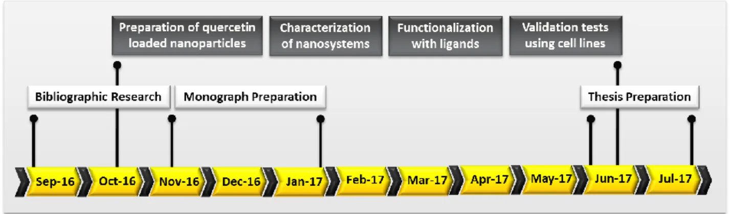
1.3.4.
T
IMELINE20
C
HAPTER
2.
M
ATERIALS AND
M
ETHODS
2.1.
P
REPARATION OF NANOPARTICLES
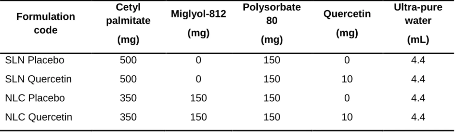
Lipid nanoparticles were produced according to an optimized hot homogenization technique followed by sonication previously developed in our group [126, 128]. SLN were prepared using cetyl palmitate as the solid lipid, while NLC were produced using cetyl palmitate as the solid lipid and miglyol-812 as the liquid lipid. Polysorbate 80 was used as surfactant in the synthesis of both SLN and NLC (Table 3). The method applied consisted in warming up the lipid and aqueous phases separately at 80°C until the lipids were melted. The lipid phase was composed by lipids, surfactant and quercetin. The aqueous phase was then added to the molten lipid mixture. In order to control the size of particles, Ultra-Turrax T25 (Janke and Kunkel IKA-Labortechnik, Staufen, Germany) was used to form microparticles, followed by sonication in a Sonics and Materials Vibra-Cell™ CV18 (Newtown, CT, USA) to produce particles in the nanometer range. SLN were stirred for 30 seconds at 12000 rpm, followed by 5 minutes of 80% intensity sonication, while NLC were homogenized for 2 minutes and then sonicated during 15 minutes at 70% intensity.
Table 3. Composition of the synthetized lipid nanoparticles (SLN - solid lipid
nanoparticles and NLC - nanostructured lipid carriers).
Formulation code Cetyl palmitate (mg) Miglyol-812 (mg) Polysorbate 80 (mg) Quercetin (mg) Ultra-pure water (mL) SLN Placebo 500 0 150 0 4.4 SLN Quercetin 500 0 150 10 4.4 NLC Placebo 350 150 150 0 4.4 NLC Quercetin 350 150 150 10 4.4
21
2.2.
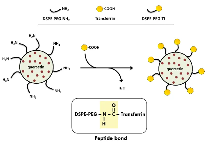
F
UNCTIONALIZATION OF NANOPARTICLES WITH TRANSFERRIN
Nanoparticles were functionalized using 1,2-distearoyl-sn-glycero-3-phosphoethanolamine (DSPE) associated with polyethylene glycol (PEG) and terminal amine groups (NH2) exposed on the nanoparticles surface. These last amine groups were conjugated with carboxyl groups of transferrin, thereby forming peptide bonds (Figure 5). Nanoparticles were prepared as previously described but incorporating 5 mg of DSPE-PEG-NH2 in the lipid phase composition (Table 4). At the same time, a transferrin solution was prepared in ultra-pure water (5 mg/mL) and then the carboxyl groups of transferrin were activated with 1-Ethyl-3-(3-dimethylaminopropyl) carbodiimide (EDC) at room temperature, with stirring, for 30 minutes. After that, the transferrin solution was mixed with the previously produced nanoparticles conjugated with DSPE-PEG-NH2 and the formulations were incubated for 2 hours, at room temperature. Finally, the Tf-functionalized nanoparticles were dialyzed in a 10 kDa MWCO SnakeSkin Dialysis Tubing against 500 mL ultra-pure water at 37°C, overnight, to remove the excess of transferrin and by-products.
Figure 5. Schematic representation of the functionalization of nanoparticles with
22
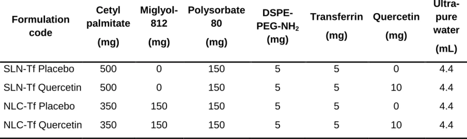
Table 4. Composition of Tf-functionalized lipid nanoparticles. Formulation code Cetyl palmitate (mg) Miglyol-812 (mg) Polysorbate 80 (mg) DSPE-PEG-NH2 (mg) Transferrin (mg) Quercetin (mg) Ultra-pure water (mL) SLN-Tf Placebo 500 0 150 5 5 0 4.4 SLN-Tf Quercetin 500 0 150 5 5 10 4.4 NLC-Tf Placebo 350 150 150 5 5 0 4.4 NLC-Tf Quercetin 350 150 150 5 5 10 4.4
2.3.
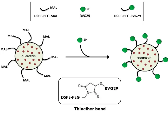
F
UNCTIONALIZATION OF NANOPARTICLES WITH
RVG29
For surface modification of nanoparticles with RVG29 peptide, DSPE-PEG-MAL was previously conjugated to RVG29. DSPE-PEG-MAL is a 1,2-distearoyl-sn-glycero-3-phosphoethanolamine (DSPE) associated with polyethylene glycol (PEG) and terminal maleimide groups (MAL). The maleimide groups can react with thiol groups of RVG29, thereby forming thioether bonds (Figure 6). RVG29 peptide is a 29 amino acid fragment derived from rabies virus glycoprotein (RVG). The peptide RVG29 with an additional cysteine on C-terminal (YTIWMPENPRPGTPCDIFTNSRGKRASNGC) was synthesized by Bachem Group (Germany). Hence, a RVG29 solution (5mg/mL) was prepared in PBS (pH 7.0) followed by the addition of 1-fold DSPE-PEG-MAL. The mixture was allowed to react at room temperature for 24 hours, thus forming the conjugate DSPE-PEG-RVG29. The conjugate was then dialyzed in a 10 kDa MWCO SnakeSkin Dialysis Tubing against 500 mL ultra-pure water at 37°C, overnight, to remove the excess of RVG29 and by-products. Finally, nanoparticles were produced as previously described but incorporating 1 mL of DSPE-PEG-RVG29 conjugate in the lipid phase composition (Table 5).
23
Figure 6. Schematic representation of the functionalization of nanoparticles with
RVG29 ligands (not to scale).
Table 5. Composition of RVG29-functionalized lipid nanoparticles. Formulation code Cetyl palmitate (mg) Miglyol-812 (mg) Polysorbate 80 (mg) DSPE-PEG-RVG29 (mL) Quercetin (mg) Ultra-pure water (mL) SLN-RVG Placebo 500 0 150 1 0 4.4 SLN-RVG Quercetin 500 0 150 1 10 4.4 NLC-RVG Placebo 350 150 150 1 0 4.4 NLC-RVG Quercetin 350 150 150 1 10 4.4
2.4.
N
UCLEAR MAGNETIC RESONANCE SPECTROSCOPY
The successuful synthesis of DSPE-PEG-Transferrin and DSPE-PEG-RVG29 conjugates were analyzed by NMR spectroscopy. NMR experiments were recorded on a Bruker Avance III 600 HD spectrometer (Bruker, Massachusetts, United States),
24
operating at 600.13 MHz for 1H, equipped with 5 mm CryoProbe Prodigy and pulse gradient units, capable of producing magnetic field pulsed gradients in the z-direction of 50 G cm1. The NMR measurements were carried out in deuterium oxide (D2O), at 300K and a spectral width of 10 000 Hz. 1H NMR experiments were performed with water suppression using excitation sculpting with gradients. The chemical shifts of the 1H NMR signals were referred to the absorption frequency of trimethylsilylpropanoic acid-d4 (TMSP-d4) as internal reference.
2.5.
F
OURIER TRANSFORM INFRARED SPECTROSCOPY
Fourier transform infrared spectroscopy (FT-IR) was used to confirm the functionalization of the nanoparticles with transferrin and RVG29 ligands. In order to perform this technique the samples were previously lyophilized in order to eliminate water bands that could mask the ones related to the sample itself. The first step of the lyophilization consisted in freezing the samples overnight in a -85°C deep freezer. After that, samples were lyophilized at -85°C and 0.76 Torr using a LyoQuest -85 freeze dryer (Telstar, Terrassa, Spain). Finally, the infrared spectra of the lyophilized nanoparticles were obtained using a Frontier FT-IR Spectrometer from PerkinElmer (Santa Clara, California, USA).
2.6.
T
RANSMISSION ELECTRON MICROSCOPY
The morphology of nanoparticles was analyzed by transmission electron microscopy (TEM). The samples were mounted on 300 mesh form var copper grids and were analyzed using a Jeol JEM 1400 transmission electron microscope (Tokyo). Uranyl acetate was used as the contrast agent. Images were digitally recorded using a Gatan SC 1000 ORIUS CCD camera (Warrendale, PA, USA).
2.7.
D
YNAMIC LIGHT SCATTERING
The mean hydrodynamic diameter of nanoparticles was measured by dynamic light scattering (DLS) using a particle size analyzer (Brookhaven Instruments, Holtsville, NY, USA). All samples were diluted in ultra-pure water by a factor of 1:400 before measuring the particles size. All determinations were made at 25°C with a light incidence angle of 90°. The hydrodynamic diameter followed a Gaussian distribution and polydispersity index was determined according to the width of particle size distribution.
25
2.8.
Z
ETA POTENTIAL ANALYZER
The zeta potential of nanoparticles was calculated by measuring the electrophoretic mobility in a zeta potential analyzer (Brookhaven Instruments, Holtsville, NY, USA). All samples were diluted in ultra-pure water by a factor of 1:400 and all the measurements were performed at 25°C.
2.9.
E
NTRAPMENT EFFICIENCY DETERMINATION
All samples were diluted in ultra-pure water by a factor of 1:200 and filtered through 3.0 μm Millipore-type SSWP membrane filters. Nanoparticles can pass through the membrane filters, while unentrapped quercetin is retained on the filters. The quercetin retained in the filter was then recovered using 2 mL of acetonitrile on disposable syringes and the absorbance was measured in a V-660 spectrophotometer (Jasco, Easton, MD, USA) at 367 nm. Quercetin entrapment efficiency was obtained by the following equation:
2.10.
P
HOTOSTABILITY STUDY
Photostability studies were performed to investigate the protection effect of lipid nanoparticles against photodegradation of quercetin. Ethanol solutions of quercetin (0.7 mg/mL) and quercetin-loaded nanoparticle formulations were exposed to UVA light (HSW 125W E27, Sylvania) in a chamber with reflecting walls and a cooling system. The UV lamp had an emission spectrum between 320 and 400 nm, with an irradiation intensity of 5.5 x 10⁻³ kJ/s/m2. After 6 h of UV light exposure, aliquots were withdrawn from each sample and diluted with acetonitrile by a factor of 1:200. In the case of nanoparticles, acetonitrile promote the disruption of lipid matrix, allowing the quantification of quercetin in solution by spectrophotometric detection at 367 nm in a V-660 spectrophotometer (Jasco, Easton, MD, USA). Quercetin photodegradation was calculated as a percentage by comparing the absorbance of quercetin at the endpoint to the maximum absorbance at the beginning of the experiment.
2.11.
H
CMEC/D3
CELL CULTURE
Immortalized human cerebral microvascular endothelial cells (hCMEC/D3) were obtained from Institut National de la Santé et de la Recherche Médicale (INSERM, Paris, France). This brain endothelial cell line is commonly used as a BBB model
26
system, since it mimics structurally and morphologically the human BBB properties [129, 130]. hCMEC/D3 cells were used between passage number 29 and 35. Cells were seeded in a concentration of 2.5 × 10⁴ cells/cm2 and grown at 37 °C, in an atmosphere of 5 % CO2 in EndoGRO medium supplemented with 1% Pen-Strep, 5% FBS, 50 μg/mL ascorbic acid, 0.75 U/mL heparin sulfate, 1.0 μg/mL hydrocortisone hemisuccinate, 10 mM L-Glutamine and 5 ng/mL rhEGF. All culture flasks were pre-coated with 0.1 mg/mL type I rat collagen, 1 h at 37 °C. Cell culture medium was changed every 2–3 days.
2.12.
LDH
CYTOTOXICITY ASSAY
Lactate dehydrogenase (LDH) assays were conducted in order to evaluate cytotoxicity and nanoparticles potential for damaging cell membranes. LDH is an enzyme that is released from cells to the surrounding cell culture supernatant when cell membranes are damaged or disrupted. Cells were seeded in 96-well plates (104 cells per well) pre-coated with type I rat collagen. After 20 h of incubation at 37 °C and 5 % CO2, different concentrations of quercetin-loaded nanoparticles (functionalized and non-functionalized) were incubated with the cells for 4 h. EndoGRO medium and TX-100 sample were also used as negative and positive controls for cytotoxicity, respectively. After the incubation time, the medium of each well was collected and centrifuged (250 g for 10 min, RT) and the supernatant separated for further LDH quantification assay (LDH cytotoxicity detection kit, Takara Bio Inc, Shiga, Japan). After treatment with catalyst and dye solutions for 20 min at RT in the dark, absorbance was read at 490 and 690 nm using a Synergy™ HT Multi-mode Microplate Reader (BioTek Instruments Inc, Winooski, VT, USA). Cytotoxicity was expressed as a percentage compared to the maximum cytotoxicity of TX-100 sample.
2.13.
T
RANSWELL PERMEABILITY ASSAY
hCMEC/D3 cells were seeded in a density of 2 x 10⁵ cells per insert on transwell devices (six-well polyester inserts, pore size 0.4 μm and a diameter of 4.67 cm2) precoated with type I rat collagen. The permeability assay was performed 7 days after seeding. The barrier integrity of hCMEC/D3 cell monolayers was previously checked using Lucifer Yellow as a small reference molecule with a well reported effective permeability coefficient (Peff) of 1.33x10⁻³ cm/min [131]. For the permeability studies, functionalized and non-functionalized nanoparticles previously loaded with FITC (0.4 mg/mL) were incubated in the apical donor compartment for 4 h at 37°C in a 5% CO2
27
atmosphere incubator. The total amount of nanoparticles in the receptor compartment was quantified after 0.5, 1, 2, 3 and 4 h by fluorescence analysis (495/519 nm).
2.14.
A
MYLOID
-
BETA PEPTIDE PREPARATION
Aβ(1–42) peptide (purity > 95%, MW 4514.14) was purchased from Selleck Chemicals. The aggregation state and the structure can vary according to the sample batch [132]. In order to solve the residual peptide aggregation and consequently improve its solubility, the peptide was previously dissolved in HFIP (1,1,1,3,3,3-hexafluoro-2-propanol) at 1 mg/mL in order to disrupt intermolecular H-bonds [133-135]. This solution was then evaporated with nitrogen flow and under vacuum and the resulting peptide film was dissolved in dimethyl sulfoxide at 9 mg/mL.
2.15.
T
HIOFLAVIN
T
BINDING ASSAY AND FLUORESCENCE
MEASUREMENTS
Thioflavin T (ThT) is a classic amyloid dye that is commonly used to probe amyloid-beta fibril formation because of its strong fluorescence emission upon binding to amyloid fibril structures [134, 136]. A ThT stock solution was prepared mixing 8 mg of ThT in 10 mL of PBS buffer. This solution was filtered through a 0.20 µm syringe filter and the stock solution was diluted in buffer (1:50 ratio). 50 μM of Aβ(1–42)peptide was incubated at 37 °C in 96 well plates with different conditions: 30 μM quercetin, loaded nanoparticles and unloaded nanoparticles. Finally, ThT solution (0.7 mg/mL) was added to each well and the fluorescence intensity was measured every 30 minutes during 24 hours using a Biotek Synergy 2 fluorescence spectrometer (Winooski, Vermont, USA). ThT conjugated with fibrils has excitation at 450 nm and maximum emission at 482 nm [137].
2.16.
S
TATISTICAL ANALYSIS
Statistical analysis was performed using SPSS software (v 24.0; IBM, Armonk, NY, USA). The measurements were repeated three times and data were expressed as mean ± SD. Data were analyzed using one-way analysis of variance (one-way ANOVA), followed by Bonferroni, Tukey and Dunnett post-hoc tests. A p value lower than 0.05 was considered statistically significant.
![Table 1. Quercetin natural sources [5, 9, 11].](https://thumb-eu.123doks.com/thumbv2/123dok_br/15724728.1070866/16.892.305.597.320.844/table-quercetin-natural-sources.webp)