http://dx.doi.org/10.1590/bjpt-rbf.2014.0173 Braz J Phys Ther. 2016 Sept-Oct; 20(5):395-404 395
Dynamometry for the measurement of grip, pinch, and
trunk muscles strength in subjects with subacute stroke:
reliability and different number of trials
Larissa T. Aguiar1, Júlia C. Martins1,Eliza M. Lara1, Julianna A. Albuquerque1,
Luci F. Teixeira-Salmela1, Christina D. C. M. Faria1
ABSTRACT | Background: Muscle strength is usually measured in individuals with stroke with Portable dynamometers (gold standard). However, no studies have investigated the reliability, the standard error of measurement (SEM) and the minimal detectable difference (MDD95%) of the dynamometry for the measurement of hand grip, pinch grip and trunk strength in subjects with subacute stroke. Objective: 1) To investigate the intra and inter-rater reliability, the SEM and the MDD95% of the portable dynamometers for the measurement of grip, pinch and trunk strength in subjects with subacute stroke, and 2) to verify whether the use of different number of trials (irst trial and the average of the irst two and three trials) affected the results. Method: 32 subjects with subacute stroke (time since stroke onset: 3.6 months, SD=0.66 months) were evaluated. Hand grip, 3 pinch grips (i.e. pulp-to-pulp/palmar/lateral) and 4 trunk muscles (i.e. lexors, extensors, lateral lexors and rotators) strength were bilaterally assessed (except trunk lexors/extensors) with portable dynamometry by two independent examiners over two sessions (1-2 weeks apart). One-way ANOVAs and intraclass correlation coeficients (ICC2,k) were used for analysis (α=0.05). SEM and MDD95% were also calculated. Results: For all muscular groups and sources of outcome values, including one trial, after familiarization, similar results were found (0.01≤F≤0.08; 0.92≤p≤0.99) with signiicant and adequate values of intra-rater (0.64≤ICC≤0.99; 0.23≤95%CI≤0.99) and
inter-rater (0.66≤ICC≤0.99; 0.25≤95%CI≤0.99) reliability. SEM and MDD95% were considered low (0.39≤EPM≤2.21 Kg; 0.96≤MMD95%≤6.12 Kg) for all outcome scores. Conclusion: Only one trial, following familiarization, demonstrated adequate intra-rater and inter-rater reliability of the portable dynamometers for the measurement of hand grip, pinch grip and trunk strength in subjects with subacute stroke.
Keywords: physical therapy; muscle strength dynamometer; reproducibility of results; stroke.
BULLET POINTS
• Portable dynamometers present adequate inter and intra-rater reliability in the subacute phase of stroke. • One trial provides similar results to those obtained with the average of the irst two and three trials. • In clinical practice, one trial can be used to measure hand and trunk muscle strength.
HOW TO CITE THIS ARTICLE
Aguiar LT, Martins JC, Lara EM, Albuquerque JA, Teixeira-Salmela LF, Faria CDCM. Dynamometry for the measurement of grip, pinch, and trunk muscles strength in subjects with subacute stroke: reliability and different number of trials. Braz J Phys Ther. 2016 Sept-Oct; 20(5):395-404. http://dx.doi.org/10.1590/bjpt-rbf.2014.0173
1 Departamento de Fisioterapia, Escola de Educação Física, Fisioterapia e Terapia Ocupacional, Universidade Federal de Minas Gerais (UFMG), Belo Horizonte, MG, Brazil
Received: May 05, 2015 Revised: Oct. 27, 2015 Accepted: Feb. 15, 2016
Introduction
Hemiparesis, a disability arising from a stroke, with muscle weakness predominant in the hemi-body contralateral to the cerebral lesion, is observed in
approximately 77% of individuals with stroke1-4.
Muscle weakness is also observed in the hemi-body ipsilateral to the brain lesion and in the torso as a whole5-8. Thus, the measurement of muscle strength
in post stroke individuals is commonly performed9
in different phases of the disease10,11.
values of validity and reliability for different muscle groups and populations tested11. The intra-rater and
inter-rater reliability of portable dynamometers in the measurement of hand grip, pinch grip, and trunk strength was recently investigated in individuals in the chronic phase of stroke13, and demonstrated adequate results, with Intraclass Correlation Coeficients (ICC)
varying from moderate to very high14 (0.58≤ICC≤0.98). Although commonly regarded as a uniform population, subjects with stroke have unique features in each recovery phase (i.e. acute (irst three months), subacute (three to six months), and chronic phases (≥ six months
of involvement by stroke))15-17, which may affect the measurement of muscular strength. Motor recovery and the ability to perform functional activities, such
as holding an object or sit-to-stand, are examples of
these features18,19. Therefore, for portable dynamometers to be employed in the subacute phase of stroke, it is necessary to assess measurement properties of these instruments in this population. To our knowledge, only
Chen et al.20 investigated the reliability of portable
dynamometers in individuals in the subacute phase of stroke. However, the authors only assessed intra-rater reliability and only for measurement of handgrip, tripod, and lateral pinch strength. Moreover, the study sample comprised 62 individuals in both subacute and chronic phases of stroke, without separation of
the results by phase. According to a recent systematic
review, no study has investigated the measurement properties of portable dynamometers to assess hand
grip, pinch grip, and trunk strength speciically in the
subacute phase of stroke20.
Because consistent and reproducible data are essential for credible evaluations, determination of the
threshold between error and true change is required
for the interpretation of the results. The measurement properties should not be intrinsic characteristics of the measuring instrument21. Furthermore, the use of only
one trial could facilitate the clinical applicability of the tests. Therefore, the objectives of this study were: (1) to investigate the intra-rater and inter-rater reliabilities of
portable dynamometers (SAEHAN Hydraulic Hand Dynamometer, SAEHAN Hydraulic Pinch Gauge,
and Microfet2 digital manual dynamometer) for the assessment of handgrip, pinch grip (i.e, pulp-to-pulp,
tripod, and lateral pinch), and trunk strength; (2) to
determine the standard error of the measurement
(SEM) and the minimal detectable change (MDC);
and (3) to assess whether a different number of trials
of measures (i.e. irst trial, the average of the irst
two trials, and the average of three trials) affects the results, in subjects in the subacute phase of stroke.
Method
Sample
Individuals from the local community participated
in this study. The following inclusion criteria were used: a diagnosis of stroke (subacute phase: between 3 and 6 months since the initial episode)17 and age
greater than 19 years. Exclusion criteria included those who presented possible cognitive deicits identiied by the Mini Mental State Examination based on cutoff
points for three levels of education as established by Bertolucci et al.22 (i.e. illiterate: 13 points; one to seven years of schooling: 18 points; eight or more years of
schooling: 26 points), or the inability to respond to the command “Raise your unaffected arm and open your hand”23, or who had pain or another health condition
that affected muscular strength of the hands or trunk. To determine the number of individuals to be assessed,
MedCalc software, version 12.7.5 (MedCalc Software, Ostend, Belgium) was used, with a sample calculation for a correlation coeficient using power=0.8, r=0.70,
and α=0.05. The result was n=14. A prerequisite
for use of statistical tests that assess the correlation between variables is the sample variability in relation to the outcome of interest21. Taking into account
the characteristics that could result in variations in muscle strength, the authors tried to achieve variability
in relation to age, sex, and the degree of motor
recovery (severe, moderate, or mild) assessed by the
Fugl-Meyer Scale for motor function of the Upper Extremity (UE-FM)24. Thus, assuming n=14 for each
of two age groups that could demonstrate variation in muscular strength (20-64 years and over 64 years),
n=28 was calculated. Assuming that approximately
15% of the participants would withdraw on the second
day of evaluation, a inal sample of 32 (28+4[15%])
participants was determined as an attempting to assure sample variability in muscular strength and improve the statistical robustness. However, it is important to point out that the sample size should be at least 14. Subjects with stroke were included in the present study even though they could not accomplish all muscle groups’ assessments. Therefore, once some subjects were not able to activate some muscle groups, the sample size for each muscle group evaluated varied.
Before data collection, the individuals read and
397
Braz J Phys Ther. 2016 Sept-Oct; 20(5):395-404 MG, Brazil (ETIC 04 92.0.203.000-10). Clinical and
demographic data such as age, sex, type and time
since stroke, paretic hand, stage of motor recovery (i.e. on Fugl-Meyer scale)24, and trunk performance (i.e. Trunk Impairment Scale)25, were collected by a physical therapist with prior experience in the
assessment of muscular strength of individuals with stroke in the chronic phase and who was trained to meet the objectives of the present study. The paretic
upper extremity (UE) was scored using the Modiied Ashworth Scale26 using lexors of the elbow, wrist, and ingers, and by the decrease in muscle strength, in comparison with the non-paretic UE. However, unlike the UE-FM, the trunk muscles were not identiied in
this study as paretic and non-paretic, but as right and left sides. This was done because these muscles receive innervation from both brain hemispheres, although predominantly from the contralateral side27, and also
because the trunk motor deiciencies in individuals
with stroke can occur on both sides, with compensation by bilateral innervation28.
Bilateral muscles strength of handgrip, pinch grip (i.e. pulp-to-pulp, tripod, and lateral), and trunk
muscles (i.e. extensors, lexors and lateral lexors,
and rotators) were measured by two trained physical therapists (rater-1 and rater-2) with one year of
experience in the assessment of muscular strength
using a dynamometer. These raters were trained for this assessment by a physical therapist with four
years of training and two years of experience in
the assessment of muscular strength of post-stroke individuals using portable dynamometers. Training
consisted of a detailed explanation and repeated
practice procedures for the assessment of strength of the chosen muscle groups using dynamometers. The evaluation of muscle strength for the collection of data was based on a previous study developed for individuals in the chronic phase of stroke, and included the muscle groups evaluated in the present study, with the same instruments and procedures13.
Instruments
The measurement of muscle strength was performed with three portable dynamometers:
SAEHAN Hydraulic Hand Dynamometer Model HS5001 (SAEHAN Corporation, Korea) was used to measure handgrip strength; SAEHAN Hydraulic Pinch Gauge Model HS5001 was used to measure the
3 pinch grip strengths (i.e.pulp-to-pulp, tripod, and lateral), and Microfet2 digital manual dynamometer
(Hoggan Health Industries, UT, USA), was used to measure trunk strength. All instruments were new and
factory-calibrated according to the manufacturer’s information manual.
Procedures
All assessments of muscle strength were performed
following the same procedures adopted by Faria et al.13, except for the interval between the two evaluation
sessions, which in this study was 1 to 2 weeks17. The range of 1 to 2 weeks was adopted as being unlikely to show changes in muscle strength during this period in subjects with subacute stroke17. Furthermore, with this interval, the rater would probably not remember
the values measured on the irst day. The data for
intra-rater reliability were obtained by a single rater (rater-1) on two evaluation days, and the data for inter-rater reliability were obtained by two raters
(rater-1 and rater-2) on the irst evaluation day. An interval of ive minutes was provided between
measurements by the two raters to prevent muscle fatigue, as previously adopted10. The reading and
recording of measurements of muscle strength were performed by a recorder, who was also trained. Thus, rater-1 and rater-2 had no access to the muscle strength data of the participants. Each rater evaluation lasted
in average 20-30 minutes. Therefore, in the irst
evaluation day the entire muscle strength evaluation lasted around 60 minutes.
The measurements of muscle strength were always obtained in the same order, in an alternating manner,
starting with the non-paretic UE and the right side of
the trunk. First, a demonstration and familiarization
with the equipment and procedures were performed
to ensure understanding of the test. Then, three trials
at maximum isometric strength were each held for ive seconds, and the peak value of the 3 trials was recorded. An interval of 15 to 20 seconds was provided
between trials. During the muscle strength test, the rater provided verbal encouragement to the participant
to attempt maximum effort.
Statistical analysis
Descriptive statistics and normality tests (i.e. Shapiro-Wilk) were performed for all outcome
measures. One-way analysis of variance (ANOVA) was used to compare all forms of operationalization of measures, taking into account the values obtained
by rater-1 on day-1. The ICC2,k and 95% conidence interval (CI) were calculated to determine the intra-rater
and inter-rater reliabilities, based on the different
results were classiied as follows: very low=0-0.25, low=0.26-0.49, moderate=0.50-0.69, high=0.70-0.89,
and very high=0.90-1.0014.
The stability of the measurements and the amount
of change needed to relect a real difference were determined by using the SEM and MDC based on 95% CI (MDC95%). Both the SEM and MDC95%
were calculated according to reliability results and the forms of operationalization of measures21.
To carry out these analyses, the following formulas were used16: SEM = standard deviation × √(1-ICC); MDC95%=1.96 × SEM × √2. The statistical package SPSS version 15.0 (SPSS Inc., Chicago, IL, USA) (α=0.05) was used for the analysis.
Results
Thirty-two individuals in the subacute phase of
stroke (18 men & 14 women), with a mean age of
63±12 years and a mean time following stroke of 3.6±0.66 months were assessed (Table 1). Of these 32, only 24 agreed to participate on the second day of evaluation for work or health reasons.
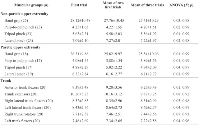
Table 2 presents the results of descriptive
statistics, as well as of the ANOVA for comparison
of all forms of operationalization of measures for all muscle groups assessed. The values found for all forms of operationalization of measures were similar (0.01≤F≤0.08; 0.92≤p≤0.99).
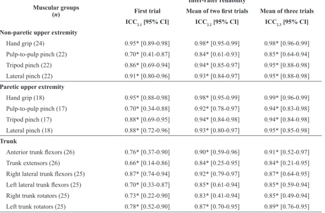
Tables 3 and 4 present the values of the ICC
with the 95% CI for the intra-rater and inter-rater
reliabilities for all muscle groups assessed and forms
of operationalization of measures. All measures
of intra-rater and inter-rater reliability displayed
signiicant values, classiied as moderate to very high
(0.64≤ICC≤0.99; p≤0.001) for all muscle groups (Table 3 and 4).
Table 5 presents the results of the SEM and MDC95%, based on the intra-rater and inter-rater reliabilities and forms of operationalization of the measures.
The values of the SEM and MDC95% were similar
between the different forms of operationalization
of the measures. All SEM (0.39≤SEM ≤2.21 Kg)
and MDC95% (0.96≤MMD95%≤6.12 Kg) values are
presented in Table 5.
Discussion
The portable dynamometers presented adequate
intra-rater and inter-rater reliabilities for measurement of grip, pinch, and trunk muscular strength in individuals in the subacute phase of stroke, with low values of
SEM and MDC. In general, the 95% CI of the ICC
for intra-rater and inter-rater reliabilities displayed
adequate values. Moreover, results obtained with one
trial were similar to those obtained with the average
of the irst two and three trials.
As the ICC is the most appropriate method for analyzing reliability, only studies that used the ICC
were used in this discussion. Faria et al.13 investigated
the intra-rater and inter-rater reliabilities of portable dynamometers in assessing the strength of the same muscle groups evaluated in this study, but with individuals in the chronic phase of stroke. The results of the reliabilities in the chronic phase of stroke were
classiied as moderate to very high (0.58 ≤ ICC ≤ 0.98)13.
These results are similar to those of the present study: (0.64 ≤ ICC ≤ 0.99). It was not possible to compare
the 95%CI of the ICC with the study of Faria et al.13 because that analysis was not reported. Chen et al.20,
with a sample of individuals in the subacute and chronic
phases grouped together, reported an ICC classiied as
very high (0.96 ≤ ICC ≤ 0.98; 0.93 ≤ 95% CI ≤ 0.99) when evaluating handgrip strength, tripod pinch, and
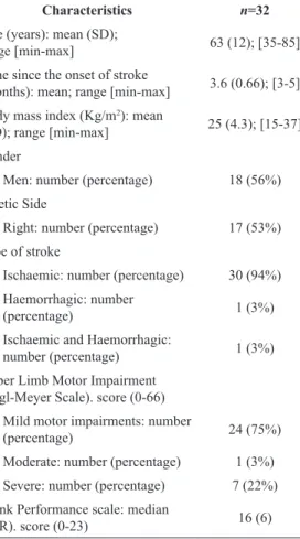
Table 1. Participant’s characteristics of 32 subacute stroke subjects.
Characteristics n=32
Age (years): mean (SD);
range [min-max] 63 (12); [35-85]
Time since the onset of stroke
(months): mean; range [min-max] 3.6 (0.66); [3-5] Body mass index (Kg/m2): mean
(SD); range [min-max] 25 (4.3); [15-37]
Gender
Men: number (percentage) 18 (56%)
Paretic Side
Right: number (percentage) 17 (53%)
Type of stroke
Ischaemic: number (percentage) 30 (94%) Haemorrhagic: number
(percentage) 1 (3%)
Ischaemic and Haemorrhagic:
number (percentage) 1 (3%)
Upper Limb Motor Impairment
(Fugl-Meyer Scale). score (0-66)
Mild motor impairments: number
(percentage) 24 (75%)
Moderate: number (percentage) 1 (3%)
Severe: number (percentage) 7 (22%)
Trunk Performance scale: median
(IQR). score (0-23) 16 (6)
399
Braz J Phys Ther. 2016 Sept-Oct; 20(5):395-404
Table 2. Results of descriptive statistics (means±SD) and ANOVA regarding the comparisons between the various sources of outcome
scores for muscular strength measurement of both the non-paretic and paretic upper extremities and trunk with portable dynamometry (Kg) for 32 subacute stroke subjects.
Muscular groups (n) First trial Mean of two irst trials Mean of three trials ANOVA (F; p)
Non-paretic upper extremity
Hand grip (25) 28.12±10.48 27.76±10.45 27.41±10.29 0.03; 0.98
Pulp-to-pulp pinch (23) 4.25±1.63 4.22±1.55 4.20±1.51 0.02; 0.98
Tripod pinch (23) 5.63±2.31 5.58±2.03 5.56±1.92 0.01; 0.99
Lateral pinch (23) 7.09±2.10 7.27±2.01 7.22±1.97 0.02; 0.98
Paretic upper extremity
Hand grip (18) 26.31±9.86 25.62±9.97 25.54±10.06 0.01; 0.99
Pulp-to-pulp pinch (17) 4.08±1.44 3.88±1.54 3.89±1.56 0.01; 0.99
Tripod pinch (17) 4.88±2.29 5.02±2.22 4.94±2.09 0.04; 0.97
Lateral pinch (19) 6.32±2.84 6.16±2.77 6.11±2.72 0.01; 0.99
Trunk
Anterior trunk lexors (20) 9.39±3.48 9.28±3.56 9.25±3.48 0.01; 0.99
Trunk extensors (20) 10.26±3.23 10.16±3.12 9.87±3.25 0.08; 0.92
Right lateral trunk lexors (20) 8.32±2.85 8.35±2.96 8.51±2.99 0.03; 0.98
Left lateral trunk lexors (20) 8.43±2.76 8.64±2.73 8.62±2.74 0.04; 0.97
Right trunk rotators (20) 7.71±2.58 7.46±2.51 7.44±2.56 0.07; 0.93
Left trunk lexors (20) 7.46±2.69 7.34±2.65 7.22±2.58 0.04; 0.96
SD: Standard deviation.
Table 3. Intra-class correlation coeficients (ICC) and 95% conidence interval (CI) for the intra-rater reliability for muscular strength
measurement of both the non-paretic and paretic upper extremities and trunk with portable dynamometers (Kg) considering various
sources of outcome scores for 24 subacute stroke patients.
Muscular groups (n)
Intra-rater reliability
First trial Mean of two irst trials Mean of three trials
ICC2.1 [95% CI] ICC2.2 [95% CI] ICC2.3 [95% CI]
Non-paretic upper extremity
Hand grip (17) 0.95* [0.87-0.98] 0.98* [0.94-0.99] 0.99* [0.96-0.99]
Pulp-to-pulp pinch (16) 0.64** [0.23-0.86] 0.83* [0.50-0.94] 0.85* [0.57-0.95] Tripod pinch (16) 0.84* [0.60-0.94] 0.93* [0.80-0.98] 0.95* [0.85-0.98]
Lateral pinch (16) 0.84* [0.60-0.94] 0.93* [0.79-0.98] 0.94* [0.84-0.98]
Paretic upper extremity
Hand grip (13) 0.97* [0.90-0.99] 0.99* [0.96-0.99] 0.98* [0.95-0.99] Pulp-to-pulp pinch (12) 0.82* [0.51-0.95] 0.89* [0.63-0.97] 0.90* [0.67-0.97] Tripod pinch (12) 0.85* [0.57-0.91] 0.95* [0.81-0.98] 0.95* [0.81-0.98]
Lateral pinch (14) 0.91* [0.74-0.97] 0.95* [0.85-0.99] 0.96* [0.88-0.99]
Trunk
Anterior trunk lexors (20) 0.87* [0.71-0.95] 0.94* [0.85-0.98] 0.94* [0.85-0.98]
Trunk extensors (20) 0.79* [0.55-0.91] 0.89* [0.70-0.96] 0.90* [0.71-0.96]
Right lateral trunk lexors (20) 0.84* [0.51-0.94] 0.94* [0.84-0.98] 0.96* [0.89-0.98]
Left lateral trunk lexors (20) 0.88* [0.72-0.95] 0.96* [0.90-0.98] 0.96* [0.90-0.98]
Right trunk rotators (20) 0.77* [0.39-0.91] 0.89* [0.69-0.96] 0.89* [0.72-0.96]
Left trunk rotators (20) 0.74* [0.46-0.89] 0.92* [0.79-0.97] 0.94* [0.84-0.98]
lateral pinch. The result of the present study was similar
to that of Chen et al.20 for the same muscle groups and
forms of operationalization (average of three trials) (0.94 ≤ ICC ≤ 0.99; 0.81 ≤ 95% CI ≤ 0.99). With regard to the use of different forms of operationalization of the measures, the results of the present study were also similar to several published studies with individuals in the chronic phase of stroke13,29-32. In these studies, only
one trial, after familiarization, presented similar results for the measurement properties investigated and for the values obtained when compared to other forms of
operationalization, except for the measurement of the plantar lexor muscle strength of the non-paretic side using the Modiied Sphygmomanometer Test (MST) measured by two raters. Nevertheless, the present study is the irst to consider a sample of individuals
in the subacute phase of stroke.
All SEM values in this study were below 2.21 Kg,
indicating that the measurement of muscular strength by portable dynamometers was able to distinguish between small real changes in muscle strength and error in individuals in the subacute phase of stroke.
In addition, the results of the SEM were similar to
those reported by Bertrand et al.33 and Boissy et al.34,
who determined the SEM of handgrip muscular
strength with a dynamometer, allowing for ICC
values of intra-rater reliability, in individuals in the
chronic phase of stroke. The MDC indicates the least
amount of change that can be interpreted as either real improvement or worsening21. The MDC values in this study varied from 0.96 to 6.12 Kg. Therefore,
for a change in muscle strength to be considered real after an intervention, when evaluated using portable
dynamometers, one must have changes equal to or greater than 0.96 to 6.12 Kg (depending on the
muscle group and the number of trials performed).
No study was found that investigated the MDC to
evaluate muscular strength with a dynamometer in
subjects with stroke. However, the values of MDC
found were similar to those of healthy individuals
(MDC=5.7 and 7.1 Kg), and were lower than the
values of individuals hospitalized in intensive care
units (MDC=7.8 and 12.5 Kg), whose handgrip strength
was evaluated using a dynamometer (average of three trials), taking into account test-test reliability35,36. The MDC values should be used in interpretation of the
Table 4. Intra-class correlation coeficients (ICC) and 95% conidence interval (CI) for the inter-rater reliability for muscular strength
measurement of both the non-paretic and paretic upper extremities and trunk with portable dynamometers (Kg) considering various
sources of outcome scores for 32 subacute stroke patients.
Muscular groups (n)
Inter-rater reliability
First trial Mean of two irst trials Mean of three trials
ICC2.1 [95% CI] ICC2.2 [95% CI] ICC2.3 [95% CI]
Non-paretic upper extremity
Hand grip (24) 0.95* [0.89-0.98] 0.98* [0.95-0.99] 0.98* [0.96-0.99]
Pulp-to-pulp pinch (22) 0.70* [0.41-0.87] 0.84* [0.61-0.93] 0.85* [0.64-0.94]
Tripod pinch (22) 0.86* [0.69-0.94] 0.94* [0.85-0.97] 0.95* [0.88-0.98]
Lateral pinch (22) 0.91* [0.80-0.96] 0.93* [0.84-0.97] 0.95* [0.88-0.98]
Paretic upper extremity
Hand grip (18) 0.95* [0.88-0.98] 0.98* [0.95-0.99] 0.99* [0.96-0.99]
Pulp-to-pulp pinch (17) 0.70* [0.34-0.88] 0.92* [0.78-0.97] 0.94* [0.83-0.98]
Tripod pinch (17) 0.88* [0.69-0.95] 0.94* [0.84-0.98] 0.94* [0.84-0.98]
Lateral pinch (18) 0.88* [0.72-0.96] 0.93* [0.80-0.97] 0.95* [0.85-0.98]
Trunk
Anterior trunk lexors (26) 0.76* [0.37-0.90] 0.90* [0.59-0.96] 0.91* [0.52-0.97]
Trunk extensors (26) 0.66* [0.14-0.86] 0.84* [0.25-0.95] 0.84* [0.21-0.95]
Right lateral trunk lexors (25) 0.87* [0.74-0.94] 0.92* [0.79-0.97] 0.87* [0.64-0.95]
Left lateral trunk lexors (25) 0.70* [0.33-0.87] 0.85* [0.61-0.94] 0.85* [0.59-0.94]
Right trunk rotators (25) 0.73* [0.22-0.90] 0.83* [0.41-0.94] 0.85* [0.49-0.94]
Left trunk rotators (25) 0.78* [0.52-0.90] 0.87* [0.70-0.95] 0.89* [0.76-0.95]
Dynamometr
y in subjects with subacut
e str
ok
e
401
Br
az J Ph
y
s Ther
. 2016 Sept-Oct; 20(5):395-404
Muscular groups
Intra-rater reliability (n=24) Inter-rater reliability (n=32)
First trial Mean of irst two trials Mean of three trials First trial Mean of irst two trials Mean of three trials
SEM MMD95% SEM MMD95% SEM MMD95% SEM MMD95% SEM MMD95% SEM MMD95%
Non-paretic upper extremity
Hand grip 2.21 6.12 1.38 3.84 0.99 2.74 2.07 5.74 1.27 3.51 1.29 3.57
Pulp-to-pulp pinch 0.79 2.20 0.54 1.51 0.50 1.38 0.80 2.21 0.58 1.62 0.56 1.56
Tripod pinch 0.84 2.33 0.50 1.39 0.41 1.15 0.73 2.01 0.45 1.23 0.41 1.12
Lateral pinch 0.82 2.27 0.54 1.48 0.50 1.38 0.60 1.68 0.52 1.44 0.44 1.22
Paretic upper extremity
Hand grip 1.68 4.64 0.95 2.63 1.33 3.69 2.18 6.03 1.33 3.69 0.93 2.58
Pulp-to-pulp pinch 0.62 1.73 0.52 1.43 0.49 1.36 0.73 2.01 0.39 1.07 0.35 0.96
Tripod pinch 0.87 2.39 0.51 1.41 0.50 1.39 0.79 2.19 0.53 1.47 0.54 1.49
Lateral pinch 0.81 2.26 0.60 1.66 0.52 1.45 0.70 1.95 0.55 1.53 0.46 1.26
Trunk
Anterior trunk lexors 1.23 3.42 0.84 2.33 0.83 2.31 1.78 4.94 1.18 3.28 1.13 3.13
Trunk extensors 1.56 4.31 1.12 3.10 1.06 2.94 2.17 6.00 1.47 4.09 1.45 4.02
Right lateral trunk lexors 1.16 3.20 0.71 1.98 0.59 1.62 1.05 2.91 0.84 2.34 1.07 2.97
Left lateral trunk lexors 0.96 2.66 0.55 1.51 0.54 1.51 1.67 4.63 1.15 3.20 1.19 3.29
Right trunk rotators 1.15 3.19 0.77 2.15 0.80 2.21 1.29 3.57 1.04 2.89 0.99 2.73
measurements of muscle strength using dynamometers in subjects with stroke.
One of the limitations of this study was the fact that
the sample comprised few individuals with moderate/ severe motor involvement, and had more individuals with the mean time following the stroke closer to
three than six months. In addition, three measures
of intra-rater reliability on the paretic side (handgrip, pulp-to-pulp, and tripod pinch) had a sample size smaller than 14, the minimum value estimated by the sample size calculation, as it was not possible to
reassess the ability of some study participants to exert
force with these muscle groups. However, the values of
intra-rater reliability for these measures were classiied as high to very high (0.82≤ICC≤0.97), and the SEM and MDC were not different. Another limitation was
the 1-2-week interval between the two evaluations days used to determine the intra-rater reliability, even
in individuals receiving rehabilitation. Although the authors did not expect changes in muscle strength
in the subacute phase of stroke within this interval, the fact that some individuals were in rehabilitation
programs could potentially have modiied muscle strength and inluenced the results of intra-rater reliability. Nevertheless, the authors emphasize that
the results were satisfactory for intra-rater reliability,
which suggests that this possible inluence did not occur or undermine the results. In addition, to ensure the
internal validity of the study, a recorder was included to perform the reading and recording of values, which is not common in clinical practice. Finally, the sample loss on the second day of evaluation was greater than
expected, and only 24 individuals were evaluated
on the second day. However, this sample size is higher than the minimum sample size value (n=14).
In addition, the values of the intra-rater reliability for all the measures were classiied as high to very high (0.74≤ICC≤0.99), except for the irst trial of the non-paretic pulp-to-pulp pinch that was classiied as moderate (ICC=0.64), which is also considered as adequate.
The portable dynamometers presented adequate
reliability in the subacute phase of stroke, and therefore, can be used in clinical practice to measure muscle strength in these individuals if the individuals are able
to do the tests. In addition, only one trial measurement of handgrip, pinch, and trunk strength is adequate. The use of only one trial has the advantage of requiring
a shorter evaluation period, reducing possible effects
of fatigue, loss of attention, and discomfort. In this speciic population, the possibility of using only one
trial after familiarization is even more important due to the characteristics of the muscle fatigue or an inability to do the test commonly observed32. Moreover, the MDC presented in this study could be used to decide
whether a change in muscle strength is real.
Conclusion
The portable dynamometers presented adequate
inter-rater and intra-rater reliability for the measurement of handgrip, pinch grip (i.e. pulp-to-pulp, tripod, and lateral pinch), and trunk muscle strength in individuals in the subacute phase of stroke, with the use of only one trial after familiarization. The values associated
with the SEM and MDC were low and should be used
for drawing conclusions when evaluating muscle strength in this population.
References
1. GoAS, Mozaffarian D, Roger VL, Benjamin EJ, Berry JD, Borden WB, et al. Heart disease and stroke statistics-2013
update: a report from the American Heart Association. Circulation. 2013;127(1):6-245. http://dx.doi.org/10.1161/
CIR.0b013e31828124ad.
2. Lawrence ES, CoshallC, Dundas R, Stewart J, Rudd AG, Howard R, et al. Estimates of the prevalence of acute stroke impairments and disability in a multiethnic population. Stroke. 2001;32(6):1279-84. http://dx.doi.org/10.1161/01.
STR.32.6.1279. PMid:11387487.
3. LeBrasseurNK, Sayers SP, Ouellette MM, Fielding RA. Muscle impairments and behavioral factors mediate functional limitations and disability following stroke. Phys Ther. 2006;86(10):1342-50. http://dx.doi.org/10.2522/ ptj.20050162. PMid:17012638.
4. Nakayama H, Jorgensen H, Raaschou H, Olsen T.
Compensation in recovery of upper extremity function after stroke: the Copenhagen Stroke Study.Arch Phys Med
Rehabil. 1994;75(8):852-7.
http://dx.doi.org/10.1016/0003-9993(94)90108-2. PMid:8053790.
5. KitsosGH, Hubbard IJ, KitsosAR, Parsons MW. The ipsilateral upper limb can be affected following stroke.
ScientificWorldJournal. 2013;26:684-860. PMid:24379748. 6. Metrot J, Froger J, Hauret I, Mottet D, van Dokkum L, Laffont
I. Motor recovery of the ipsilateral upper limb in subacute stroke. Arch Phys Med Rehabil. 2013;94(11):2283-90. http://
dx.doi.org/10.1016/j.apmr.2013.05.024. PMid:23796686.
7. Silva P, Franco J, GusmãoA, Moura J, Teixeira-Salmela
L, Faria C. Trunk strength is associated with sit-to-stand perfomance in both stroke and healthy subjects. Eur J Phys Rehabil Med. 2015;51(6):717-24. PMid:25673183.
8. Karthikbabu S, Chakrapani M, Ganeshan S, Rakshith KC,
Nafeez S, Prem V. A review on assessment and treatment
403
Braz J Phys Ther. 2016 Sept-Oct; 20(5):395-404
9. Tyson S, Watson A, Moss S, Troop H, Dean-LofthouseG,
Jorritsma S, et al. Development of a framework for the evidence-based choice of outcome measures in neurological physiotherapy. Disabil Rehabil. 2008;30(2):142-9. http://
dx.doi.org/10.1080/09638280701216847. PMid:17852285. 10. Martins J, Teixeira-SalmelaL, AguiarL, Souza L, Lara E, Faria CDCM. Assessment of the strength of the trunk and upper limb muscles in stroke subjects with portable dynamometry: a literature review. Phys Ther Movement. 2015;28(1):169-86.
11. Souza LAC, Martins JC, Teixeira-SalmelaLF, Godoy MR,
AguiarLT, Faria CDCM. Evaluation of muscular strength with the modified sphygmomanometer test: a literature review. Fisioter Mov. 2013;26(2):437-52. http://dx.doi. org/10.1590/S0103-51502013000200021.
12. Stark T, Walker B, Phillips J, Fejer R, Beck R. Hand-held dynamometry correlation with the gold standard isokinetic dynamometry: a systematic review. PM R. 2011;3(5):472-9.
http://dx.doi.org/10.1016/j.pmrj.2010.10.025. PMid:21570036. 13. Faria CDCM, AguiarL, Lara E, Souza L, Martins J, Teixeira-Salmela L. Dynamometry for the assessment of grip, pinch, and trunk strength in subjects with chronic stroke: reliability and various sources of outcome values. Int J Phys Med Rehabil. 2013;1(8):1-5.
14. Munro B. Correlation. In: Munro B, editor. Statistical methods for health care research. 5th ed. Philadelphia: Lippincott
Williams & Wilkins; 2005. p. 239-58.
15. CramerSC. Repairing the human brain after stroke:
I. Mechanisms of spontaneous recovery.Ann Neurol.
2008;63(3):272-87. http://dx.doi.org/10.1002/ana.21393.
PMid:18383072.
16. KrakauerJ. Motor learning: its relevance to stroke recovery and neurorehabilitation. Curr Opin Neurol. 2006;19(1):84 -90. http://dx.doi.org/10.1097/01.wco.0000200544.29915.cc.
PMid:16415682.
17. KwakkelG, KollenBJ. Predicting activities after stroke: what is clinically relevant? Int J Stroke. 2013;8 (1):25-32. http://dx.doi.org/10.1111/j.1747-4949.2012.00967.x.
PMid:23280266.
18. Mirbagheri MM, Rymer WZ. Time-course of changes in arm impairment after stroke: variables predicting motor recovery over 12 months. Arch Phys Med Rehabil. 2008;89(8):1507-13.
http://dx.doi.org/10.1016/j.apmr.2008.02.017. PMid:18586221. 19. Guimarães RM, Pereira JS, Batista LA. Soleus muscle
strengthening: impact on gait kinematics of hemiparetic subjects. Fisioter Mov. 2007;20(3):11-6.
20. Chen HM, ChenCC, Hsueh I, Huang SL, Hsieh CL. Test-retest reproducibility and smallest real difference of 5 hand function tests in patients with stroke. Neurorehabil Neural Repair. 2009;23(5):435-40. http://dx.doi.org/10.1177/1545968308331146.
PMid:19261767.
21. Portney LG, Watkins MP. Foundations of clinical research: applications to practice. 3rd ed. New Jersey: Prentice-Hall; 2009.
22. Bertolucci PHF, Brucki SMD, Campacci SR, Juliano Y. The
Mini-Mental State Examination in a general population:
impact of educational status. Arq Neuropsiquiatr. 1994;
52(1):1-7. http://dx.doi.org/10.1590/S0004-282X1994000100001.
PMid:8002795.
23. Teixeira-SalmelaLF, Devaraj R, OlneySJ. Validation of the
human activity profile in stroke: a comparison of observed, proxy
and self-reported scores. Disabil Rehabil. 2007;29(19):1518-24.
http://dx.doi.org/10.1080/09638280601055733. PMid:17852225. 24. Michaelsen S, Rocha A, Knabben R, Rodrigues L, Fernandes
C. Tradução, adaptação e confiabilidade interexaminadores do manual de administração da escala de Fugl-Meyer. Braz
J Phys Ther. 2011;15(1):80-8. http://dx.doi.org/10.1590/ S1413-35552011000100013.
25. VerheydenG, NieuwboerA, Mertin J, Preger R, KiekensC, De Weerdt W. Trunk Impairment Scale: a new tool to measure motor impairment of the trunk after stroke. Clin Rehabil. 2004;18(3):326-34. http://dx.doi.org/10.1191/0269215504cr733oa.
PMid:15137564.
26. Brashear A, Zafonte R, Corcoran M, Galvez-JimenezN,
GraciesJ, Gordon M, et al. Inter- and intrarater reliability of the ashworth scale and the disability assessment scale in patients with upper-limb poststroke spasticity. Arch Phys Med Rehabil. 2002;83(10):1349-54. http://dx.doi.org/10.1053/
apmr.2002.35474. PMid:12370866.
27. Hsieh CL, Sheu CF, Hsueh I, Wang CH. Trunk control as an early predictor of comprehensive activities of daily living function in stroke patients. Stroke. 2002;33(11):2626-30.
http://dx.doi.org/10.1161/01.STR.0000033930.05931.93. PMid:12411652.
28. Karthikbabu S, Chakrapani M, Ganeshan S, Rakshith KC,
Nafeez S, Prem V. A review on assessment and treatment
of the trunk in stroke: a need or luxury.Neural Regen Res. 2012;7(25):1974-7. PMid:25624827.
29. Faria CD, Teixeira-SalmelaLF, Gomes M No, Rodrigues-de-Paula F. Performance-based tests in subjects with stroke: outcome scores, reliability and measurement erros. Clin Rehabil. 2012;26(5):460-9. http://dx.doi.
org/10.1177/0269215511423849. PMid:22008883. 30. Martins J, Teixeira-SalmelaL, Souza L, AguiarL, Lara
E, Moura J, et al. Validity and reliability of the modified sphygmomanometer test to assess strength of the upper limbs after stroke. J Rehabil Med. 2015. http://dx.doi.
org/10.2340/16501977-1978.
31. Souza LA, Martins JC, Teixeira-Salmela LF, Lara EM, Moura JB, AguiarLT, et al. Validity and reliability of the modified sphygmomanometer test to assess strength of the lower limb and trunk muscles after stroke. J Rehabil Med. 2014;46(7):620-8. http://dx.doi.org/10.2340/16501977-1823.
PMid:24849895.
32. Pinheiro MB, Scianni AA, AdaL, Faria CDCM, Teixeira-Salmela LF. Reference values and psychometric properties
of the lower extremity motor coordination test.Arch Phys
Med Rehabil. 2014;95(8):1490-7. http://dx.doi.org/10.1016/j. apmr.2014.03.006. PMid:24681388.
33. Bertrand AM, Mercier C, Bourbonnais D, Desrosiers J, Gravel D. Reliability of maximal static strength measurements of the arms in subjects with hemiparesis. Clin Rehabil.
2007;21(3):248-57. http://dx.doi.org/10.1177/0269215506070792.
PMid:17329282.
34. Boissy P, Bourbonnais D, Carlotti MM, Gravel D,
Arsenault BA. Maximal grip force in chronic stroke
subjects and its relationship to global upper extremity
function. Clin Rehabil. 1999;13(4):354-62. http://dx.doi.
35. Baldwin CE, Paratz JD, Bersten AD. Muscle strength assessment in critically ill patients with handheld dynamometry: an investigation of reliability,minimal detectable change, and time to peak force generation. J Crit Care. 2013;28(1):77-86.
http://dx.doi.org/10.1016/j.jcrc.2012.03.001. PMid:22520490. 36. Horstman AM, GerritsKH, Beltman MJ, KoppePA, Janssen TW, de Haan A. Intrinsic properties of the knee extensor muscles after subacute stroke. Arch Phys Med Rehabil. 2010;91(1):123-8. http://dx.doi.org/10.1016/j.apmr.2009.09.008. PMid:20103406.
Correspondence
Christina Danielli Coelho de Morais Faria Universidade Federal de Minas Gerais
Departamento de Fisioterapia
Avenida Antonio Carlos, 6627, Campus Pampulha CEP 31270-901, Belo Horizonte, MG, Brazil