Printed in Brazil - ©2004 Sociedade Brasileira de Química 0103 - 5053 $6.00+0.00
Article
* e-mail: [email protected]
Alkaloids and Flavone Acyl Glycosides from
Acanthus arboreus
Masouda E. Amera, Mohamed I. Abou-Shoera, Maged S. Abdel-Kader*,a, Amina M.S. El-Shaibanyb and Nabil A. Abdel-Salama
a
Department of Pharmacognosy, Faculty of Pharmacy, Alexandria University, Alexandria, Egypt b
Department of Pharmacognosy, Faculty of Medicine, Sana’a University, Sana’a, Yemen
O estudo fitoquímico de Acanthus arboreus resultou no isolamento de 3 novos alcalóides: 6-hidróxi-2-benzoxazolinona, 4-hidróxi-acantamina (3,4-di-hidróxi-1,4-benzoxazino-2-ona) e acantaminosídeo (3-O-glicopiranosídeo-1,4-benzoxazino-2-ona). Além destes alcalóides foi também isolado o novo flavonóide 7-O-β-D(6”-trans-p-cumaroil)3”-O-acetilglicopiranosídeo apigenina e os seguintes compostos de estruturas já conhecidas: apigenina, 7-O-β-D(6”-trans -p-cumaroil)3”-O-glicopiranosídeo apigenina, ácido vanílico, lupeol, estigmasterol e 3-β-glicopiranosídeo sitosterol. As estruturas dos compostos foram determinadas por métodos espectroscópicos e transformações químicas.
Phytochemical study of Acanthus arboreus resulted in the isolation of three novel alkaloids: 6-hydroxy-benzoxazolinone, 4-hydroxyacanthamine and acanthaminoside. In addition, a new acyl flavonoid apigenin-7-O-β-D-(6”-trans-p-coumaroyl)-3”-O-acetyl glucopyranoside was also isolated. The known compounds were identified as apigenin, apigenin-7- O-β-D-(6”-trans-p -coumaroyl)-glucoside, vanillic acid, lupeol, stigmasterol and sitosterol glucoside. The structures were determined by physical, chemical and spectral techniques.
Keywords: Acanthus arboreus, Acanthaceae, alkaloids, flavone acyl glycosides, antimicrobial activity
Introduction
The Acanthaceae is a large family with more than 250
genera and 2700 species.1 Chemical investigation of genus
Acanthus resulted in the isolation of flavonoids, alkaloids,
triterpenoids and sterols.2-5 A. ilicifolius is used as
anticonvulsant, hypnotic and skeletal muscle relaxant due to the presence of benzoxazolinone; an alkaloid with CNS depressant activity.6,7
Benzoxazolinone also exhibited antiprotozoal activity
against Leishmania donovani in vitro; while its ribose
derivatives were active as anticancer and antiviral agents.5,8
Results and Discussion
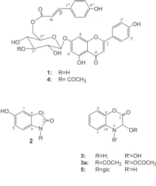
The HRCI-MS of 1 showed an M++1 at m/z 579.151 for
the molecular formula C30H26O12. Physical and spectral data
of 1 were identical with those reported for apigenin O-β
-D-(6”-trans-p-coumaroyl)-glucoside isolated from
Pogostemon cablin.9 However, Singh et al in 1986 reported
the same compound as a novel product from Echinops
echinatus under the name echinacin.10 Based on COSY
and HMQC experiments, complete assignments for the sugar protons and all carbons were achieved. Our assignments were in complete agreement with first
publication.9 However, some of the 13C-NMR assignments
in the latter publication10 must be revised.
The HRCI-MS (M++1 at m/z 621.161 for the molecular
formula C32H28O13) and other spectral data of 4 (see
experimental) indicated an additional acetyl group compared
with 1. Complete assignments of the sugar protons could be
achieved by a combination of COSY and HMQC experi-ments. A major difference was observed in the chemical shift of H-3″ (δ 4.97, t) compared to that of 1 (δ 3.30, m), indicating that the acetyl group is located at C-3″. Further evidence for
the position of the acetyl group was obtained from the 13
C-NMR, where a substantial downfield shift ∆δC-3” = 1.20 ppm
and the upfield shift of C-2″ and C-4″ (∆δC-2” =1.66 ppm and
(∆δC-4” = 1.67 ppm) relative to those of 1 were observed. 11 A
literature search revealed that 4 (apigenin 7-O-β-D(6″-
trans-p-coumaroyl)3″-O-acetylglucopyranoside) is a previously
unreported natural compound.
The new alkaloid 2 gave positive reactions with FeCl3,
nitrogenous nature. HREI-MS (experimental) showed an
M+ at m/z 151.027 for the molecular formula C
7H5O3N. It
also showed characteristic mass fragments at m/z 105, 95,
91 and 77, suggesting the presence of 2-benzoxazolinone.12
In the 13C-NMR spectrum (Table 1) the seven carbons were
resolved as three aromatic methines (δ 99.1, 111.0 and
111.6) assigned to C-7, C-5 and C-4 respectively, three
quanternary aromatic carbons (δ 129.9, 146.1 and 154.8)
attributed to C-9, C-8 and C-6 respectively, and a fourth
quaternary carbon at δ 157.7 assigned to the lactone
carbonyl. The 1H-NMR spectrum (Table 1) showed an ABX
system at δ 6.68 (1H, d, J 2.3 Hz), 6.85 (1H, d, J 8.5 Hz) and
6.60 (1H, dd, J 2.3, 8.5 Hz) indicating a trisubstituted
aromatic system. Compound 2 was identified as the
demethyl derivative of 6-methoxy-benzoxazolinone i.e. 6-hydroxy-benzoxazolinone. Even the known 6-methoxy-benzoxazolinone is a rare natural product isolated only
once from Corn plants.13
Chemical reactions and solubility in NaOH, followed
by recovery after acidification, suggested that 3 is a
nitrogenous compound with a lactone function. This was confirmed by the IR absorption bands for carbonyl
(1670 cm-1), hydroxy and/or NH groups (3325 cm-1). The
HRCI-MS showed an M++1 at m/z 182, and an M+ at m/z
181.144 for the molecular formula C8H7O4N. The 13C-NMR
spectrum (Table 1) showed five methine signals and three
quaternary carbons. In the 1H-NMR spectrum the coupling
pattern of the 4 aromatic protons suggested the presence
of an O-disubstituted benzene ring. In the 1H-NMR
spectrum the singlet at δ 5.68, diagnostic for a proton
flanked by two electronegative atoms, was assigned for
H-3. This was supported by a 13C-NMR signal which
appeared at δ 93.7 and was attributed to the oxymethine
carbon. The remaining three quaternary carbon absorptions
at δ 129.7, 142.5 and 160.1 were assigned to C-10, C-9
and the carbonyl group respectively. The violet colour
with FeCl314 as well as the M+ of the diacetate derivative
3a at m/z 265 indicated the presence of a hydroxylamine
group. The exact positions of the carbonyl and hydroxyl groups were established from alkaline hydrolysis and by
the inability of 3 to give O-aminophenol after fusion with
KOH. This clearly distinct the new alkaloid 3
(3,4-dihydroxy-1,4-benzoxazine-2-one) from blephari-genin (2-hydroxy-1,4-benzoxazine-3-one) a compound
with very close 1H-NMR data.15
The HRCI-MS of the third new alkaloid 5 showed M+
at m/z 327.095 for the molecular formula C14H17O8N. The
Table 1. 1H- and 13C-NMR spectral data of compounds 2, 3 and 5 (coupling constants in Hz)a
2b 3b 5c
# 1H 13C 1H 13C 1H 13C
2 - 157.7 160.1 - 162.6
3 - - 5.68 s 93.7 5.78 s 96.4
4 6.85 d J 8.5 111.6 - - -
-5 6.60 dd, J 2.3, 8.5 111.0 7.36 dd J 1.6,7.8 114.4 7.12 m 119.1
6 - 154.8 7.08 m 125.6 7.05 m 124.9
7 6.68 d J 2.3 99.1 7.08 m 123.8 7.05 m 124.2
8 - 146.1 7.01 dd J 1.6, 7.8 118.5 6.99 m 116.9
9 - 129.9 142.5 142.0
1 0 129.7 127.4
1 ‘ 4.70 d J 7.8 104.0
2 ‘ 3.28 m 74.9
3 ‘ 3.30-3.34 m 78.0
4 ‘ 3.30-3.34 m 71.1
5 ‘ 3.30-3.34 m 78.6
6 ‘ 3.83 d J 11.9 62.5
3.68 dd J 3.6, 11.9
a Assignments are based on HMQC experiments; b Spectra were measured in CD 3OD;
c Spectra were measured in CD
14 carbon signals were clear in the 13C-NMR spectrum
(Table 1), of which 8 signals were accounted for the aglycone part, while the remaining 6 carbon signals were assigned for the sugar moiety. The 1,4-benzoxazine-2-one
skeleton was assigned to 5 rather than the
1,4-benzoxazine-3-one as indicated from its reaction with KOH solution
and fusion test.15 The spectral data of 5 aglycone (Table 1
and Experimental) showed a close similarity to 3. However,
the CI-MS and the negative reaction with FeCl3 suggested
the absence of a hydroxyl group attached to nitrogen atom. Consequently, the only possible site for glycosylation is the C-3 hydroxyl. The identity of the sugar moiety was established as glucose by spectral evidences and by TLC comparison after acid hydrolysis. On the basis of the chemical shift and coupling constant of the anomeric
proton (J1‘,2‘7.8 Hz), the glucosidic linkage should have
the β-orientation. The identity of 5 was therefore
established as 1,4-benzoxazine-2-one-3-O-glucoside.
The known compounds were identified by direct comparison with reference materials (Aldrich).
Compounds 1 and 3-5 were subjected to antimicrobial
testing using 10 microorganisms. Only compounds 1 and
4 were active against Bacillus subtilis with an MIC 64 and
128 µg/mL respectively.
Experimental
General procedure
The CI-MS of 3a was measured on a Finnigan SSQ7000
mass spectrometer. NMR spectra were recorded on a JEOL
500 NMR instrument at 500 MHz for 1H and 125 MHz for
13C. Other experimental conditions were as previously
described.16
Plant material
The whole plants of Acanthus arboreus Forssk.
growing wild in Wadi Dhar, Sana’a, Yemen was collected during the flowering stage in August 1998 and was identified by Prof. Nabil El-Hadidy, Department of Plant Taxonomy, Faculty of Science, Cairo University. A voucher sample (YA1) is preserved in the Department of Pharmacognosy, Faculty of Pharmacy, University of Alexandria, Egypt.
Extraction and isolation
The air-dried powdered whole plant of Acanthus
arboreus Forssk (3.5 kg) were extracted by 95% ethanol at room temperature. The concentrated ethanolic extract was
partitioned between CHCl3 (1L) and water (1L). The CHCl3
fraction (80 g) was again partitioned between 90% MeOH (1L) and petroleum ether (1L). The aqueous fraction was
extracted with EtOAc (3x500 mL), then with n-butanol
(3x500 mL).
A sample (10 g) of the 90% methanolic extract (26 g) was chromatographed on silica gel column (200 g, 4 cm)
eluted with petroleum ether-CH2Cl2) (1:1) with increasing
content of CH2Cl2, then methanol. Fractions of 250 ml
each were collected, screened by TLC and similar fractions were combined. Fractions 6-15 (1.9 g, petroleum ether/
CH2Cl2 40:60) afforded lupeol (800 mg) upon
crystalliza-tion from petroleum ether. Crystallizacrystalliza-tion of fraccrystalliza-tions
16-20 (0.8 g, petroleum ether/CH2Cl2 25:75) from methanol
gave stigmasterol (100 mg). Fractions 36-42 (0.9 g, CH2Cl2/
MeOH 92.5:7.5) were rechromatographed on silica gel column (30 g, 1 cm) eluted with a mixture of EtOAc/MeOH with increasing proportion of MeOH. Fractions of 50 ml each were collected. Fractions 5-10 were further purified
by PTLC on silica gel plates developed with CHCl3/MeOH
(9:1)(double development) and the zone with an Rf value
of 0.54 was scraped off, eluted with a mixture of chloroform
and methanol (1:1) to afford 1 (160 mg). Fractions 43-47
(1.1 g, CH2Cl2/MeOH 90:10) gave sitosterol glucoside
(230 mg) on crystallization from methanol.
The EtOAc extract (12 g) was fractionated on silica gel
column (400 g, 3 cm) eluted with CH2Cl2 and CH2Cl2/
MeOH mixtures with gradual increase of methanol content. Ninety fractions (150 mL each) were collected. Repeated
crystallization of fraction 12 (0.85 g, CH2Cl2/MeOH 98:2)
from methanol afforded vanillic acid (200 mg).
Crystallization of fractions 25-29 (0.78 g, CH2Cl2/MeOH
97:3) from methanol afforded apigenin (10 mg); while PTLC of the supernatant on silica gel plates developed
with CHCl3/MeOH (8:2) gave 5 mg of 2 (Rf= 0.58).
Fractions 36-45 (2.1 g, CH2Cl2/MeOH 96:4) were
crystallized from methanol to give 3 (850 mg) (Rf = 0.34
EtOAc/MeOH/H2O 30:5:2). Crystallization of fractions
49-55 (1.6 g, CH2Cl2/MeOH 94:6) from methanol gave 4
(620 mg) (Rf = 0.30 EtOAc/MeOH/H2O 30:5:2). Additional
quantity of 1 (210 mg) was obtained by crystallization of
fractions 78- 81(1.4 g, CH2Cl2/MeOH 90:10) from MeOH.
A sample (8 g) of the n-butanol extract (30 g) was
chromatographed of a silica gel column (160 g, 2 cm)
eluted with CH2Cl2 and CH2Cl2/MeOH mixtures with a
gradual increase of methanol content. Twenty five fractions (100 mL each) were collected. Fractions 13-16 (0.7 g,
CH2Cl2/MeOH 80:20) afforded 5 (120 mg) (Rf = 0.47
EtOAc/MeOH/H2O 30:5:4) on crystallization from MeOH.
Apigenin 7-O-β-D-(6″-trans-p-coumaroyl) glucoside
CI-MS m/z (rel. Int.): 579 (6, [M+1]+), 489 (2), 433 (4), 417
(16), 416 (8), 350 (2), 311 (6), 309 (18), 299 (30), 271 (100), 270 (25) 192 (8), 165 (78), 147 (40), 121 (18), 99
(5). ). HRCI-MS m/z 579.151 (M++1), calcd for C
30H26O12,
579.150. 1H-NMR of sugar protons (ppm, DMSO-d
6) δ 3.30
(1H, m, H-3”), 3.32 (1H, m, H-4”), 3.36 (1H, m, H-2”), 3.83 (1H, dd, J 7.8, 9.4 Hz, H-5”), 4.17 (1H, dd, J 4.8, 10.3 Hz,
H-6”b), 4.45 (1H, d, J 10.3 Hz, H-6”a), 5.16 (1H, d, J 8.0
Hz, H-1”). 13C-NMR of sugar carbons (ppm, DMSO-d
6) δ
63.4 6”), 70.0 4”), 72.9 2”), 73.8 5”), 76.2 (C-3”), 99.5 (C-1”).
6-Hydroxy-2-benzoxazolinone (2). Yellowish white
crystals, mp 260 oC, UV λ
max/nm (MeOH): 340, 305, 302,
268. EI-MS m/z (rel. Int.) 152 (6, [M+1]+), 151 (25, M+),
149 (38), 142 (28), 130 (5), 122 (3, [M-(HCO)]), 117 (9),
107(5) 105 (21), 95 (21, [M+ -2CO]), 91 (14), 84 (22), 77
(14), 66 (30), 55 (38). HREI-MS m/z 151.027 (M+); Calc.
for C7H5O3N, 151.026. 1H- and 13C-NMR (Table 1).
4-Hydroxyacanthamine (3,4-dihydroxy-1,4-benzoxazine-2-one) (3). Yellow needles, mp 135oC. IR
(KBr): νmax/ cm
-1 : 3325 (OH), 3050, 2823, 1670 (CO), 1550,
1345. UV λmax/nm (MeOH): 290, 279, 253. EI-MS m/z (rel.
Int.): 182 (46, [M+1]+), 181 (14, M+), 165 (12), 164 (100,
[M+-OH]), 136 (25), 108 (10). HRCI-MS m/z 181.144 (M+);
Cald. for C8H7O4N, 181.145. 1H-and 13C-NMR (Table 1).
Alkaline hydrolysis of 3.A sample (5 mg) of 3 was dissolved in MeOH/3N NaOH (1:1) and heated for fifteen minutes on water bath. The resulting solution was first extracted with EtOAc, acidified with dil. HCl and extracted with EtOAc. The EtOAc fraction, after acidification, showed
a TLC spot with same Rf value of material 3.
Potassium hydroxide fusion test of 3. A mixture of 3
(3 mg) and KOH (15 mg) was fused in an oil bath for 30 min. The reaction mixture was then allowed to cool, diluted with water and filtered. The filtrate was neutralized with dil. HCl and extracted with EtOAc. TLC revealed that the EtOAc extract resulting from the KOH fusion test was
devoid of an ortho-aminophenol spot.
Acetylation of 3. A sample (4.0 mg) of 3 in pyridine
(2.0 mL) was treated with Ac2O (0.2 mL) for 24 h at room
temperature. Evaporation of the mixture under a stream of
nitrogen yielded chromatographically homogeneous 3a
(4.0 mg). CI-MS m/z (rel. Int.): 266 (4, [M+1]+), 265 (10,
M+), 223 (16), 206 (100) 164 (169), 136 (20), 79 (5).
Apigenin 7-O-β-D-(6″-trans-p-coumaroyl)3″ -O-acetylglucopyranoside (4). Pale yellow crystals, mp 223oC.
IR (KBr): νmax/cm
-1: 3395 (OH), 3050, 2820, 1670 (CO),
1560, 1320. UV λmax/nm (MeOH): 316, 268, (NaOMe) 368,
300, 272, (AlCl3), 375, 325, 319, 298, 276, (AlCl3/HCl)
375, 325, 318, 298, 276, (NaOAc) 382, 318, 268. CI-MS
m/z (rel. Int.): 621 (4, [M++1]), 517 (1), 475 (2), 417 (3),
414 (3), 351 (51), 313 (7), 299 (15), 271 (52), 267 (6), 187
(13), 165 (35), 121 (100), 99 (18), 61 (84), HREIMS m/z
621.161 (M++1); Calç. for C
32H28O13, 621.160.
1H-NMR
(ppm, DMSO-d6): δ 2.07 (3H, s, COCH3), 3.48 (1H, m,
H-2”), 3.98 (2H, m, H-4”, 5”), 4.20 (1H, dd, J 5.3, 11.7 Hz,
H-6”b), 4.43 (1H, H-6”a), 4.97 (1H, t, J 9.6 Hz, H-3”), 5.30
(1H, d, J 7.8 Hz, H-1”), 6.35 (1H, d, J 16 Hz, H-α), 6.50 (1H,
d, J 1.9 Hz, H-6), 6.66 (2H, d, J 8.5 Hz, H-3”’, 5”’), 6.84
(1H, d, J 1.9 Hz, H-8), 6.85 (1H, s, H-3), 6.92 (2H, d, J 8.9 Hz, H-3’, 5’),7.39 (2H, d, J 8.5 Hz, H-2”’, 6”’), 7.49 (1H, d, J 16 Hz, H-β), 7.94 (2H, d, J 8.9 Hz, H-2’, 6’). 13C-NMR
(ppm, DMSO-d6): δ 21.1 (COCH3), 63.3 (C-6”), 68.3 (C-4”), 71.3 (C-2”), 73.9 (C-5”), 77.4 (C-3”), 95.1 (C-8), 99.8 (C-1”,
C-6), 103.4 (C-3), 105.9 (C-10), 114.0 (C-α), 115.9 (C-3”’,
5”’), 116.2 (C-3’, 5’), 121.4 (C-1’), 125.4 (C-1”’), 128.8
(C-6’), 130.3 (C-2”’, 6”’), 145.4 (C-β), 157.3 (C-9), 160.1
(C-4’), 161.4 (C-4”’), 161.7 (C-5), 162.8 (C-7), 164.7
(C-2), 166.8 (C=O), 170.2 (COCH3), 182.3 (C-4).
Acanthaminoside (1,4-benzoxazine-2-one-3-O-glucoside) (5). Colourless needles, mp 213-214 oC. IR (KBr)
νmax/cm
-1: 3475 (OH), 3125, 2960, 1700 (CO), 1600, 1375.
UV λmax/nm (MeOH): 283, 275, 250. CI-MS m/z (rel. Int.):
328 (14, [M+1]+), 327 (14, M+), 166 (100), 165 (15.5), 164
(16, aglycone), 163 (13, glucosyl), 148 (100); 145 (30),
136 (18). HRCI-MS m/z 327.095 (M+), calcd for C
14H17O8N,
327.095. 1H-and 13C-NMR (Table 1).
Acid hydrolysis of 5.A sample (6 mg) of 5 was dissolved in methanol (10 mL)/2N HCl (1 mL) and heated under reflux for 1 hour. After cooling, the aqueous solution was extracted with EtOAc (3x5 mL) and neutralized with 5%
Na2CO3. TLC identified the sugar in the aqueous layer as
glucose using CHCl3/MeOH (6:4) as developing system
and thymol/H2SO4 as spray reagent.
Antimicrobial testing
The MIC were determined for compounds 1 and 3-5
using a procedure described elsewhere.17 Twelve
microorganisms: Bacillis subtilis, Micrococcus luteus,
Sarcina lutea, Staphylococcus aureus, Bordetella bronchiseptica, Eschirichia coli, Klebsiella pneumoniae, Proteus mirabilis, Pseudomonas aeruginosa, Salmonella typhi, Serratia marcescens and Shigella sonnie were used in the study. The antibiotics ampicillin, ciprofloxacin, erythromycin and gentamicin were used as controls.
Acknowledgments
References
1. Airy Shaw, H.K.; Willis, J.C.; A Dictionary of the Flowering
Plants and Ferns, 8th ed., Cambridge University Press:
Cam-bridge, 1973.
2. Reunaud, J.; Couble, A.; Rayanaud, J.; Pharmazie1988, 43, 378.
3. D’Souza, L.; Wahidulla, S.; Mishra, P.D.; Ind. J. Chem. Sect.
B: Org. Chem. Incl. Med. Chem.1997, 36, 1079.
4. Anam, E.M.; Ind. J. Chem. Sect. B: Org. Chem. Incl. Med.
Chem.1997, 36, 109.
5. Kokpol, U.; Chittawong, V.; J. Nat. Prod.1986, 49, 355. 6. Sam, J.; Valantine, J.L.; J. Pharm. Sci. 1969,58, 1043. 7. Sam, J.; Plampin, J.N.; J. Pharm. Sci.1964, 53, 538. 8. Kapil, A.; Sharma, S.; Wahidulla, S.; Planta Med.1994, 60,
187.
9. Itokawa, H.; Suto, K.; Takeya, K.; Chem. Pharm. Bull.1981,
29, 254.
10. Singh, K.N.; Pandey, V.B.; Banerjee, S.; Bohlmann. F.; Keinan,
E.; Chem. Ind.(London, U. K.)1986, 20, 713.
11. Markham, K.R.; Temai, B.; Stanley, R.; Geigen, H.; Mabry, T.J.; Tetrahedron1978, 34, 1389.
12. Thomson, M.L.; DeJongh, D.C.; Can. J. Chem. 1973, 51, 13313.
13. Sissman, E.E.; LaPidus, J.B.; Beck, S.D.; J.Am. Chem. Soc.
1957, 79, 4697.
14. Fieser, L.F.; Williamson, K.L.; Organic Experiments, 4th ed.,
D.C. Health and Company, Washington DC, 1979.
15. Chatterjee, A.; Basa, S.C.; Chem. Ind. (London, U. K.)1969,
15, 328.
16. Abdel-Kader, M.S.; Wisse, J.; Evans, R.; Werff, H.; Kingston, D.G.L.; J. Nat. Prod. 1997, 60, 1294.
17. Lorian, V.; Antibiotics in Laboratory Medicine, William’s and Wilkin’s: London, 1980.