Article
Printed in Brazil - ©2017 Sociedade Brasileira de Química 0103 - 5053 $6.00+0.00
*e-mail: [email protected]
Photoelectroanalytical Detection of Adrenaline Based on DNA and TiO
2Nanoparticles
Sensitized with Bis(ethylenedithio)tetrathiafulvalene Exploiting LED Light
Thiago Augusto D. Santos, Sakae Yotsumoto Neto, Cleidivan S. Macena, Rita de Cássia S. Luz and Flávio S. Damos*
Departamento de Química, Universidade Federal do Maranhão, 65080-805 São Luís-MA, Brazil
In this study, a photoelectroanalytical sensor for determination of adrenaline based on deoxyribonucleic acid (DNA) and anatase titanium dioxide (TiO2) nanoparticles sensitized with bis(ethylenedithio)tetrathiafulvalene (BEDT-TTF) was developed, which we henceforward call BEDT-TTF/DNA/TiO2/ITO. The photoelectroanalytical sensor showed high photocurrent to adrenaline under visible light emitting diode (LED) light irradiation in comparison to each component of the composite material. Under optimized conditions, the BEDT-TTF/DNA/TiO2/ITO sensor shows a linear response range from 10 nmol L-1 up to 100 µmol L-1 with a sensitivity of 8.1 nA L µmol-1 and limit of detection of 1 nmol L-1 for the detection of adrenaline. The photoelectrochemical sensor showed high photocurrent to adrenaline in comparison to photocurrent response to ascorbic acid and uric acid. The BEDT-TTF/DNA/TiO2/ITO photoelectrochemical sensor was successfully applied to urine samples, with recovery values between 96 and 106%.
Keywords: adrenaline, photoelectrochemical sensor, visible LED light, deoxyribonucleic acid
Introduction
Adrenaline, also known as epinephrine, is one of the most important message transfer compound in the mammalian central nervous system, which exist as an organic cation in the nervous tissue and the biological body
fluid.1 The level of adrenaline in the body affects a series
of actions of the nervous system including the regulation of blood pressure, heart rate, lipolysis, immune response
and glycogen metabolism.2 In addition, adrenaline has been
advocated during early cardiopulmonary resuscitation, early defibrillation and early advanced care in cardiac
arrest for decades.3
Therefore, the quantification of adrenaline in biological samples is of high interest nowadays. Urine represents one of the most easily attainable and, consequently, very common samples in adrenaline clinical analysis and diagnostics. The levels of adrenaline in biological fluids such as urine depends on the age and condition of the
patient.4 For healthy people, for example, the physiological
concentration of adrenaline found in urine samples are in
nanomolar level (22-109 nmol L-1).5
A number of analytical methodologies have been proposed for determination of adrenaline such as
fluorimetric,6 spectrophotometric,7 enzymatic
radio-immunoassay,8 gas chromatography,9 high performance
liquid chromatography,10 capillary electrophoresis11 and
electrochemical methods.12-15 Despite the high number of
analytical methods for adrenaline determination, most of them suffer from some disadvantages such as high cost, long analysis time, extensive sample pretreatment based on derivatization, extraction and purification as well as demands for highly trained users. The fluorometric and radioenzymatic assays are presently the most widely used techniques for the estimation of plasma, urine and tissue adrenaline. The fluorometric assay lacks specificity and sensitivity. The radioenzymatic assay is significantly more sensitive and specific but is technically very complex, time
consuming and expensive.12
On the other hand, the electrochemical methods are cheaper, simple and fast in comparison to several analytical methods. However, the most of electrochemical sensors for adrenaline show low linear response range or lack with the limit of detection and sensitivity necessary to adrenaline
detection in biological samples.13-15
Nowadays, the photoelectroanalytical devices have emerged as a potentially useful and sensitive system
with advantage of high linear response range.16-18 The
material. Since the excitation source and detection apparatus are from distinct nature, the photoelectroanalytical devices
show high signal-to-noise ratio.18,19
The anatase titanium dioxide (TiO2) has attracted
high attention in the development of photocatalytic and photoelectrochemical areas due to its nontoxicity,
hydrophilicity, low cost and stability against photocorrosion.19
In addition, the anatase TiO2 shows a band gap with a quite
deep valence band allowing that generated holes tend to locates on the surface of the particle, which makes it active
to be harvested by free electrons from molecules.19
However, anatase TiO2 shows some limitations as
photocatalyst including the recombination of photo-generated charge carriers and wide bandgap (3.22 eV)
limiting anatase TiO2 to almost only ultraviolet (UV) light
absorption.20 The wide bandgap of anatase TiO
2 limits
their potential applications in photoelectroanalytical fields
because UV light may damage the biologic samples.21
A number of strategies have been proposed to improve the
photoelectrochemical performance of TiO2 under visible light
excitation including the doping with transition-metal ions or
oxygen defects into TiO2 lattice as well as dye sensitization.22,23
Thus, the anatase TiO2 has been sensitized with several
compounds in order to make the titanium oxide properties more attractive to development of photoelectroanalytical
devices including phthlocyanines,24,25 conducting polymers,26
porphyrins,27 lithium tetracyanoethylenide,28 quantum dots,29
among others.
Tetrathiafulvalene is a strong π-electron donor which
has attracted particular attention in the development
of chemical sensors,30 superconducting materials,31
ferromagnets,32 non-linear optic devices,33 biofuel cell,34
and recently in development of dye sensitized solar cells.35
The aim of the present work is the development of a novel photoelectrochemical sensor based on indium tin oxide
modified with anatase TiO2 nanoparticles sensitized with
bis(ethylenedithio)tetrathiafulvalene (BEDT-TTF/TiO2)
for adrenaline detection with visible light emitting diode (LED) light. The photoelectroanalytical sensor showed a wide linear range, good stability and selectivity to adrenaline. To the best of our knowledge, this is the first photoelectrochemical sensor for adrenaline determination exploiting the interaction between BEDT-TTF and anatase
TiO2 nanoparticles under visible LED light.
Experimental
Materials
All chemicals were of analytical grade and used as received without any further purification steps.
Bis(ethylenedithio)tetrathiafulvalene (BEDT-TTF), double-stranded deoxyribonucleic acid (ds-DNA), adrenaline and indium tin oxide coated glass slides (ITO) were purchased from Sigma-Aldrich (Saint Louis, USA). 2-[4-(2-Hydroxyethyl)piperazin-1-yl]ethanesulfonic acid (HEPES), boric acid, citric acid, phosphoric acid, disodium
and monosodium phosphates (Na2HPO4 and NaH2PO4),
were acquired from Synth, São Paulo, Brazil. All solutions were prepared with water purified in an OS100LXE system from GEHAKA Company (Gehaka Ltd., São Paulo, SP, Brazil). The actual pH of the buffer solutions was determined with a Quimis pH/Ion Analyser Q400AS model.
The anatase TiO2 nanopowder with nominal particle size
of 25 nm were acquired from Sigma-Aldrich (Saint Louis, USA). The double-stranded template deoxyribonucleic
acid with UV absorbance ratio (A260nm/A280nm) of 1.9 were
isolated from the calf thymus. The double strand DNA used in present work is a double helix of a chain of nucleotides constituted of about 41.9 mole% G-C (guanine-cytidine) and 58.1 mole% A-T (adenosine-thymine).
Apparatus
Photoelectroanalytical measurements were carried out with an Autolab PGSTAT 128N potentiostat/galvanostat from Metrohm Autolab B.V., Utrecht, the Netherlands controlled by GPES software. All photoelectrochemical measurements were carried out in a three-electrode system positioned in a box, which was used for control of the light incidence in the photoelectrochemical cell. The photoelectrochemical measurements were performed with a cheap commercial 20 W white LED light as the source of irradiation energy with emission between 380 and 680 nm. The bare or
modified SnO2/In2O3 coated glass slide (ITO) with surface
resistivity of 8-12 Ω sq-1 was used as the working electrode.
The ITO photoactive area was 0.5 cm2. The ITO glass slides
were of 1.1 mm of thickness and nominal transmittance of 84% (nominal at 550 nm). The photoelectrochemical measurements were performed by front side illumination.
A Pt wire was used as counter electrode and Ag/AgCl(saturated)
was used as reference electrode.
Construction of the BEDT-TTF/DNA/TiO2
photoelectro-chemical sensor
First, an ITO coated glass slide was sonicated and copiously washed with ethanol and water to remove any adsorbed species. After that, a suspension was prepared
by mixing 50 mg of anatase TiO2 nanoparticles and 5 mg
solid washed with water and it was let to dry at 70 ºC for
2 h to give BEDT-TTF/TiO2 composite.
Then, 5 mg of the BEDT-TTF/TiO2 composite
was dispersed in aqueous solution of single-stranded DNA (ss-DNA), and the resultant mixture was stirred for 1 h. The ss-DNA solution was prepared heating ds-DNA solution at 90 °C for 2 h. After cooling to room temperature, the resulting materials were then centrifuged and washed three times with distilled water to remove excess ss-DNA. The as-prepared
BEDT-TTF/DNA/TiO2 composite was obtained.
Then, 1.0 mg of the BEDT-TTF/DNA/TiO2 composite
was mixed with 50 µL of water with the aid of sonication for 10 min. Finally, 10 µL of this suspension was placed directly onto the photoelectrode substrate and allowed to dry at 50 ºC for 10 min to form BEDT-TTF/ds-DNA/
TiO2/ITO photoelectrochemical sensor. The amount of
BEDT-TFF/DNA/TiO2 dissolved in water was chosen
taking into account the capability of the paste to cover ITO surface and produce more stable films. Films prepared with
amounts of BEDT-TFF/DNA/TiO2 composite higher than
1 mg dissolved in water produced surfaces that are more fragile. On the other hand, films prepared with amounts of composite lower than 1 mg did not produce films with homogenous surfaces.
Preparation and analysis of urine samples
Urine samples were collected into the plastic tubes
and 6 mol L-1 HCl was added to each one to give solutions
with 1% of HCl.36 The acidified samples were centrifuged
for 30 min at 2500 rpm.37 The supernatant was filtered and
then diluted 10-times with the phosphate buffer solution (pH 7.0). The solution was transferred into the voltammetric cell to be analyzed with the photoelectroanalytical sensor in three replicates. The standard addition method was used for the determination of adrenaline in real samples.
Results and Discussion
Electrochemical characteristics of the BEDT-TTF/DNA/TiO2
composite photoelectrochemical sensor
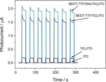
Figure 1 shows the amperometric response of 1 mmol L-1
of adrenaline (ADR) in 0.1 mol L-1 phosphate buffer
solution (pH 7.0) on ITO, TiO2/ITO, BEDT-TTF/TiO2/ITO,
BEDT-TTF/TiO2/ITO and BEDT-TTF/DNA/TiO2/ITO
under LED light on and off. The values of the photocurrents to each one of photosensors previously presented were 0.0011 ± 0.0002, 0.17 ± 0.01, 1.69 ± 0.05 and 1.91 ± 0.02 µA, respectively.
As can be seen, the BEDT-TTF/DNA/TiO2/ITO shows
higher mean value of the photocurrent as well as presented lower standard deviation to seven measurements of
photocurrent. The BEDT-TTF/DNA/TiO2/ITO photosensor
presents a photocurrent to adrenaline about 11-fold higher
than that to TiO2/ITO, indicating the high performance of the
BEDT-TTF/DNA/TiO2/ITO photosensor in photo-generate
electrons and holes improving the photoelectrochemical efficiency. It is probable that the capability of DNA to form stable biocompatible films can improve the stability and sensitivity of the platform. In addition, the negative character of DNA due to phosphate groups in their chains can favor the interaction between the photocatalyst and adrenaline molecule, since the last is positively charged at physiological pH.
Figure 2 shows schematic diagram for the proposed mechanism to photoelectrochemical oxidation of adrenaline
on BEDT-TTF/DNA/TiO2/ITO. Under white LED light
irradiation, the BEDT-TTF complex adsorbed on anatase
TiO2 nanoparticles surface can absorb the LED light such
as electrons of the dye are excited from highest occupied molecular orbital (HOMO) to the lowest unoccupied molecular orbital (LUMO) state since the BEDT-TTF complex shows an absorption band at visible region of the electromagnetic spectrum with maximum at about 460 nm justifying its ability to harvest photons from LED
light.38 The excited electron can then be transferred to the
conduction band of the TiO2 and finally be transferred to
ITO electrode to originate the photocurrent.
On the other hand, the addition of adrenaline to photoelectrochemical cell can improve the spatial separation
of photogenerated charges such as the holes from TiO2 can
localize on adrenaline and electrons localize within the
lattice of TiO2, which suppress the recombination of charges and consequently improve the photoelectrochemical performance of the composite to adrenaline oxidation. Thus, a higher response to photoelectrochemical adrenaline oxidation is achieved, when more surface complexes are
formed between the adrenaline and TiO2.
After that, the electron in LUMO state of dye is
transferred to the conduction band of anatase TiO2
nanoparticles and the holes in HOMO state promote the adrenaline oxidation. In addition, adrenaline can act as a
scavenger of holes generated at TiO2 nanoparticles improving
the spatial separation of charges at valence and conduction
bands of TiO2 nanoparticles. Therefore, the response of the
photosensor depends on adrenaline concentration.
Optimization of the BEDT-TTF/DNA/TiO2
photoelectro-analytical sensor response
The influences of buffer solution, solution pH and
applied potential on the BEDT-TTF/DNA/TiO2 sensor
response were evaluated in order to found the best experimental conditions for adrenaline determination. In order to evaluate the photocurrent response, the amperometric response was recorded while the LED light was turned on and off. Initially, the effects of solution buffer
on the photocurrent of the BEDT-TTF/DNA/TiO2/ITO
obtained for adrenaline was investigated. Figure 3 shows
the response of the BEDT-TTF/DNA/TiO2 modified
photoelectrode to adrenaline in different buffer solutions, such as, phosphate, HEPES and Britton-Robinson at
0.1 mol L-1 and pH 7.
The values of photocurrents to 100 µmol L-1 of adrenaline
on BEDT-TTF/DNA/TiO2 modified photoelectrode in
phosphate, HEPES and Britton-Robinson buffers were 0.80 ± 0.04, 0.61 ± 0.02 and 0.45 ± 0.02 µA, respectively. The best response to adrenaline at phosphate buffer solution may be due to the high ionic mobility of the phosphate
and potassium ions making possible a better charge transportation in solution, which may favor the better charge compensating during the adrenaline oxidation. In this sense, the phosphate buffer solution was chosen for further experiments.
After that, the response of the BEDT-TTF/DNA/TiO2
photoelectrochemical sensor to adrenaline was investigated in phosphate buffer solution with pH ranging from 5.0 up
to 8.0 under an applied potential of 0.25 V vs. Ag/AgCl(sat)
(Figure 4).
The phototocurrent to adrenaline oxidation on
BEDT-TTF/DNA/TiO2/ITO increased from pH 5.0 up to
pH 7.0. The changes in the solution pH could significantly affect the relative energies of the sensitizer excited states and the ITO acceptor states, and accordingly sensitized photocurrents. It is possible that the LUMO energy levels
Figure 2. Proposed mechanism for the photoelectrochemical detection of adrenaline.
Figure 3. Influence of buffer solution on the photosensor response for 100 µmol L-1 of adrenaline. Measurements were carried out in 0.1 mol L-1 phosphate buffer solution. Eapp = 0.25 V vs. Ag/AgCl.
of the dye increase with increasing solution pH, being favorable to the anodic photocurrent generation. After
that, the response of the BEDT-TTF/DNA/TiO2/ITO to
adrenaline decreased from pH 7.0 until pH 8.0 (Figure 4), which can be due to the adrenaline decomposition at higher pH values. Therefore, all subsequent measurements were carried out in phosphate buffer solution at pH 7.0.
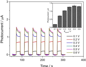
The applied potential was varied from 0.1 up to 0.6 V in order to evaluate the effect of the electric potential on the photosensor response to adrenaline (Figure 5). The photoelectrochemical response of BEDT-TTF/DNA/
TiO2/ITO to adrenaline were 0.21 ± 0.01, 0.72 ± 0.01,
1.04 ± 0.02, 1.27 ± 0.01, 1.34 ± 0.05 and 1.30 ± 0.03 µA to applied potential of 0.1, 0.2, 0.3, 0.4, 0.5 and 0.6 V, respectively.
As can be seen, the photoelectrochemical response of
BEDT-TTF/DNA/TiO2/ITO to adrenaline showed a high
increase until an applied potential of 0.4 V. Therefore, the measurements with the proposed photoelectrochemical
sensor were carried out in 0.1 mol L-1 phosphate
buffer solution at pH 7.0 under an applied potential of
0.4 V vs. Ag/AgCl(sat) for all subsequent determinations
of adrenaline.
Analytical performance of the BEDT-TTF/DNA/TiO2/ITO
sensor
Figure 6 shows the amperometric response of
BEDT-TTF/DNA/TiO2/ITO sensor under an applied
potential of 0.4 V to successive additions of adrenaline into
0.1 mol L-1 in phosphate buffer solution (pH 7.0) under light
off and on, respectively.
The photoelectrochemical sensor showed a linear
response range to adrenaline from 10 nmol L-1 to 100 µmol L-1,
which can be expressed according to equation 1 (inset of Figure 6):
Iphotocurrent (nA) = (25 ± 3) + (8.1 ± 0.1)[Adrenaline] (µmol L-1) (1)
with a correlation coefficient of 0.999 (for n = 7).
A detection limit (LOD) of 1 nmol L-1 was determined
using the equation LOD = 3 σbl/slope, where σbl is the
standard deviation of the blank response which is obtained from 10 replicate measurements of the blank solution and slope is the sensitivity of the analytical photosensor. The linear range of response and limit of detection were analyzed in comparison to electrochemical sensors to
adrenaline reported in the literature (Table 1).39-51
As can be seen, the BEDT-TTF/DNA/TiO2/ITO
photoelectroanalytical sensor shows a linear range of response of four orders of magnitude and a limit of
detection of 1 nmol L-1, which were as good as the best
electrochemical sensors to adrenaline. The precision of
measurements using the BEDT-TTF/DNA/TiO2/ITO
photoelectroanalytical sensor was investigated from intra-day and inter-day repeatability studies. The relative
standard deviation for 10 determinations of 100 µmol L-1
adrenaline carried out in the same working day was 3.0%. The inter-day reproducibility was performed by comparing the analytical response of the photosensor for
10 determinations of 100 µmol L-1 adrenaline. The relative
standard deviation of photosensor response for adrenaline at ten different days was 5.3%. This set of results indicates a high precision in terms of repeatability and reproducibility of the measurements obtained using the BEDT-TTF/DNA/
TiO2/ITO photoelectroanalytical sensor.
Figure 5. Influence of applied potential on the photocurrent for 100 µmol L-1 of adrenaline. Measurements were carried out in 0.1 mol L-1 phosphate buffer solution.
In order to evaluate the selectivity of proposed photosensor, the influence of potential interfering agents commonly existing in the human urine was investigated (Figure 7).
Thus, the effects of substances frequently found in urine samples, such as ascorbic acid, uric acid, urea, glucose, folic acid and barbituric acid on the response of
BEDT-TTF/DNA/TiO2/ITO photoelectroanalytical sensor
were investigated. Solutions of 100 µmol L-1 of these
compounds were freshly prepared under the same conditions
of adrenaline (0.1 mol L-1 phosphate buffer solution, pH 7.0)
at four different concentrations. The photocurrent response of foreign species was monitored and compared with the signal obtained to adrenaline. The variation in the photosensor response was evaluated by amperometry under dark/light
conditions at +0.4 V vs. Ag/AgCl. It was interesting to note
that high concentrations of these foreign compounds showed very low photoelectrochemical response under the same conditions of adrenaline (Figure 7).
The stability of the photoelectrochemical sensor evaluated by successive measurements of the photocurrent
of the BEDT-TTF/DNA/TiO2/ITO sensor to 100 µmol L-1
adrenaline in 0.1 mol L-1 phosphate buffer solution at
pH 7.0. After 100 measurements the photocurrent shows a decrease of only 6% in respect to the first measurement of the photocurrent.
Application of BEDT-TTF/DNA/TiO2/ITO
photoelectro-analytical sensor in urine samples
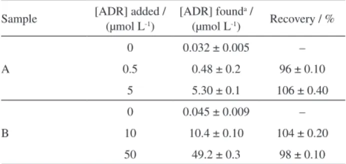
The standard addition method was applied for analysis of human urine samples spiked with adrenaline for evaluation of the practical usefulness of proposed photoelectrochemical sensor. The average results of three replicate measurements of adrenaline with the
BEDT-TTF/DNA/TiO2/ITO photoelectroanalytical sensor
are summarized in Table 2.
Table 1. Comparison of some analytical parameters of sensors for determination of adrenaline
Electrode Technique Linear range / (µmol L-1) LOD / (µmol L-1) Reference
DTT-DDT/AuNP/AuE CV 0.1-8 0.06 39
Pt-AuNPs/GCE DPV 63-400 57 40
PtNP/BMI.PF6/LAC/CPE SWV 0.99-210 0.29 41
Poly(methyl-Py)/GCE SWV 0.75-200 0.17 42
P(tau)/GCE DPV 2-600 0.3 43
PR/PIGE DPV 3-90 0.8 44
Au/PP/GCE DPV 0.3-21 0.03 45
PP/MWCNT/GCE DPV 0.1-8 0.04 46
CHIT/IL/SWCNT/GCE DPV 1-580 0.09 47
IL/CNT/CPE DPV 0.3-450 0.09 48
HT/MWCNT/GCE DPV 0.078-0.2 0.02 49
CuFe2O4/ILs/CPE SWV 0.1-400 0.07 50
BDDFE SWV 0.7-60 0.21 51
BEDT-TTF/DNA/TiO2/ITO PEC 0.01-100 0.001 this work
LOD: limit of detection; DTT: dithiothreitol; DDT: dodecanethiol; AuNP: gold nanoparticle; CV: cyclic voltammetry; GCE: glassy carbon electrode; DPV: differential pulse voltammetry; PtNP: platinum nanoparticle; BMI.PF6: ionic liquid 1-butyl-3-methylimidazolium hexafluorophosphate; LAC: laccase; CPE: carbon paste electrode; SWV: square-wave voltammetry; Poly(methyl-Py): polymethoxyphenol; P(tau): polytaurine; PR: polyrutin; PIGE: paraffin-impregnated graphite electrode; PP: polypyrrole; ; MWCNT: multi-walled carbon nanotube; CHIT: chitosan; ; IL: ionic liquid; SWCNT: single-walled carbon nanotube; HT: hematoxylin; BEDT-TTF: bis(ethylenedithio)tetrathiafulvalene.
Figure 7. Photocurrent obtained for proposed photoelectrochemical sensor
The recovery values between 96 and 106% using
the BEDT-TTF/DNA/TiO2/ITO photoelectroanalytical
sensor indicate that there are no significant interferences of matrix of the human urine as well as that the method is sufficiently accurate and suitable for quantification of adrenaline. Taking into account that the proposed sensor exhibited wider linear response range and lower LOD compared to previously reported electrochemical sensors, it is cost effective and exhibits satisfactory applicability for adrenaline determination. In this sense, the proposed sensor could be directly applied to the determination of adrenaline in urine samples without prior complex sample preparation or separation.
Conclusions
To the best of our knowledge, the present work describes the first photoelectrochemical sensor for determination
of adrenaline based on BEDT-TTF/DNA/TiO2/ITO
composite material exploiting visible LED light. The proposed sensor exhibited low limit of detection, wide linear range, high stability and repeatability for the determination
of adrenaline. The BEDT-TTF/DNA/TiO2/ITO
photoelectrochemical sensor was able to detect adrenaline
at 0.4 V vs. Ag/AgCl without the interference of ascorbic
acid, uric acid, urea, glucose, folic acid and barbituric
acid. In this sense, the BEDT-TTF/DNA/TiO2/ITO
photo-electrochemical sensor is a sensitive, precise, robust and stable sensor for adrenaline determination in urine samples.
Acknowledgments
The authors are grateful to Fundação de Amparo à Pesquisa do Estado do Maranhão (FAPEMA), Conselho Nacional de Desenvolvimento Científico e Tecnológico (CNPq), and Instituto Nacional de Ciência e Tecnologia de Bioanalítica for financial support.
References
1. Zare, H. R.; Ghanbari, Z.; Nasirizadeh, N.; Benvidi, A.; C. R. Chim.2013, 16, 287.
2. Tavana, T.; Khalilzadeh, M. A.; Karimi-Maleh, H.; Ensafi, A. A.; Beitollahi, H.; Zareyee, D.; J. Mol. Liq. 2012, 168, 69.
3. Machida, M.; Miura, S.; Matsuo, K.; Ishikura, H.; Saku, K.;
J. Cardiol.2012, 60, 503.
4. Goodall, McC.; Stone, C.; Haytens Jr., B. W.; Ann. Surg. 1957,
145, 479.
5. Szeponik, J.; Moller, B.; Pfeiffer, D.; Lisdat, F.; Wollenberger, U.; Makower, A.; Scheller, F. W.; Biosens. Bioelectron. 1997, 9-10, 947.
6. Adeniyi, W. K.; Wright, A. R.; Spectrochim. Acta, Part A2009, 74, 1001.
7. Bulatov, A. V.; Petrova, A. V.; Vishnikin, A. B.; Moskvin, A. L.; Moskvin, L. N.; Talanta2012, 96, 62.
8. Raum, W. J.; Methods Enzymol. 1987, 142, 550.
9. Gyllenhaal, O.; Johansson, L.; Vessman, J.; J. Chromatogr. A 1980, 190, 347.
10. Carrera, V.; Sabater, E.; Vilanova, E.; Sogorb, M. A.;
J. Chromatogr. B: Anal. Technol. Biomed. Life Sci. 2007, 847, 88.
11. Li, T.; Wang, Z.; Xie, H.; Fu, Z.; J. Chromatogr. B: Anal. Technol. Biomed. Life Sci. 2012, 911, 1.
12. Raum, W. J.; Am. J. Physiol. 1984, 247, E4.
13. Ding, M.; Zhou, Y.; Liang, X.; Zou, H.; Wang, Z.; Wang, M.; Ma, J.; J. Electroanal. Chem.2016, 763, 25.
14. Dorraji, P. S.; Jalali, F.; Sens. Actuators, B2014, 200, 251.
15. Thomas, T.; Mascarenhas, R. J.; D’Souza, O. J.; Detriche, S.; Mekhalif, Z.; Martis, P.; Talanta2014, 125, 352.
16. Zhang, X.; Xu, F.; Zhao, B.; Ji, X.; Yao, Y.; Wu, D.; Gao, Z.; Jiang, K.; Electrochim. Acta2014, 133, 615.
17. Shen, Q.; Jiang, J.; Liu, S.; Han, L.; Fan, X.; Fan, M.; Fan, Q.; Wang, L.; Huang, W.; Nanoscale 2014, 6, 6315.
18. Devadoss, A.; Sudhagar, P.; Terashima, C.; Nakata, K.; Fujishima, A.; J. Photochem. Photobiol., C2015, 24, 43.
19. Zhao, W.-W.; Xu, J.-J.; Chen, H.-Y.; TrAC, Trends Anal. Chem. 2016, 82, 307.
20. Etacheri, V.; di Valentin, C.; Schneider, J.; Bahnemann, D.; Pillai, S. C.; J. Photochem. Photobiol., C2015, 25, 1.
21. Okoth, O. K.; Yan, K.; Liu, Y.; Zhang, J.; Biosens. Bioelectron.
2016, 86, 636.
22. Dahl, M.; Liu, Y.; Yin, Y.; Chem. Rev.2014, 114, 9853. 23. Asahi, R.; Morikawa, T.; Irie, H.; Ohwaki, T.; Chem. Rev. 2014,
114, 9824.
24. Yotsumoto Neto, S.; Luz, R. C. S.; Damos, F. S.; Electrochem. Commun. 2016, 62, 1.
25. Yotsumoto Neto, S.; Luz, R. C. S.; Damos, F. S.; Electroanalysis 2016, 28, 1.
26. Wang, Y.; Wang, W.; Wang, S.; Chu, W.; Wei, T.; Tao, H.; Zhang, C.; Sun, Y.; Sens. Actuators, B2016, 232, 448.
Table 2. Recovery values of adrenaline (ADR) obtained for two urine samples (n = 5)
Sample [ADR] added / (µmol L-1)
[ADR] founda /
(µmol L-1) Recovery / %
A
0 0.032 ± 0.005 –
0.5 0.48 ± 0.2 96 ± 0.10
5 5.30 ± 0.1 106 ± 0.40
B
0 0.045 ± 0.009 –
10 10.4 ± 0.10 104 ± 0.20
50 49.2 ± 0.3 98 ± 0.10
27. Wei, M.; Wan, J.; Hu, Z.; Peng, Z.; Wang, B.; Appl. Surf. Sci.
2016, 377, 149.
28. Monteiro, T. O.; Yotsumoto Neto, S.; Damos, F. S.; Luz, R. C. S.; J. Electroanal. Chem. 2016, 774, 36.
29. Pang, X.; Pan, J.; Wang, L.; Ren, W.; Gao, P.; Wei, Q.; Du, B.;
Biosens. Bioelectron.2015, 71, 88.
30. Zamfira, L.-G.; Rotariua, L.; Bala, C.; Biosens. Bioelectron. 2013, 46, 61.
31. Nishida, Y.; Isono, T.; Ueda, A.; Mori, H.; Eur. J. Inorg. Chem. 2014, 24, 3845.
32. Nishijo, J.; Enomoto, M.; Inorg. Chem.2013, 52, 13263. 33. Konuma, T.; Akutagawa, T.; Yumoto, T.; Nakamura, T.;
Kawamata, J.; Inoue, K.; Nakamura, T.; Tachibana, H.; Matsumoto, M.; Ikegami, H.; Horiuchi, S.; Yamochi, H.; Saito, G.; Thin Solid Films1998, 327-329, 348.
34. Koo, M. H.; Yoon, H. H.; J. Nanosci. Nanotechnol. 2013, 13,
7434.
35. Wenger, S.; Bouit, P.-A.; Chen, Q.; Teuscher, J.; di Censo, D.; Humphry-Baker, R.; Moser, J. E.; Delgado, J. L.; Martín, N.; Zakeeruddin, S. M.; Gratzel, M.; J. Am. Chem. Soc.2010, 132,
5164.
36. Davletbaeva, P.; Falkova, M.; Safonova, E.; Moskvin, L.; Bulatov, A.; Anal. Chim. Acta2016, 911, 69.
37. Bavandpour, R.; Karimi-Maleh, H.; Asif, M.; Gupta, V. K.; Atare, N.; Abbasghorbanif, M.; J. Mol. Liq. 2016, 213, 369. 38. Shen, Y.; Cosquer, G.; Breedlove, B. K.; Yamashita, M.;
Magnetochemistry2016, 44, 1.
39. Wang, L.; Bai, J.; Huang, P.; Wang, H.; Zhang, L.; Zhao, Y.;
Electrochem. Commun.2006, 8,1035.
40. Thiagarajan, S.; Chen, S. M.; J. Solid State Electrochem. 2009,
13, 445.
41. Brondani, D.; Scheeren, C. W.; Dupont, J.; Vieira, I. C.; Sens. Actuators, B2009, 140, 252.
42. Aslanoglu, M.; Kutluay, A.; Karabulut, S.; Abbasoglu, S.;
J. Chin. Chem. Soc. 2008, 55, 794.
43. Wang, Y.; Chen, Z. Z.; Colloids Surf., B2009, 74, 322. 44. Jin, G. P.; Chen, Q. Z.; Ding, Y. F.; He, J. B.; Electrochim. Acta
2007, 52, 2535.
45. Li, J.; Lin, X. Q.; Anal. Chim. Acta2007, 596, 222.
46. Shahrokhian, S.; Saberi, R. S.; Electrochim. Acta2011, 57, 132. 47. Babaei, A.; Babazadeh, M.; Afrasiabi, M.; Chin. J. Chem.2011,
29, 2157.
48. Tavana, T.; Khalilzadeh, M. A.; Karimi-Maleh, H.; Ensafi, A. A.; Beitollahi, H.; Zareyee, D.; J. Mol. Liq.2012, 168, 69. 49. Zare, H. R.; Nasirizadeh, N.; Sens. Actuators, B2010, 143, 666.
50. Bavandpour, R.; Karimi-Maleh, H.; Asif, M.; Gupta, V. K.; Atar, N.; Abbasghorbani, M.; J. Mol. Liq. 2016, 213, 369.
51. Sochr, J.; Švorc, L.; Rievaj, M.; Bustin, D.; Diamond Relat. Mater. 2014, 43, 5.
Submitted: November 2, 2016