Article
J. Braz. Chem. Soc., Vol. 22, No. 3, 468-477, 2011. Printed in Brazil - ©2011 Sociedade Brasileira de Química 0103 - 5053 $6.00+0.00
A
*e-mail: [email protected]
Electroless Deposition of bis(4’-(4-Pyridyl)-2,2’:6’,2’’-terpyridine)iron(II) Thiocyanate
Complex onto Carbon Nanotubes Modiied Glassy Carbon Electrode: Application to
Simultaneous Determination of Ascorbic Acid, Dopamine and Uric Acid
M. A. Kamyabi,* O. Narimani and H. H. Monfared
Department of Chemistry, Zanjan University, P. O. Box 45195-313 Zanjan, Islamic Republic of Iran
Um eletrodo de carbono vítreo modiicado por nanotubos de carbono de paredes múltiplas (NTCPM) e pelo complexo tiocianato de bis(piterpi) ferro(II) (piterpi = 4’-(4-piridil)-2,2’:6’,2”-terpiridina) foi investigado através de métodos voltamétricos em solução de tampão acetato (pH 5). A performance do eletrodo modiicado pelo complexo de ferro(II)/NTCPM foi avaliada por voltametria de pulso diferencial e microscopia eletrônica de varredura (MEV). O eletrodo de carbono vítreo modiicado apresentou uma excelente resposta eletroquímica para ácido ascórbico (AA), dopamina (DA) e ácido úrico (AU). Os picos de oxidação em voltametria de pulso diferencial para AA e DA, DA e AU, AA e AU são separados por 210, 136 e 346 mV, respectivamente. Esta separação permite a determinação simultânea de AA, DA e AU. As correntes de pico anódico para
AA, DA e AU aumentam linearmente com a concentração na faixa 1,10×10⁻⁵-1,50×10⁻³ mol L-1,
9,0×10⁻⁷-1,20×10⁻³ mol L-1 e 2,00×10⁻⁶-1,50×10⁻³ mol L-1, respectivamente, com um coeiciente de
correlação (r) sempre maior do que 0.998. Além disto, o eletrodo modiicado também apresentou boa sensibilidade e estabilidade. Resultados satisfatórios foram alcançados para a determinação de AA em tabletes de vitamina C, DA em solução de dopamina de injeção, e AU em amostras de sangue humano.
A glassy carbon (GC) electrode modiied by multi-walled carbon nanotubes (MWNTs) and bis(pyterpy)iron(II) thiocyanate complex (pyterpy = 4’-(4-pyridyl)-2,2’:6’,2’’-terpyridine) was investigated by voltammetric methods in acetate buffer solution (pH 5). Performances of the iron(II)-complex/MWNTs modiied electrode were evaluated with differential pulse voltammetry and scanning electron microscopy (SEM). The modiied glassy carbon electrode shows an excellent electrochemical response for ascorbic acid (AA), dopamine (DA) and uric acid (UA). The differential pulse voltammetry oxidation peaks for AA and DA, DA and UA, AA and UA are separated by 210, 136 and 346 mV, respectively. This separation permits the simultaneous determination of AA, DA and UA. The anodic peak currents of AA, DA and UA increase linearly with concentration in the range of 1.10×10⁻⁵-1.50×10⁻³ mol L-1, 9.0×10⁻⁷-1.20×10⁻³ mol L-1 and
2.00×10⁻⁶-1.50×10⁻³ mol L-1, respectively, with a correlation coeficient (r) always higher than
0.998. In addition, the modiied electrode also shows good sensitivity and stability. Satisfactory results were achieved for the determination of AA in vitamin C tablets, DA in dopamine injection solution and UA in human blood serum samples.
Keywords:multi-wall carbon nanotubes, bis(pyterpy)iron(II) thiocyanate complex, ascorbic
acid, dopamine, uric acid
Introduction
Recently there has been a considerable effort in the development of voltammetric methods for the determination of ascorbic acid (AA), dopamine (DA) and uric acid (UA) in biological samples. Ascorbic acid is a water-soluble vitamin, and is a compound that takes part
in many important life processes. It is one of the most important vitamins, due to its antioxidant and pH regulator properties often being added to various food products and pharmaceuticals.1,2 Ascorbic acid exists in mammalian brain
in the presence of several other neurotransmitter amines including dopamine.
disease and Schizophrenia.3-5 As a result, there is a
continuing interest in the development of fast, sensitive and selective methods for the detection of DA. In particular, there has been much interest in the electrochemical methods because DA is electrochemically active and these methods are inexpensive and easy to employ. However, the determination of DA is complicated by the coexistence of many interfering compounds such as AA and UA, with AA being of particular signiicance.6 AA is the most
problematic because it is oxidized at similar potentials to DA and AA is present in concentrations that are several hundred times higher than DA in the living organism. Furthermore, the oxidized dopamine product, dopamine-quinone, can be chemically reduced by AA to DA, which can be reoxidised at the electrode surface.7
Uric acid is a primary product of purine metabolism. Its abnormal concentration level in the human body causes many diseases, such as gout, hyperuricaemia and Lesch-Nyan disease. Elevated UA concentration in serum causes kidney damage and cardiovascular disease. Therefore, the research of UA determination is of great importance in reality.
AA, DA and UA usually coexist in physiological samples,8 but there is an overlapping oxidation potential
on the solid electrodes. Therefore it is essential to develop simple and rapid methods for their determination in routine analysis.9-14
Among many methods for determination of UA, DA and AA in biological samples, voltammetric method has been shown to be a powerful tool. It is generally believed that direct redox reactions of these species on the bare electrodes are irreversible and high overpotentials are usually required for their amperometric detections. Moreover, the direct redox reactions of these species at the bare electrodes take place at very similar potentials and often suffer from a pronounced fouling effect, which results in rather poor selectivity and reproducibility. Correspondingly, various approaches have been developed to solve the problems.15-22
Carbon nanotubes (CNTs) with single or multiple walls have attracted considerable attention of the researchers since they were discovered by Iijima in 1991.23 They are
molecular-scale wires with high electrical conductivity, high chemical stability, and extremely high mechanical strength and modulus.24
Utilization of these properties has lead to application of carbon nanotubes as scanning probes,25 electron ield
emission sources,26 actuators,27 nanoelectronic devices,28
batteries,29 nanotubes-reinforced materials,30 potential
hydrogen storage material31 and chemical sensors.32
Due to its good electronic properties and electric conductivity, carbon nanotube has also been applied in
electrochemical research.33-38 The direct electron transfer of
enzymes such as cytochrome C,39 glucose oxidase (GOx),40
catalase,41 horseradish peroxidase, myoglobin42 and
hemoglobin43,44 can be observed in the presence of carbon
nanotube. In addition, due to its ability of fast electron transfer, carbon nanotube also shows electrocatalytic activities towards many substances such as hydrogen peroxide (H2O2), NADH, ascorbic acid, dopamine, catechol, homocysteine and thiocytosine.45-51 The good
catalytic activities of these molecules as well as their good biocompatibility make possible to apply it in the fabrication of excellent biosensors.
In the current study, we report the electrochemical behavior of ascorbic acid, dopamine and uric acid on the surface of the iron(II) complex-MWNT modified GC electrode. It has been found that the modiied glassy carbon electrode shows an excellent electrochemical response for ascorbic acid, dopamine and uric acid. This capability of the modiied glassy carbon electrode makes it possible to determine AA, DA and UA simultaneously.
Experimental
Reagents
The ligand 4’-(4-pyridyl)-2,2’:6’,2’’-terpyridine (pyterpy) and [Fe(pyterpy)2](SCN)2 complex (Scheme 1) were synthesized, purified and characterized as reported.52 A brief methodology for the synthesis of
this ligand is as follows: i) 1,5-bis(2-pyridyl)-3-(4-pyridyl)-1,5-pentanedione 2-acetylpyridine (8.4 mL) and 4-pyridinecarboxaldehyde (3.0 mL) are dissolved in ethanol (35 mL) in a 100 mL round-bottomed lask and a solution of sodium hydroxide (2.0 g) in water (25 mL) added. The mixture is stirred for 1 h at room temperature and then 30 mL of water added. This should give an off-white precipitate of 1,5-bis(2-pyridyl)-3-(4-pyridyl)-1,5-pentanedione. Collect this by iltration, wash well with water and a little cold ethanol, and dry in a desiccator. ii) 4’-(4-pyridyl)-2,2’: 6’,2”-terpyridine (pytpy) Heat a solution of 1,5-bis(2-pyridyl)-3-(4-pyridyl)-1,5-pentanedione (0.40 g) and ammonium acetate (5.0 g) in 50 mL of ethanol to relux for 2 h. Allow the solution to cool and add 50 mL of water to precipitate the product. Recrystallise the product from a small amount of ethanol.
Multiwall carbon nanotubes, (MWNTs) (1-8 nm inner diameter, 2-25 nm outer diameter and BET of 285 m2 g-1),
used to prepare buffer and reagent solutions. Stock solutions of AA, DA and UA were freshly prepared as required in 0.10 mol L-1 of appropriate buffer solution. In these
experiments, 0.10 mol L-1 acetate was used for pH 4 and 5,
and 0.10 mol L-1 phosphate for pH 3, 6 and 7. The prepared
solutions were purged with pure nitrogen gas (99.999%) for 5 min before the voltammetric measurements. Voltammetric experiments were carried out in the buffered solutions of AA, DA and UA, deoxygenated by purging the pure nitrogen. During the experiments, nitrogen gas was passed over the surface of the test solutions in order to avoid entrance of oxygen into the solution.
Apparatus
Voltammetric experiments were performed with a Metrohm computrace voltammetric analyzer model 757 VA. A conventional three-electrode system including glassy carbon electrode modified with MWNTs and bis(pyterpy)iron(II) thiocyanate complex as working electrode, an Ag/AgCl (saturated KCl) and a platinum wire were used as reference and counter electrode, respectively. A digital pH/mV meter model 780 Metrohm was applied for the preparation of the buffer solution. A Carl Zeiss CEM 902A, Germany, device was used for transmission electron microscopy (TEM) and scanning electron microscopy (SEM) was carried out on a Philips device (XL30). Electrochemical experiments were carried out at room temperature 25.0 ± 0.1 °C.
Preparation of the modiied electrode
The GC (glassy carbon) electrode (2 mm diameter) was carefully polished with aluminum on polishing cloth. The electrode was placed in ethanol and subjected to vibration to remove adsorbed particles. Then 15 cycle scans were carried out in the potential range of −2.0 to +2.0 V versus
reference electrode in a solution of 1.0 mol L-1 sulfuric acid
(H2SO4).39 Finally, the electrode was heated for 5 min at
50 °C in oven. MWNTs (4 mg) were dispersed in 10 mL dimethyl sulfoxide (DMSO) by ultrasonic agitation for about 30 min to give a 0.4 mg mL-1 black suspension. The
MWNTs modiied electrode was prepared by casting 30 μL of the MWNTs on the surface of GC electrode, which was dried in air for 24 h at room temperature. When the DMSO was volatilized, a MWNTs ilm was formed. The iron(II) complex-MWNTs-modiied GC electrode was prepared by the following procedure. The MWNTs-modiied GC electrode was dipped in 1 mmol L-1 acetonitrile solution
of bis(pyterpy)iron(II) thiocyanate complex for 60-120 s. The modiied electrode, then, was rinsed with distilled water and was dried for 1 h at room temperature. The iron(II) complex-MWNTs-modiied GC electrode was then dipped in 0.1 mol L-1 acetate buffer solution (pH 5),
and consecutive cyclic potential scan ( 15-20 cycles) were performed in the potential range −1.0 to +1.0 V with a scan rate of 100 mV s-1 to obtain a stable cyclic voltammogram.
The pretreatment of activation is believed to result in the increase in surface roughness by removal of the surface contaminants and inhibitory layers which hinder electron transfer.39,53,54 This activation also breaks the end caps of
carbon nanotubes, exposing new carbon plane and leading to electrocatalytic activity.55 The modiied electrode was
stored in pH 5 buffer solution at 4 °C before use.
Results and Discussion
Characterization of the iron(II)-complex/MWNTs-modiied GC electrode
The iron(II)-complex/multi-walled nanotubes modiied GC electrode was irstcharacterized by TEM and SEM. Figure 1 (A and B) shows a typical TEM image of the MWNTs which were dispersed in DMF by sonication. It is clear that MWNTs arehighly entangled with the diameter of several tens of nanometers. A typical scanningelectron
N N
N N
+ Fe(SCN)2
MeOH
N N
N
N
N N
N N
Fe
2+
2SCN−
micrograph image of the iron(II)-complex/MWNTs-modiied GC ilmelectrode surface, is shown in Figure 1c. It is clearly seen that the glassy carbon disk surface is completely and homogeneously coated by the iron(II)-complex/MWNTscomposite ilm.
Electrochemical behavior of AA, DA and UA at the iron(II)-complex/MWNTsmodiied GC electrode
The cyclic voltammograms of ascorbic acid, dopamine and uric acid at a bare GC and the iron(II)-complex/ MWNTs-modiied GC electrodes are shown in Figure 2.
The oxidation peak potentials of AA, DA and UA are very close to each other at a bare GC electrode (Figure 2a). The electrochemical reactions of AA, DA and UA are also quasireversible or irreversible. This inding indicates the slow electron transfer rates for the oxidation of these biomolecules at a bare GC electrode. Such sluggish electron
Figure 1. TEM images of (A, B) MWNT in DMF and (C) SEM image of the iron(II)-complex/MWNTs composite ilm over GC electrode.
transfer kinetics may be related to the electrode fouling caused by the deposition of AA, DA, UA and their oxidation products on the electrode surface.56 For dopamine, the
cathodic and anodic peaks appeared on GCE at 192 mV and 420 mV, respectively. The peak potential separation is about 280 mV. Moreover, on the electrode surface, DA is oxidized at about the same potential as AA; and the homogeneous catalytic oxidation of AA with DA in the vicinity of the electrode surface causes the main interference of AA for the detection of DA. Therefore, the AA oxidation peak is not distinguishable in the presence of DA.8 In a comparison
with the GC electrode, the reversibility of DA at the iron(II)-complex/MWNTs8 modiied GC electrode is considerably
improved, so that the anodic and cathodic peak potentials separation becomes 70 mV, and the ipa/ipc ratio becomes near unity (Figure 2b). The peak potential due to the oxidation of AA occurs at 180 mV (vs. Ag/AgCl, KCl, 3 mol L-1) which is about 200 mV more negative than the
GC electrode. In the case of UA, a sharp oxidation peak at 465 mV and a small reduction peak at 418 mV occurred at the iron(II)-complex/MWNTs-modiied GC electrode, i.e., the negative shift with much enhanced anodic peak current in comparison with the GC electrode.
These findings showed high enhancement in the electron transfer rates of AA, DA and UA on the iron(II)-complex/MWNTs-modiied GC electrode (Figure 2b). Apparent peak shapes for AA, DA and UA at the iron(II)-complex/MWNTs-modiied GC electrode are improved against those of the GC electrode. So the well-shaped peaks of these species could be observed in the presence of iron(II)- complex/MWNTs-modiied GC and provided an excellent electrochemical reactivity. Moreover, no fouling of the modiied electrode was observed due to the oxidation of AA, DA or UA.
Dependence of the voltammetric resolution of AA, DA and UA at the iron(II)-complex/MWNTs modiied GC electrode on pH
To optimize the electrochemical response of the modiied electrode for the electrocatalytic oxidation of AA, DA and UA, the effects of pH on the electrode response and the oxidation potential were investigated by cyclic voltammetry in the solution containing 1.0 mmol L-1 of
analyte. With increasing pH (3-9), the peak potentials of all three species AA, DA and UA were shifted to more negative values (not shown). This is a consequence of a deprotonation step involved in the oxidation processes and is facilitated at higher pH. For AA and DA, the anodic peak potential difference (Epa) increased up to pH 5 with increasing in pH and then decreased with more increase in
pH (Figure 3). As Figure 3 shows, in the pH range 3-9 the anodic peak potential difference (Epa) in the instance of DA and UA, was almost constant. However, the iron(II)-complex showed a better electrocatalytic activity for the oxidation of all three components in solution with a pH equal to 5 which had the maximum anodic peak potential differences and the sensitivities were also enhanced by working at this pH where the currents are maximum.
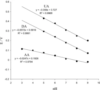
The pH effects on peak potentials (Epa) for the oxidation of AA, DA and UA on the iron(II)-complex/ MWNTs-modiied GC electrode are shown in Figure 4. All species Epa shifted to lower potential by increasing
Figure 3. Anodic peak potential differences (Epa) between AA-DA and DA-UA of ascorbic acid (0.5 mmol L-1), dopamine (100 μmol L-1) and uric acid (0.5 mmol L-1) at the iron(II)-complex/MWNTs modiied electrode in 0.1 mol L-1 acetate buffer solution (pH 5.0). Scan rate was 100 mV s-1.
pH. The calculated [∂Epa/∂pH] = −24.7 for AA, suggests a two-electron, one-proton oxidation process.57,58 However,
the calculated [∂Epa/∂pH] for DA and UA are −57.2, −59.0 mV pH-1, respectively. These are close to that of
expected for monoelectronic/monoprotonic electrode reaction (−59.2 mV pH-1 at 25 °C) and shows the electron
transfer was accompanied by an equal number of protons. The stability of electrocatalytic activity of the modiied GC electrode towards oxidation of AA, DA and UA were checked separately by repetitive scanning in the acetate buffer pH 5.0 solutions of each of species, between 0.0 to 0.7 V at scan rate of 100 mV s-1. The peak current was considered as
a factor in indicating the stability of the modiied electrode at various conditions of operation. The anodic peak current showed a small decrease (about 2-3%, not shown) after twenty cyclic voltamograms in the pH 5.0 acetate buffer solution. In addition, the electrode could also withstand if stored in solution for a period of time (at least 2 week) and the electrocatalytic currents remained almost unchanged. These phenomena show that the modiied electrode is stable in aqueous solution. The high stability of the adsorbed iron(II)-complex against desorption in aqueous solution, is related to the chemical and mechanical stability of nanotube ilm. This stability probably is due to the strong interaction of aromatic groups of the iron(II)-complex with π-staking of carbon nanotubes and the possible interaction between the iron(II)-complex and activated carbon nanotubes.52
Differential pulse voltammetry
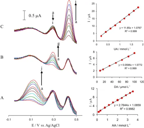
Differential pulse voltammetry (DPV) was used for simultaneous determination of AA, DA and UA at the iron-complex/MWNTs modiied GC electrode because of its higher current sensitivity and better resolution than the cyclic voltammetry. For this purpose, DPV was carried out in the potential range of −50 to 550 mV. Figure 5 shows the DPV of a mixture of AA (0.5 mmol L-1), DA (0.1 mmol L-1)
and UA (0.5 mmol L-1) at a bare GC electrode,
MWNTs-modiied GC electrode and the iron-complex/MWNTs-modiied GC electrode in 0.1 mol L-1 acetate buffer solution
(pH 5). It is obvious that the oxidation peak potentials of AA, DA and UA are very close to each other at a bare GC electrode and MWNTs-modiied GC electrode (Figure 5, curves A and B). Three well-deined peaks at about 72, 298, and 460 mV vs. Ag/AgCl (KCl, 3.0 mol L-1) were observed
at the iron(II)-complex/MWNTs modified electrode (Figure 5, curve C), which correspond to the differential pulse voltammograms of AA, DA, and UA, respectively. Peak separations of 226, 162 and 388 mV between DA and AA, DA and UA, and UA and AA, respectively, allow us to detect AA, DA and UA simultaneously by using DPV.
Figure 5. Differential pulse voltammograms of mixture of AA (0.5 mmol L-1), DA (100 μmol L-1) and UA (0.5 mmol L-1) at (A) a bare GC electrode, (B) MWNTs modiied GC electrode, (C) the iron(II)-complex/MWNTs modiied GC electrode in 0.1 mol L-1 acetate buffer solution (pH 5.0). Scan rate was 100 mV s-1.
The linear ranges for the determination of AA, DA and UA using DPV were 1.10×10⁻⁵-1.50×10⁻³,
9.00×10⁻⁷-1.20×10⁻³ and 2.00×10⁻⁶-1.50×10⁻³ mol L-1,
respectively. The limit of detection equals 3 sb m-1,
where sb is the standard deviation of the blank signal and mol L-1 is the slope of the calibration curve for
the proposed method. The theoretical detection limits, of the proposed method, for AA, DA and UA were 8.00×10⁻⁶, 2.00×10⁻⁷, and 1.0×10⁻⁶ mol L-1, respectively.
Table 1. Comparison of analytical parameters of several modiied electrodes for AA, DA and UA determination
Electrode Method Analyte Linear range
(μmol L-1)
Limit of detection (μmol L-1)
Sensitivity (μA μmol-1 L)
Reference
Modiied carbon paste electrode by tetrabromo-p
benzoquinone DPV AA DA UA 10-600 10-100 10-100 0.62 -0.005 0.0074 0.0022 19 Poly (3-(5-chloro-2-hydroxyphenylazo)-4,5 dihydroxynaphthalene-2,7-disulfonic acid) ilm
DPV AA DA UA 5-240 5-280 0.1-180 1.43 2.9 0.16 0.013 0.057 0.353 34
Oxidation in mild acidic media CV AA DA UA 197-988 1.97-9.78 19.7-97.8 -33
Dopamine solutions-phosphate buffer DPV AA DA UA 25-500 1-20 2.5-20 13 0.11 1.4 0.007 0.006 0.09 39
Novel choline and acetylcholine modiied glassy carbon DPV DA AA 0.7-5 7-90 0.3 0.9 -14 I
CPE/CNF/Pdnano DPV AA
DA UA 50-4000 0.5-160 2-200 15 0.2 0.7 -54
Pt/PF/Pdnano DPV AA in presence
of ACOP DA in presence
of ACOP 50-1000 0.5-100 7.1 0.5 5.92 0.0213 59
Iron(II)-complex / MWNTs/GC DPV AA
DA UA 11-1500 0.9-1200 2-1500 8 0.2 1 0.0118 0.059 0.0027 (This work)
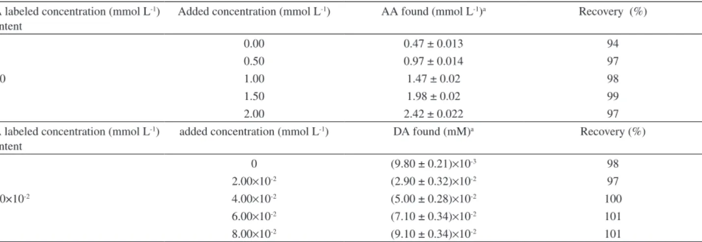
Table 3. Recovery results obtained for determination of AA and DA and the spiked AA and DA in injection solutions (n = 5)
AA labeled concentration (mmol L-1) Content
Added concentration (mmol L-1) AA found (mmol L-1)a Recovery (%)
0.00 0.47 ± 0.013 94
0.50 0.97 ± 0.014 97
0.50 1.00 1.47 ± 0.02 98
1.50 1.98 ± 0.02 99
2.00 2.42 ± 0.022 97
DA labeled concentration (mmol L-1) Content
added concentration (mmol L-1) DA found (mM)a Recovery (%)
0 (9.80 ± 0.21)×10-3 98
2.00×10⁻² (2.90 ± 0.32)×10-2 97
1.00×10⁻² 4.00×10⁻² (5.00 ± 0.28)×10-2 100
6.00×10⁻² (7.10 ± 0.34)×10-2 101
8.00×10⁻² (9.10 ± 0.34)×10-2 101
aResults are expressed as mean value ± SD, based on ive replicate.
Table 4. Recovery results obtained for determination of UA and the spiked UA in human blood serum (n = 5)
Sample detected UA (mmol L-1) Content
Added concentration (mmol L-1) UA Found (mmol L-1)a Recovery (%)
5.00×10⁻¹ (9.10 ± 0.21)×10-1 101
4.00×10⁻¹ 1.00 1.43 ± 0.32 102
1.50 1.86 ± 0.28 98
2.00 2.42 ± 0.34 101
aResults are expressed as mean value ± SD, based on ive replicate. modiied GC electrode. As can be seen the proposed modiied electrode shows somewhat similar (or worse) performances, in some cases, or superior ones, in most cases, than the previously reported modiied electrodes.
Interferences
For investigating the interference, several compounds were selected. If the tolerance limit was taken as the maximum concentration of the foreign substances, which causes an approximately 5% relative error, for 0.5 mmol L-1
AA, 0.1 mmol L-1 DA, and 0.5 mmol L-1 UA, no interference
was observed for the following compounds (μ mol L-1): K+,
Ca2+, Mg2+, Zn2+, starch, citric acid, cysteine, glucose. The
results are listed in Table 2.
Determination of AA in vitamin C injection, DA in dopamine hydrochloride injection solutions and UA in human blood serum
In order to demonstrate the capability of this modiied electrode for the catalytic oxidation of AA, DA and UA in real samples, we examined thisability in the voltammetric determination of AA, DA and UA in some pharmaceutical preparations, such as vitamin C injection solution (standard content 100 mg mL-1 AA, 5mL per injection), (Daro Pakhsh
Co.), dopamine hydrochloride injection (DHI) solution (standard content of 40 mg mL DA, 5 mL per injection), (Rasht Co.) andhuman blood serum. Human blood serum samples were centrifuged before the experiment. All samples were diluted with acetate buffer (pH 5.0) and then appropriate amounts of these diluted samples were transferred to the electrochemical cell to determine each species using DPV. The standard addition technique was employed for AA, DA and UA determination. The results of AA, DA and UAdeterminations in the real samples and the spiked samples with AA, DA or UAstandard solutions are shown in Tables 3 and 4. The recovery and precision wereacceptable, revealing that the modiied electrode could be eficiently applied fordetermination of AA, DA and UA in pharmaceutical samples.
Table 2. Interferences of some foreign substances for 0.5 mmol L-1 AA, 100 μmol L-1 DA, and 0.5 mmol L-1 UA
Foreign substances Tolerance level (μmol L-1)
Starch 400
K+, Mg2+, Ca2+, Zn2+ 250
Citric acid 100
Cysteine 40
Conclusions
We have demonstrated the possibility of using the iron(II)-complex/MWNTs modified electrode for the simultaneous determination of UA, DA and AA. The modiied electrode showed excellent sensitivity, selectivity and anti-fouling properties. The differential pulse voltammetry oxidation peaks for AA and DA, DA and UA, AA and UA are separated by 210, 136 and 346 mV, respectively. Therefore, simultaneous or independent measurements of the three analytes are possible without any interference. The proposed methods can be applied to the determination of DA, AA and UA in real samples with satisfactory results.
Acknowledgments
The authors wish to express their gratitude to the Zanjan University Research Council for the support of this work.
References
1. Martin, D. W. In Harper’s Review of Biochemistry, Martin, D. W.; Mayes, D. A.; Rodwell, V. W.; 19th ed., Lange: Los Altos, CA, 1983, p. 112.
2. Dryhurst, G.; Kadish, K. M.; Scheller, F.; Renneberg, R.;
Biological Electrochemistry, Academic Press: New York, 1982, v. 1.
3. Martin, C.; Chem. Br.1998, 34, 40.
4. Heinz, A.; Przuntek, H.; Winterer, G.; Pietzcker, A.; Nervenarzt 1995, 66, 662.
5. Wightman, R. M.; May, L. J.; Michael, A. C.; Anal. Chem. 1988,
60, 769A.
6. Safavi, A.; Maleki, N.; Moradlou, O.; Tajabadi, F.; Anal. Biochem. 2006, 359, 224.
7. Dayton, M. A.; Ewing, A. G.; Wightman, R. M.; Anal. Chem. 1980, 52, 2392.
8. Stamford, J. A.; Justice Jr., J. B.; Anal. Chem.1996, 68, 359A. 9. Zare, H. R.; Nasirzadeh, N.; Ardakani, M. M.; J.Electroanal.
Chem.2005, 577, 25.
10. Thiagarajan, S.; Tsai, T.-H.; Chen, S.-M.; Biosens. Bioelectron. 2009, 24, 2712.
11. Da Silva, R. P.; Lima, A. W. O.; Serrano, S. H. P.; Anal. Chim. Acta 2008, 612, 89.
12. Jin, G. P.; Lin, X. Q.; Gong, J. M.; J. Electroanal. Chem. 2004,
569, 135.
13. Huang, J.; Liu, Y.; Hou, H.; You, T.; Biosens. Bioelectron.2008,
24, 632.
14. Atta, N. F.; El-Kady, M. F.; Galal, A.; Sens. Actuators, B 2009,
141, 566.
15. Kalimuthu, P.; John, S. A.; Talanta2010, 80, 1686.
16. Noroozifar, M.; Khorasani-Motlagh, M.; Taheri, A.; Talanta 2010, 80, 1657.
17. Atta, N. F.; El-Kady, M. F.; Galal, A.; Anal. Biochem.2010,
400, 78.
18. Zhu, S.; Li, H.; Niu, W.; Xu, G.; Biosens. Bioelectron. 2009,
25, 940.
19. Kalimuthu, P.; John, S. A.; Bioelectrochemistry2009, 77, 13. 20. Kumar, S. A.; Cheng, H. W.; Chen, S.-M.; Electroanalysis2009,
21, 2281.
21. Ensai, A. A.; Taei, M.; Khayamian, T.; J. Electroanal. Chem. 2009, 633, 212.
22. Kamyabi, M. A.; Asgari, Z.; Hosseini-Monfared, H.; Morsali, A.; J. Electroanal. Chem. 2009, 632, 170.
23. Iijima, S.; Nature1991, 354, 56.
24. Ajayan, P. M.; Chem. Rev. 1999, 99, 1787.
25. Wong, S.; Joselevich, E.; Woolley, A.; Cheung, C.; Lieber, C.;
Nature 1998, 394, 52.
26. De Heer, W. A.; Chatelain, A.; Ugarte, D.; Science1995, 270, 1179.
27. Baughman, R. H.; Cui, C. C.; Zakhidov, A. A.; Iqbal, Z.; Barisci, J. N.; Spinks, G. M.; Wallace, G. G.; Mazzoldi, A.; De Rossi, D.; Rinzler, A. G.; Jaschinski, O.; Roth, S.; Kertesz, M.; Science 1999, 284, 1340.
28. Tans, S.; Verschueren, A.; Dekker, C.; Nature1998, 393, 49. 29. Che, G. L.; Lakschmi, B. B.; Fisher, E. R.; Martin, C. R.; Nature
1998, 393, 346.
30. Dresselhaus, M. S.; Nature1992, 358, 195.
31. Chen, P.; Wu, X.; Lin, J.; Tan, K. L.; Science1999, 285, 91. 32. Kong, J.; Franklin, N. R.; Zhou, C. W.; Chapline, M. G.; Peng,
S.; Cho, K.; Dai, H. J.; Science2000, 287, 622.
33. Antiochia, R.; Lvagnini, I.; Magno, F.; Valentini, F.; Palleschi, G.; Electroanalysis2004, 16, 1451.
34. Poh, W. C.; Loh, K. P.; Zhang, W. D.; Triparthy, S.; Ye, J. S.; Shen, F. S.; Langmuir2004, 20, 5484.
35. Hrapovic, S.; Liu, Y. L.; Mall, K. B.; Luong, J. H. T.; Anal. Chem.2004, 76, 1083.
36. Luo, H. X.; Shi, Z. J.; Li, N. Q.; Gu, Z. N.; Zhuang, Q. K.; Anal. Chem. 2001, 73, 915.
37. Lin, Y. H.; Lu, F.; Tu, Y.; Ren, Z. F.; Nano Lett.2004, 4, 191. 38. Xu, Z.; Gao, N.; Dong, S.; Talanta2006, 68, 753.
39. Wang, J. X.; Li, M. X.; Shi, Z. J.; Li, N. Q.; Gu, Z. N.; Anal. Chem.2002, 74, 1993.
40. Cai, C. X.; Chen, J.; Anal. Biochem.2004, 332, 75.
41. Wang, L.; Wang, J. X.; Zhou, F. M.; Electroanalysis2004, 16, 627.
42. Zhao, G. C.; Zhang, L.; Wei, X. W.; Yang, Z. S.; Electrochem. Commun.2003, 5, 825.
43. Cai, C. X.; Chen, J.; Anal. Biochem.2004, 335, 285. 44. Zhao, Y. D.; Bi, Y. H.; Zhang, W. D.; Luo, Q. M.; Talanta2005,
45. Salimi, A.; Hallaj, R.; Talanta2005, 66, 967.
46. Musameh, M.; Wang, J.; Merkoci, A.; Lin, Y. H.; Electrochem. Commun.2002, 4, 743.
47. Xu, Z. A.; Chen, X.; Qu, X. H.; Dong, S.; J. Electroanalysis 2004, 16, 684.
48. Gong, K. P.; Dong, Y.; Xiong, S. X.; Chen, Y.; Mao, L. G.;
Biosens. Bioelectron.2004, 20, 253.
49. Zhao, Y.; Gao, Y.; Zhan, D.; Liu, H.; Zhao, Q.; Kou, Y.; Shao, Y.; Li, M.; Talanta2005, 66, 51.
50. Wang, J.; Musameh, M.; Lin, Y. H.; J. Am. Chem. Soc.2003,
125, 2408.
51. Salimi, A.; Noorbakhsh, A.; Ghadermarz, M.; Anal. Biochem. 2005, 344, 16.
52. Salimi, A.; Mamkhezri, H.; Mohebbi, S.; Electrochem. Commun.2006, 8, 688.
53. Constable, E. C.; Thompson, A. M. W. C.; J.Chem. Soc., Dalton Trans. 1992, 2947.
54. Wang, J.; Peng, T.; Vince, V.; J. Electroanal. Chem.1987, 234, 119.
55. Musameh, M.; Lawrence, N. S.; Wang, J.; Electrochem. Commun. 2005, 7, 14.
56. Salimi, A.; Mam-Khezri, H.; Hallaj, R.; Talanta2006, 70, 823. 57. Bard, A. J.; Faulkner, L. R.; Electrochemical Methods,
Fundamentals and Applications, Wiley: New York, 2001. 58. Fei, J.; Wu, K.; Yi, L.; Li, J.; Bull.Korean Chem. Soc.2005,
26, 1403.
Submitted: March 15, 2010