Article
Printed in Brazil - ©2014 Sociedade Brasileira de Química0103 - 5053 $6.00+0.00
A
*e-mail: [email protected], [email protected]
New Phenolic Glycosides from
Phyllanthus cochinchinensis
Jian-Qiang Zhao,a,b Yan-Ming Wang,a,b Jun-Jiang Lv, a Hong-Tao Zhu,a Dong Wang,a
Chong-Ren Yang,a Min Xu*,a and Ying-Jun Zhang*,a
aState Key Laboratory of Phytochemistry and Plant Resources in West China, Kunming Institute of Botany, Chinese Academy of Sciences, 650201 Kunming, P. R. China
bUniversity of Chinese Academy of Sciences, 100049 Beijing, P. R. China
A investigação química das plantas Phyllanthus cochinchinensis (Euphorbiaceae) levou ao isolamento de três novos glicosídeos fenólicos, filantuosídeos A-C, juntamente com 12 compostos conhecidos. Suas estruturas foram determinadas com base em extensivas análises espectroscópicas e métodos químicos. Dentre eles, filantuosídeos A e B são dois glicosídeos fenólicos raros, com um esqueleto C6-C3-C6. A configuração absoluta dos filantuosídeos A e B foi estabelecida por dicroísmo circular elétrico calculado (ECD) usando teoria do funcional da densidade e a sua abordagem dependente do tempo (TDDFT). Os compostos isolados também tiveram sua citotoxicidade e atividade antimicrobial testadas.
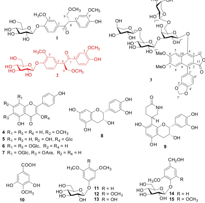
Chemical investigation of the whole plants of Phyllanthus cochinchinensis (Euphorbiaceae) led to the isolation of three new phenolic glycosides, phyllanthuosides A-C, together with 12 known compounds. Their structures were established on the basis of extensive spectroscopic analysis and chemical methods. Among them, phyllanthuosides A and B are two of the rare phenolic glycosides, featuring with a C6-C3-C6 skeleton. The absolute configurations of phyllanthuosides A and B were established by calculated electric circular dichroism (ECD) using time dependent density functional theory (TDDFT). The isolates were also tested for their cytotoxicity and antimicrobial activity.
Keywords: Phyllantus cochinchinensis, Euphorbiaceae, phenolic glycosides, cytotoxicity, antimicrobial activity
Introduction
The genus Phyllanthus composing of approximate 600 species is one of the largest genera in the family Euphorbiaceae, which is distributed mainly in tropical and subtropical regions throughout the world. Thirty-three Phyllanthus species and four varieties are growing in China, particularly in the south of the Yangtze River. Among them, most species have been used in folk medicine to treat kidney and urinary bladder disturbances, intestinal infections, diabetes, and hepatitis B.1 Phytochemcial investigation of this genus have revealed the occurrence of a large number of bioactive constituents including tannins, alkaloids, flavonoids, lignans, phenols, and terpenes.1-7 Some of them showed potential therapeutic actions as antinociception, treating the genitourinary disturbances, antiviral activity and other pharmacological effects.1-6
Phyllanthus cochinchinensis mainly grows in the southern parts of China. Previous phytochemical studies carried out by our group on this plant have afforded antifeedant limonoids and sucrose esters.5,7 As a part of our continuing study on bioactive compounds from Phyllanthus species,4-7 three new phenolic glycosides, phyllanthuosides A-C (1-3), together with twelve known compounds were
isolated from the whole plants of P. cochinchinensis. Their structures were established by means of extensive spectroscopic analysis and chemical methods. Most of the isolates were tested for their cytotoxicity and antimicrobial activity. The results obtained are discussed herein.
Experimental
General experimental procedures
were measured on a Bio-Rad FTS-135 series spectrometer. UV spectra were recorded on a Shimadzu UV2401A ultraviolet-visible spectrophotometer. Electrospray ionization mass spectrometry (ESIMS) and high-resolution electrospray ionization mass spectrometry (HRESI-MS) were run on an API QSTAR Pular-1 spectrometer. Nuclear magnetic resonance (NMR) spectra were measured in methanol-d4 or DMSO-d6 solution and recorded on a Bruker AV-400, DRX-500 or AV III-600 spectrometer, using tetramethylsilane (TMS) as internal standard. Chemical shifts were reported in units of d (ppm) and coupling constants (J) were expressed in Hz. Column chromatography (CC) was carried out over Diaion HP20SS (Mitsubishi Chemical Industry, Ltd.), silica gel (200-300 mesh, Qingdao Marine Chemical Factory), MCI-gel CHP-20P (75-150 µm, Mitsubishi Chemical Industry, Ltd), and Sephadex LH-20 (25-100 µm, Pharmacia Fine Chemical Co., Ltd. Uppsala, Sweden). Pre-coated silica gel plates (Qingdao Haiyang Chemical Co.) were used for thin layer chromatography (TLC). Detection was done under UV light (254 nm and 365 nm) and by spraying the plates with 10% sulfuric acid followed by heating. Electric circular dichroism (ECD) spectra were tested on a Chirascan Circular Dichroism spectrometer. An Agilent series 1260 (Agilent Technologies) was used for high-performance liquid choromatography (HPLC). An Agilent ZORBAX SB-C18 column 5 µm 143 Å column (250 mm × 9.4 mm) was used for semi-preparative HPLC separation. Gas chromatography (GC) analysis was run on Agilent Technologies HP5890 gas chromatography equipped with an H2 flame ionization detector. The column was 30QC2/AC-5 quartz capillary column (30 m × 0.32 mm) with the following conditions: column temperature: from 180 °C to 280 °C; programmed increase, 3 °C min−1; carrier gas N2 (1 mL min−1); injection and detector temperature at 250 °C; injection volume of 4 µL, split ratio 1/50.
Plant material
The whole plant of P. cochinchinensis was collected from Guangdong Province, People’s Republic of China, on December 2011. A voucher specimen (KUN-1215860) was deposited at the State Key Laboratory of Phytochemistry and Plant Resources in West China, Kunming Institute of Botany, Chinese Academy of Sciences, and identified by Mr. Xiao-Ming Fang from South China Botanical Garden, Chinese Academy of Sciences.
Extraction and isolation
The air-dried and powdered whole plants of P. cochinchinensis (3.2 kg) were extracted with MeOH
(3 times, 3 h each time) under reflux at 60 °C. Evaporation of the solvent under vacuum gave a residue (270 g), which was suspended in water and then extracted sequentially with chloroform and butanol. The butanol extract (90 g) was chromatographed on Diaion HP20SS eluting with a gradient of MeOH-H2O (1:9 → 9:1, finally MeOH), to give 6 fractions F1-F6. F1 (5.0 g) was fractionated through silica gel CC (CHCl3/MeOH/H2O, 9:1:0.1 → 8:2:0.2) to obtain three fractions. The first fraction was loaded on a column of Sephadex LH-20 (eluted with MeOH/H2O, from 1:9 → 5:5), and then the residue was purified by semi-preparative HPLC (MeCN/H2O, 14:86) to yield 13 (3.0 mg) and 14 (6.0 mg) and 15 (3.0 mg). F2 (6.0 g) was fractionated
by silica gel CC (CHCl3/MeOH/H2O, 9:1:0.1 → 8:2:0.2) to afford four fractions (F21-F24). F21 was chromatographed over a column of Sephadex LH-20 (eluted with MeOH/H2O, 1:9 → 5:5), then subjected to MCI-gel CHP-20P CC (eluted with MeOH/H2O, 1:9 → 5:5), and finally purified by semi-preparative HPLC (MeCN/H2O, 15:85) to yield 11 (8.0 mg) and 12 (8 mg). F22 was
chromatographed over Sephadex LH-20 CC (eluted with MeOH/H2O, from 1:9 → 2:8), and then subjected to MCI-gel CHP-20P CC and then purified by semi-preparative HPLC (MeCN/H2O, 14:86) to yield 6 (62.0 mg), 7 (10.0 mg) and
8 (17.0 mg). F3 (11.0 g) was subjected to silica gel CC
(CHCl3/MeOH/H2O, 9:1:0.1 → 8:2:0.2) to afford three fractions (F31-F33). Fraction F33 was chromatographed over a column of Sephadex LH-20 CC (eluted with MeOH/H2O, from 1:9 → 5:5), and then subjected to MCI-gel CHP-20P CC (eluted with MeOH/H2O, 3:7 → 5:5) to afford
9 (6.0 mg) and 10 (6.0 mg). F4 (6.0 g) was loaded on a silica
gel CC (CHCl3/MeOH/H2O, 9:1:0.1 → 8:2:0.2) to afford four fractions (F41-F44). F43 was subjected to Sephadex LH-20 CC (eluted with MeOH/H2O, from 1:9 → 2:8), and then purified by semi-preparative HPLC (MeCN/H2O, 14:86) to yield 1 (4.0 mg), 2 (6.0 mg) and 4 (10.0 mg).
F6 (16 g) was subjected to Sephadex LH-20 CC eluted with a gradient MeOH-H2O (1:9 → 6:4) to attain five fractions (F61-F65). F62 was fractionated through silica gel CC, using CHCl3-MeOH-H2O (90:10:1 and 80:20:2) to give two fractions F621 and F622. F622 was chromatographed through semi-preparative HPLC (MeCN/H2O, 14:86) to yield 3 (2 mg). F65 was fractionated through silica gel
CC, using CHCl3-MeOH-H2O (90:10:1 and 80:20:2) to obtain 5 (3 mg).
Phyllanthuoside A (1): colorless amorphous powder;
[α]D20 −38.2° (c 0.25, MeOH); UV (MeOH) λmax (log ε) 202 (4.38), 224 (4.14), 272 (3.87); CD (0.08 mM, MeOH)
positive ESIMS m/z 517 [M + Na]+; positive HRESIMS (m/z 517.1686 [M + Na]+, calcd. for C
24H30O11Na 517.1680). Phyllanthuoside B (2): colorless amorphous powder;
[α]D20 −19.7° (c 0.35, MeOH); UV (MeOH) λmax (log ε) 202 (4.56), 227 (4.29), 280 (4.02), 307 (3.93); CD (0.41 mM, MeOH)λmax nm (Dε): 204 (−3.0), 235 (1.7), 250 (−0.6), 302 (1.1); IR (KBr) νmax 3424, 2934, 1665, 1592, 1514, 1463, 1424, 1269, 1074, 1031; 1H and 13C NMR data, see Table 1; positive ESIMS m/z 517 [M + Na]+; positive HRESIMS (m/z 517.1688 [M + Na]+, calcd. for C
24H30O11Na 517.1680). Phyllanthuoside C (3): colorless amorphous powder;
[α]D22 −14.93° (c 0.25, MeOH); UV (MeOH) λmax (log ε) 203 (4.36), 221 (4.16), 259 (4.46), 294 (3.78), 313 (3.79); IR (KBr) νmax 3425, 2925, 1681, 1625, 1508, 1480, 1436, 1386, 1346, 1264, 1211, 1138, 1040; 1H and 13C NMR data, see Table 2; negative HRESIMS (m/z 1003.2509 [M + Cl]−, calcd. for C43H52O25Cl, 1003.2486).
Acid hydrolysis of compounds 1 and 2
Compounds 1 and 2 (each 3 mg) in 2 mol L-1
HCl-dioxane (1:1, v/v, 5 mL) were heated at 85 °C in a water bath for 8 h, respectively. The reaction mixtures were partitioned between H2O and CHCl3 (2 mL × 3) four times. The aqueous layer was neutralized with 2 mol L−1 NaOH and then dried to give a saccharide mixture. Solutions of the sugar residues of these compounds in pyridine (2 mL) were added to L-cysteine methyl ester hydrochloride (1.5 mg) and kept at 60 °C for 1 h. Trimethylsilylimidazole (1.5 mL) was added to the reaction mixtures, which were kept at 60 °C for 30 min. The supernatants (4 µL) were analyzed by GC, and determined as D-glucose trimethylsilylated L-cysteine derivatives by comparison with a standard (retention time 21.7 min).
Quantum chemical calculations
The conformation analysis was carried out using Monte Carlo searching with molecular mechanics MMFF in Sparton’06 (Wavefunction Inc. Irvine, CA). The resulted conformers were re-optimized using DFT at the B3LYP /6-31G (d) level in vacuo. The free energies and vibrational frequencies were calculated at the same level to confirm their stability, and no imaginary frequencies were found. The optimized low energy conformers with energy < 2 kcal mol−1 were considered for ECD calculation. The TD-DFT/ B3LYP /6-311G (d, p) method was applied to calculate the ECD data in vacuo. All the calculations were run with Gaussian 09.8 The excited energies and rotational strength were used to simulate ECD spectra of each conformer by introducing the Gaussian Function. The final
ECD spectra of each compound were obtained by averaging all the simulated ECD spectra of all conformers according to their excited energies and Boltzmann distribution.6 The band shape of the calculated ECD curves were all 0.5 eV.
Cytotoxicity assay
Five human cancer cell lines, human myeloid leukemia HL-60, hepatocellular carcinoma SMMC-7721, lung cancer A-549 cells, breast cancer MCF-7, and colon cancer SW480, were used in the cytotoxic assay. All the cells were cultured in RPMI-1640 or DMEM (Dulbecco’s Modified Eagle Medium) medium (Hyclone, USA), supplemented with 10% fetal bovine serum (Hyclone, USA). The cytotoxicity assay was performed according to the MTT [3-(4,5-dimethylthiazol-2-yl)-2,5-diphenyltetrazolium bromide] method in 96-well microplates.9 Briefly, adherent cells (100 µL) were seeded into each well of 96-well cell culture plates and allowed to adhere for 12 h before drug addition, while suspended cells were seeded just before drug add with an initial density of 0.5 × 10⁵ - 1 × 10⁵ cells mL−1. Each tumor cell line was exposed to the test compound dissolved in dimethyl sulfoxide (DMSO) in triplicates for 48 h at 37 °C, with cisplatinum (DDP) and taxol (Sigma, USA) as positive controls. Then, MTT (50 µL) was added to each well, and the tumor cells were incubated for another 4 h at 37 °C. After the supernatant liquor was removed, SDS (200 µL) was added to each well. The optical density was measured at 595 nm on a microplate reader. Cell viability was detected and a cell growth curve was graphed. IC50 values were calculated by Reed and Muench’s method.10
Antimicrobial bioassay
Susceptibility testing was performed using a modified version of the NCCLS methods11,12 using organisms obtained from the American type culture collection (Manassas, VA) including Candida albicans ATCC 90028, Candida glabrata ATCC 90030, Candida krusei ATCC 6258, Cryptococcus neoformans ATCC 90113, and Aspergillus fumigatus ATCC 90906. Detailed procedures have been described in a previous paper.13
Results and Discussion
Diaion HP20SS, silica gel, Sephadex LH-20 and MCI-gel CHP-20P, followed by semi-preparative HPLC purification of the MeOH extract yielded three new compounds (1-3),
together with twelve known ones. The known compounds were elucidated as rhamnocitrin (4),14 shannaifen (5),15
vicenin-2 (6),16 schaftoside (7),17 (−)-epicatechin (8),18 8-(2-pyrrolidinone-5-yl)-(−)-epicatechin (9)19 gallic
acid 4-methyl ether (10),20 3,4-dimethoxyphenyl-β -D-glucopyranoside (11),21 3,4,5-trimethoxy-phenyl-β-D- glucopyranoside (12),22 koaburaside (13),23 vanilloloside (14),24 di-O-methylcrenatin (15),25 by comparison of their
spectroscopic data with reported literature values. Compound 1 was isolated as a colorless amorphous
powder. Its molecular formula was determined to be
C24H30O11, on the basis of positive HRESIMS (m/z 517.1686 [M+Na]+, calcd. 517.1680) and its 13C NMR (DEPT) spectra. The IR spectrum showed the occurrence of hydroxyl (3425 cm−1) and benzene rings (1600-1400 cm−1) groups. Twenty-four carbon resonances were well resolved in the 13C NMR spectrum (Table 1) and further assigned by DEPT and HSQC experiments as one carbonyl (dC 199.3), six quaternary aromatic carbons (dC 111-155), one aliphatic methine (dC 53.8) and six aromatic methines, an oxygenated methylene at dC 75.9 (C-9), and three methoxyls, in addition to one hexosyl moiety. The 1H NMR spectrum of 1 (Table 1) displayed two sets of characteristic ABX
coupled aromatic protons at dH 7.16 (1H, d, J = 8.6 Hz), 7.69 (1H, dd, J = 8.6, 1.8 Hz) and 7.59 (1H, d, J = 1.8 Hz), and
dH 6.71 (1H, d, J = 8.2 Hz), 6.75 (1H, dd , J = 8.2, 1.9 Hz) and 6.90 (1H, d, J = 1.9 Hz), suggesting the existence of two 1,3,4-trisubstituted benzene rings. Additionally, two aromatic methoxys (dH 3.82, 3.87, each 3H, s), one aliphatic methoxy (dH 3.34, 3H, s), and an anomeric proton signal (dH 5.01, 1H, d, J = 7.6 Hz) were observed apparently. The aforementioned NMR data for 1 were closely resembled
to those of evofolin B, 4-hydroxy-1,2-bis(4-hydroxy-3-methoxyphenyl)butan-1-one, a phenolic compound isolated from Tetradium glabrifolium.26 The differences were merely the presence of an additional aliphatic methoxy [dH 3.34,
dC 59.3] and one more hexosyl moiety in 1, related to evofolin B. Acid hydrolysis of 1 afforded D-glucose as
sugar residue, which was confirmed by GC analysis of its corresponding trimethylsilylated L-cysteine adduct. In the HMBC spectrum of 1 (Figure 1), correlation of
the glucosyl anomeric proton (dH 5.01, H-1”) with C-4 (dC 152.3) indicated the additional β-D-glucosyl unit located on C-4 position. Moreover, the HMBC correlations of the aliphatic methoxy at dH 3.34 with dC 75.9 (C-9) and H-9 (dH 4.12) with C-7 (dC 199.3, carbonyl carbon)/C-8 (dC 53.8)/9-OMe (dC 59.3) confirmed the location of the aliphatic methoxy on C-9. The ROESY correlations of the glucosyl H-1” with H-5 (dH 7.16/7.17), the 3-OMe with H-2 (dH 7.59) and the 3’-OMe with H-2’ (dH 6.90) further confirmed the positions of the β-D-glucosyl and the aromatic methoxy. The absolute configuration of 1 was
determined by comparison of the time dependent density functional theory (TDDFT)6 calculated ECD curve with the experimental results (Figure 3). The positive Cotton effects at 200 nm, 250 nm, and 260 nm of 1 agreed well
with the calculated ECD curve of the aglycon of 1 with
8S configuration. Thus, the structure of compound 1
was established as shown in Figure 1 and named as phyllanthuoside A.
Compound 2, a colorless amorphous powder, had the
same molecular formula C24H30O11 as 1, on the basis of the positive HRESIMS (m/z 517.1688 [M+Na]+, calcd. 517.1680) and 13C NMR (DEPT) spectra (Table 1). Acid
hydrolysis of 2 afforded D-glucose as sugar residue,
which was confirmed by GC analysis of its corresponding trimethylsilylated L-cysteine adduct. The 1H and 13C NMR spectroscopic data (Table 1) of 2 showed close resemblance
to those of 1. Extensive analysis of the 1D and 2D NMR
data suggested that 2 and 1 had the same C6-C3-C6
skeleton, while the only difference was the position of the
β-D-glucosyl group. All the proton and carbon signals of compound 2 could be assigned unambiguously by HSQC,
and HMBC analysis (Figure 2). In the HMBC experiment, the glucosyl H-1’’ (dH 4.85/4.86) of 2 was correlated with the aromatic carbon at dC 147.38/147.41 (C-4’), indicating that the β-D-glucosyl group in 2 was located on C-4’.
This was further confirmed by the ROESY correlation between H-1” with H-5’ (dH 7.08/7.09). Other key 1H-1H COSY, HMBC, and ROESY (Figure 2) correlations confirmed the planar structure of 2 as shown in Figure 1.
The experimental ECD curve of 2 displayed the mirror
image of that of compound 1 with 8S configuration (Figure 3), which suggested its 8R configuration. Thus, the structure of compound 2 was constructed and named
as phyllanthuoside B.
Compound 3, a colorless amorphous powder, possessed
a molecular formula C43H52O25, as deduced by the Figure 2. Key 1H-1H COSY, HMBC and ROESY correlations of 1 and 2.
HRESIMS (m/z 1003.2509 [M+Cl]−, calcd for C
43H52O25Cl, 1003.2486). The IR spectrum showed the presence of hydroxyl group (3425 cm−1), lactone carbonyl (1736 cm−1) and benzene ring (1625-1400 cm−1). The 13C NMR (DEPT) spectra displayed 43 carbon singals including one carbonyl (dC 169.3), 16 aromatic carbons (dC 101-152), one methylenedioxy (dC 101.3), one oxymethylene (dC 67.2), two methoxys (dC 55.3 and 56.1), and 22 saccharide carbons. The 1H NMR spectrum of 3 exhibited the presence of two aromatic singlets at dH 8.12 (1H, s) and 6.95/6.97 (1H, s), three ABX coupled aromatic protons at
dH6.92/6.94 (1H, d, J = 1.5 Hz), 7.02 (1H, d, J = 7.8 Hz), and 6.79/6.80 (1H, dd, J = 7.8, 1.5 Hz), two non-equivalent
γ-lactone methylene protons at dH 5.47 and 5.60 (each 1H, d, J = 14.6 Hz), and a methylenedioxy group with two characteristic protons at dH 6.12 (2H, brs). Furthermore,
four anomeric protons at dH 4.83 (1H, d, J = 7.8 Hz), 4.52 (1H, d, J = 7.9 Hz), and 4.18 (2H, d, J = 5.6 Hz) were observed in 1H NMR spectrum. The aforementioned data indicated that 3 is an arynaphthalene lignan with four
sugar units.27 The extensive comparison and analysis of 1D and 2D NMR data suggested that 3 shared the same
aglycon and saccharide moiety with those of mananthoside J,28 except for the absence of an acetyl group in 3. And the 13C NMR resonances due to the galactosyl unit of 3 were different from that of mananthoside J. Although acid hydrolysis of 3 was not done due to limited amount,
the tentative assignments of a galactosysl,29 a glucossyl,30 and two arabinosyls,31 for sugar units of 3 were confirmed on the basis of literature comparison,28 and HSQC, HMBC, 1H-1H COSY, and 1H-1H-TOCSY experiments (SI - Supplementary Information).
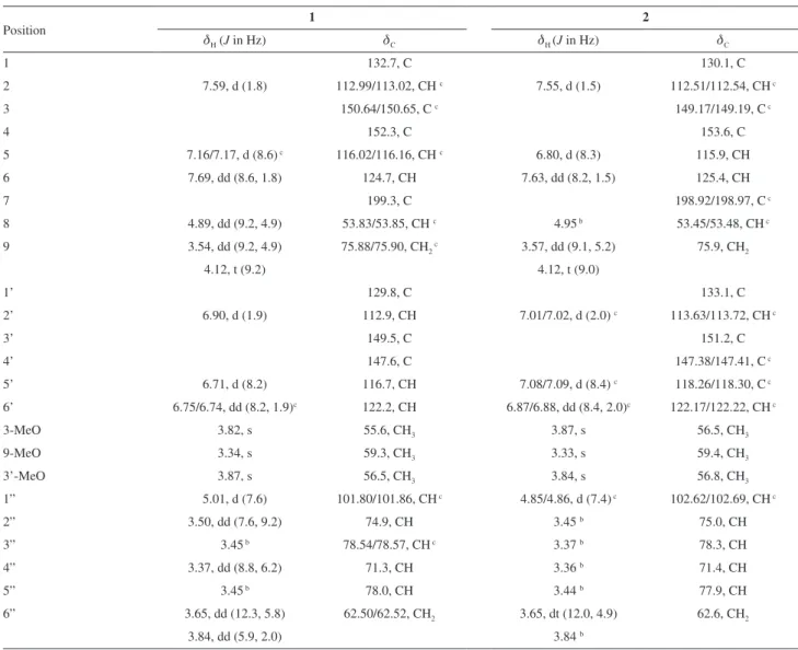
Table 1.1H NMR and 13C NMR spectroscopic data of compounds 1 a and 2a (CD
3OD, d in ppm)
Position 1 2
dH (J in Hz) dC dH (J in Hz) dC
1 132.7, C 130.1, C
2 7.59, d (1.8) 112.99/113.02, CH c 7.55, d (1.5) 112.51/112.54, CH c
3 150.64/150.65, C c 149.17/149.19, C c
4 152.3, C 153.6, C
5 7.16/7.17, d (8.6) c 116.02/116.16, CH c 6.80, d (8.3) 115.9, CH
6 7.69, dd (8.6, 1.8) 124.7, CH 7.63, dd (8.2, 1.5) 125.4, CH
7 199.3, C 198.92/198.97, C c
8 4.89, dd (9.2, 4.9) 53.83/53.85, CH c 4.95 b 53.45/53.48, CH c
9 3.54, dd (9.2, 4.9) 75.88/75.90, CH2 c 3.57, dd (9.1, 5.2) 75.9, CH2
4.12, t (9.2) 4.12, t (9.0)
1’ 129.8, C 133.1, C
2’ 6.90, d (1.9) 112.9, CH 7.01/7.02, d (2.0) c 113.63/113.72, CH c
3’ 149.5, C 151.2, C
4’ 147.6, C 147.38/147.41, C c
5’ 6.71, d (8.2) 116.7, CH 7.08/7.09, d (8.4) c 118.26/118.30, C c
6’ 6.75/6.74, dd (8.2, 1.9)c 122.2, CH 6.87/6.88, dd (8.4, 2.0)c 122.17/122.22, CH c
3-MeO 3.82, s 55.6, CH3 3.87, s 56.5, CH3
9-MeO 3.34, s 59.3, CH3 3.33, s 59.4, CH3
3’-MeO 3.87, s 56.5, CH3 3.84, s 56.8, CH3
1” 5.01, d (7.6) 101.80/101.86, CH c 4.85/4.86, d (7.4) c 102.62/102.69, CH c
2” 3.50, dd (7.6, 9.2) 74.9, CH 3.45 b 75.0, CH
3” 3.45 b 78.54/78.57, CH c 3.37 b 78.3, CH
4” 3.37, dd (8.8, 6.2) 71.3, CH 3.36 b 71.4, CH
5” 3.45 b 78.0, CH 3.44 b 77.9, CH
6” 3.65, dd (12.3, 5.8) 62.50/62.52, CH2 3.65, dt (12.0, 4.9) 62.6, CH2
3.84, dd (5.9, 2.0) 3.84 b
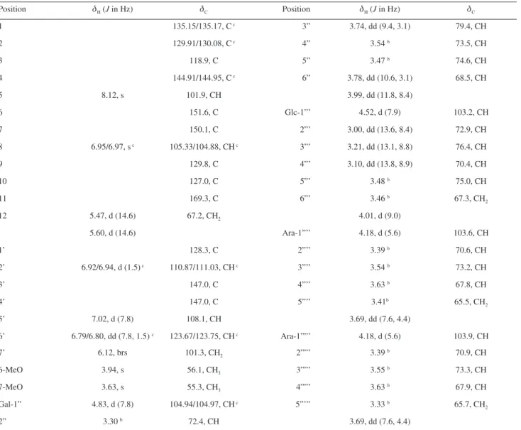
Based on the coupling constants of anomeric protons of galactosyl (7.8 Hz), glucosyl (7.9 Hz), and arabinosyl (5.6 Hz and 5.6 Hz) units, the anomeric configurations of saccharides were determined as β, β, α and α, respectively. The galactosyl moiety was directly linked to the aglycone C-4 by the HMBC correlation between H-1” (dH 4.83, 1H, d, J = 7.8 Hz) and C-4 (dC 144.91/144.95). The ROESY correlation between H-1” (dH 4.83, 1H, d, J = 7.8 Hz) and H-5 (dH 8.12, 1H, s) further confirmed the suggestion. Moreover, the HMBC correlation between glucosyl H-1”’(dH 4.52, 1H, d, J = 7.9 Hz) and galactosyl C-3” (dC 79.4) indicated that glucosyl moiety was linked to the galactosyl C-3”. Two arabinosyl groups were located on the galactosyl C-6” and glucosyl C-6”’, respectively, as deduced from the HMBC correlations of arabinosyl H-1””
and H-1””’ (dH 4.18, 2H, d, J = 5.6 Hz) with galactosyl C-6” (dC 68.5) and glucosyl C-6”’ (dC 67.3), respectively. Based on the above evidence, the structure of 3 was determined
as shown in Figure 1 and named as phyllanthuoside C. It was noted that some aromatic protons and carbons of the new phenolic glycosides 1-3 appeared to be very close
pairs of signals in the 1H and 13C NMR spectra acquired at room temperature, due to the equilibrium between two conformational isomers resulting from the slow rotation of the sugar unit around the glycosidic linkage.32 The energy barrier around the glycosidic bond was sufficiently high to prevent fast exchange between the two rotamers at room temperature.33
All of the isolated compounds, except compounds
7 and 9, were evaluated for their cytotoxicity against
Table 2. 1H and 13C NMR spectroscopic data of compound 3 a (DMSO-d
6, d in ppm)
Position dH (J in Hz) dC Position dH (J in Hz) dC
1 135.15/135.17, C c 3” 3.74, dd (9.4, 3.1) 79.4, CH
2 129.91/130.08, C c 4” 3.54 b 73.5, CH
3 118.9, C 5” 3.47 b 74.6, CH
4 144.91/144.95, C c 6” 3.78, dd (10.6, 3.1) 68.5, CH
5 8.12, s 101.9, CH 3.99, dd (11.8, 8.4)
6 151.6, C Glc-1”’ 4.52, d (7.9) 103.2, CH
7 150.1, C 2”’ 3.00, dd (13.6, 8.4) 72.9, CH
8 6.95/6.97, s c 105.33/104.88, CH c 3”’ 3.21, dd (13.1, 8.8) 76.4, CH
9 129.8, C 4”’ 3.10, dd (13.8, 8.9) 70.4, CH
10 127.0, C 5”’ 3.48 b 75.0, CH
11 169.3, C 6”’ 3.46 b 67.3, CH
2
12 5.47, d (14.6) 67.2, CH2 4.01, d (9.0)
5.60, d (14.6) Ara-1”’’ 4.18, d (5.6) 103.6, CH
1’ 128.3, C 2”’’ 3.39 b 70.6, CH
2’ 6.92/6.94, d (1.5) c 110.87/111.03, CH c 3”’’ 3.54 b 73.2, CH
3’ 147.0, C 4”’’ 3.63 b 67.8, CH
4’ 147.0, C 5”’’ 3.41b 65.5, CH
2
5’ 7.02, d (7.8) 108.1, CH 3.69, dd (7.6, 4.4)
6’ 6.79/6.80, dd (7.8, 1.5) c 123.67/123.75, CH c Ara-1””’ 4.18, d (5.6) 103.9, CH
7’ 6.12, brs 101.3, CH2 2””’ 3.39 b 70.9, CH
6-MeO 3.94, s 56.1, CH3 3””’ 3.55 b 73.3, CH
7-MeO 3.63, s 55.3, CH3 4””’ 3.63 b 67.9, CH
Gal-1” 4.83, d (7.8) 104.94/104.97, CH c 5”’” 3.33 b 65.7, CH
2
2” 3.30 b 72.4, CH 3.69, dd (7.6, 4.4)
five human cancer cell lines (breast cancer MCF-7, hepatocellular carcinoma SMMC-7721, human myeloid leukemia HL-60, colon cancer SW480, and lung cancer A-549). None of them showed cytotoxic activity at a concentration of 40 µmol L−1. These compounds were also tested for their antimicrobial effects, but none displayed activity.
Conclusions
The present phytochemical investigation of the whole plants of Phyllanthus cochinchinensis led to the isolation and identification of three new phenolic glycosides, phyllanthuosides A-C (1-3), together with twelve known
compounds. Phyllanthuosides A and B (1 and 2) are two
phenolic glycosides with a rare C6-C3-C6 skeleton. All of the isolated compounds, except compounds 7 and 9 were
tested for their cytotoxicity and antimicrobial activity but none of them showed any activity.
Supplementary Information
1D NMR and 2D NMR spectra for compounds 1-3 are
available free of charge at http://jbcs.sbq.org.br as a PDF file.
Acknowledgments
We are grateful to the members of the analytical group of our institute for the measurement of spectroscopic data. This work was supported by the NSFC 21002105, the 973 Program of Science and Technology of P. R. China (2011CB915503), the 12th Five Year National Science & Technology Supporting Program (2012BAI29B06), the Fourteenth Batch Candidates of the Young Academic Leaders of Yunnan Province (Min. XU, 2011CI044) and by West Light Foundation of the Chinese Academy of Sciences.
References
1. Calixto, J. B.; Santos, A. R. S.; Cechinel-Filho, V.; Yunes, R. A.;
Med. Res. Rev. 1998, 18, 225.
2. Zhang, Y. J.; Tanaka, T.; Iwamoto, Y.; Yang, C. R.; Kouno, I.;
J. Nat. Prod. 2000, 63, 1507.
3. Zhang, Y. J.; Tanaka, T.; Iwamoto, Y.; Yang, C. R.; Kouno, I.;
J. Nat. Prod. 2001, 64, 870.
4. Liu, Q.; Wang, Y. F.; Chen, R. J.; Zhang, M. Y.; Wang, Y. F.; Yang, C. R.; Zhang, Y. J.; J. Nat. Prod. 2009, 72, 969.
5. Zhao, J. Q.; Wang, Y. M.; He, H. P.; Li, S. H.; Li, X. N.; Yang, C. R.; Wang, D.; Zhu, H. T.; Xu, M.; Zhang, Y. J.; Org. Lett.
2013, 15, 2414.
6. Zhao, J. Q.; Lv, J. J.; Wang, Y. M.; Xu, M.; Zhu, H. T.; Wang, D.; Yang, C. R.; Wang, Y. F.; Zhang, Y. J.; Tetrahedron Lett. 2013,
54, 4670.
7. Zhao, J. Q.; Wang, Y. M.; Wang, D.; Yang, C. R.; Xu, M.; Zhang, Y. J.; Nat. Prod. Bioprospect. 2013, 3, 61.
8. Frisch, M. J.; Trucks, G. W.; Schlegel, H. B.; Scuseria, G. E.; Robb, M. A.; Cheeseman, J. R.; Scalmani, G.; Barone, V.; Mennucci, B.; Petersson, G. A.; Nakatsuji, H.; Caricato, M.; Li, X.; Hratchian, H. P.; Izmaylov, A. F.; Bloino, J.; Zheng, G.; Sonnenberg, J. L.; Hada, M.; Ehara, M.; Toyota, K.; Fukuda, R.; Hasegawa, J.; Ishida, M.; Nakajima, T.; Honda, Y.; Kitao, O.; Nakai, H.; Vreven, T.; Montgomery, J. A.; Peralta, J. E.; Ogliaro, F.; Bearpark, M.; Heyd, J. J.; Brothers, E.; Kudin, K. N.; Staroverov, V. N.; Kobayashi, R.; Normand, J.; Raghavachari, K.; Rendell, A.; Burant, J. C.; Iyengar, S. S.; Tomasi, J.; Cossi, M.; Rega, N.; Millam, J. M.; Klene, M.; Knox, J. E.; Cross, J. B.; Bakken, V.; Adamo, C.; Jaramillo, J.; Gomperts, R.; Stratmann, R. E.; Yazyev, O.; Austin, A. J.; Cammi, R.; Pomelli, C.; Ochterski, J. W.; Martin, R. L.; Morokuma, K.; Zakrzewsk, V. G.; Voth, G. A.; Salvador, P.; Dannenberg, J. J.; Dapprich, S.; Daniels, A. D.; Farkas, O.; Foresman, J. B.; Ortiz, J. V.; Cioslowski, J.; Fox, D. J.; Gaussian 09, Revision A.02; Gaussian, Inc., Wallingford CT, 2009. 9. Chang, S. L.; Chang, C. L.; Chiang, Y. M.; Hsieh, R. H.; Tzeng,
C. R.; Wu, T. K.; Sytwu, H. K.; Shyur, L. F.; Yang, W. C.; Planta Med. 2004, 70, 1045.
10. Reed, L. J.; Muench, H.; Am. J. Hygiene 1938, 27, 493. 11. National Committee for Clinical Laboratory Standards
(NCCLS); Reference Method for Broth Dilution Antifungal Susceptibility Testing of Yeasts; approved standard M27-A2,
22(15); 2002.
12. National Committee for Clinical Laboratory Standards (NCCLS); Method for Broth Dilution Antifungal Susceptibility Testing of Filamentous Fungi; approved standard M38-A, 22
(16); 2002.
13. Li, X. C.; Hala, N. E.; Alison, C. N.; Alice, M. C.; J. Nat. Prod.
1999, 62, 767.
14. Barberá, O.; Sanz, J. F.; Sánchez-Parareda, J.; Marco, J. A.;
Phytochemistry 1986, 25, 2361.
15. Hu, X. L.; Zhu, H.; Liu, C. R.; Tu, P. F.; Chin. Tradit. Pat. Med.
2003, 25, 833.
16. Lu, Y.; Yeap-Foo, L.; Phytochemistry 2000, 55, 263.
17. Besson, E.; Chopin, J. R.; Markham, K.; Mues, R.; Wong, H.; Bouillant, M. L.; Phytochemistry 1984, 23, 159.
18. Zhang, W. J.; Liu, Y. Q.; Li, X. C.; Yang, C. R.; Acta Bot. Yunnanica 1995, 17, 204.
19. Jang, D. S.; Lee, G. Y.; Lee, Y. M.; Kim, Y. S.; Sun, H.; Kim, D. H.; Kim, J. S.; Chem. Pharm. Bull.2009, 57, 397.
20. Saxena, G.; McCutcheon, A. R.; Farmer, S.; Towers, G. H. N.; Hancock, R. E. W.; J. Ethnopharmacol. 1994, 42, 95.
22. Shimomura, H.; Sashida, Y.; Oohara, M.; Tenma, H.;
Phytochemistry 1988, 27, 644.
23. Geweli, M. B.; Hattori, M.; Namba, T.; Shoyakugaku Zasshi
1988, 42, 247.
24. Koike, Y.; Fukumura, M.; Hirai, Y.; Hori, Y.; Usui, S.; Atsumi, T.; Toriizuka, K.; J. Nat. Med. 2010, 64, 245.
25. Li, X. C.; ElSohly, H. N.; Walker, L. A.; Clark, A. M.; Planta Med. 2005, 71, 977.
26. Wu, T. S.; Yeh, J. H.; Wu, P. L.; Phytochemistry 1995, 40, 121. 27. Siani, A. C.; Zoghbi, M. G.; Wolter, E. L. A.; Vencato, I.; J. Nat.
Prod. 1998, 61, 796.
28. Tian, J. M.; He, H. P.; Di, Y. T.; Yang, X. W.; Gao, Z. L.; Hao, X. J.; J. Asian Nat. Prod. Res. 2008, 3, 228.
29. Zhou, L. Y.; Hua, Y.; Ni, W.; Chen, C. X.; Acta Bot. Yunnanica
2004, 26, 349.
30. Li, N.; Jia, A. Q.; Liu, Y. Q.; Zhou, J.; Acta Bot. Yunnanica 2003,
25, 241.
31. Chen, B.; Liu, Y.; Feng, C.; Li, B. G.; Zhang, G. L.; Chinese Chem. Lett. 2002, 13, 959.
32. Alabed, Y.; Sabri, S.; Zarga, M. A.; Shah, Z.; J. Nat. Prod.1990,
53, 1152.
33. Lewis, K. C.; Maxwell, A. R.; McLean, S.; Reynolds, W. F.; Enriquez, R. G.; Magn. Reson. Chem. 2000, 38, 771.
Submitted on: February 13, 2014