O
RIGINALA
RTICLE Revista Brasileira de FisioterapiaComparison of active and passive forces of
the pelvic floor muscles in women with and
without stress urinary incontinence
Comparação das forças ativa e passiva dos músculos do assoalho pélvico de
mulheres com e sem incontinência urinária de estresse
Carla C. M. Chamochumbi1, Fabiana R. Nunes2, Rinaldo R. J. Guirro3, Elaine C. O. Guirro3
Abstract
Background: The reduction of the pelvic floor muscles (PFM) strength is a major cause of stress urinary incontinence (SUI). Objective:
To compare active and passive forces, and vaginal cavity aperture in continent and stress urinary incontinent women. Method: The study included a total of thirty-two women, sixteen continent women (group 1 - G1) and sixteen women with SUI (group 2 - G2). To evaluate PFM passive and active forces in anteroposterior (sagittal plane) and left-right directions (frontal plane) a stainless steel specular dynamometer was used. Results: The anteroposterior active strength for the continent women (mean±standard deviation) (0.3±0.2 N) was greater compared to the values found in the evaluation of incontinent women (0.1±0.1 N). The left-right active strength (G1=0.43±0.1 N; G2=0.40±0.1 N), the passive force (G1=1.1±0.2 N; G2=1.1±0.3 N) and the vaginal cavity aperture (G1=21±3 mm; G2=24±4 mm) did not differ between groups 1 and 2. Conclusion: The function evaluation of PFM showed that women with SUI had a lower anteroposterior active strength compared to continent women.
Keywords: muscle strength; pelvic floor; physical therapy; urinary incontinence.
Resumo
Contextualização: A redução da força dos músculos do assoalho pélvico (MAP) é a maior causa da incontinência urinária de estresse (IUE). Objetivo: Comparar as forças ativa e passiva e a abertura da cavidade vaginal em mulheres continentes e com IUE. Método: O estudo incluiu um total de 32 mulheres, 16 mulheres continentes (grupo 1 - G1) e 16 mulheres com IUE (grupo 2 - G2). Um espéculo dinamométrico de aço inoxidável foi usado para avaliar as forças ativa e passiva dos MAP nas direções ântero-posterior (plano sagital) e látero-lateral (plano frontal). Resultados: A força ativa ântero-posterior nas mulheres continentes (0,3±0,2 N) foi maior que nas mulheres com IUE (0,1±0,1 N). Não houve diferença entre o G1 e o G2 nos valores de força ativa látero-lateral (G1=0,43±0,1 N; G2=0,40±0,1 N), força passiva (G1=1,1±0,2 N; G2=1,1±0,3 N) e abertura da cavidade vaginal (G1=21±3 mm; G2=24±4 mm).
Conclusão: A avaliação da função dos MAP mostrou que mulheres com IUE tiveram uma menor força ativa ântero-posterior quando comparadas com as mulheres continentes.
Palavras-chave: força muscular; assoalho pélvico; fisioterapia; incontinência urinária.
Received: 01/23/2012 – Revised: 01/24/2012 – Accepted: 01/24/2012
1Department of Gynecology and Obstetrics, Faculty of Medicine of Ribeirão Preto, Universidade de São Paulo (USP), Ribeirão Preto, SP, Brazil 2Department of Obstetrics and Gynecology, School of Medicine, Universidade Estadual de Campinas (UNICAMP), Campinas, SP, Brazil
3Department of Biomechanics, Medicine and Rehabilitation of the Locomotor System, School of Medicine of Ribeirão Preto, USP, Ribeirão Preto, SP, Brazil
Correspondence to: Elaine Caldeira de Oliveira Guirro, Faculdade de Medicina de Ribeirão Preto, Universidade de São Paulo, Curso de Fisioterapia, Av. dos Bandeirantes, 3900, Monte Alegre,
CEP 14049-900, Ribeirão Preto, SP, Brasil, e-mail: [email protected]
Introduction
Urinary incontinence (UI) is a highly prevalent condition1-3.
Its evaluation and treatment are costly and afects the indi-vidual’s self-esteem, social and sexual activities4. Although
sur-gery for stress urinary incontinence (SUI) is an option to treat this dysfunction5, the conservative treatment prioritizes pelvic
loor muscle (PFM) strengthening, which highlights the need for assessment methods before and after intervention6-8.
Strength evaluation of PFM plays a decisive role in the physical therapy treatment for SUI. In addition, it represents an important tool to monitor the clinical results, and on occasions, it can be a method to demonstrate and motivate women to contract the PFM9-12. Morin et al.13 studied anteroposterior PFM force and
Verelst and Leivseth14 studied left-right force in dynamometers
de-veloped by each research group of authors. In another study, Peng et al.15 measured the pressure exerted by the PFM on vaginal
cav-ity in four directions (anterior, posterior, left and right) in women with SUI and continence, and found a diference in PFM contrac-tion pressure only in anterior and posterior direccontrac-tion. Based on this proposition and on the fact that no studies were found which would assess PFM strength in the anteroposterior and left-right directions concomitantly, the presented study proposed to ap-praise a new method in order to support the evaluation and the physical therapy treatment in a broad and more reliable way.
herefore, the aim of this study was to compare active and passive ( force at rest) anteroposterior and left-right strengths and vaginal cavity aperture in women with and without SUI using this equipment proved to be reliable16.
Method
A total of 32 women participated in this study, sixteen continent women - group 1 (G1) – mean age of 37±8 and body mass index (BMI) of 23±1 kg/m2; and sixteen women with stress
urinary incontinence - group 2 (G2) – mean age of 48±7 and BMI of 26±3 kg/m2. he women from G1 were recruited at the
university where the study was developed and an evaluation including some questions was performed to make sure that they did not have any symptoms of urinary incontinence. he women from G2 were recruited in a center specialized in urol-ogy and the diagnosis of stress urinary incontinence of these volunteers was obtained by clinical (questions about symptoms) and urodynamic tests (to exclude women with an overactive bladder). Exclusion criteria were: pregnant women, vaginal and urinary tract infection, history of urogynecological surgery, dis-eases which could interfere with the PFM force measurement, allergy to latex, neurological diseases, had previously performed exercises for the PFM, prolapse of pelvic organs, urge urinary
incontinence, had taken pain killers or muscle relaxants in a week before the data colection16-18. his study was approved by
the Research Ethics Committee of the Universidade Metodista de Piracicaba (UNIMEP), Piracicaba, SP, Brazil number 56/07, and
all women signed an informed consent form.
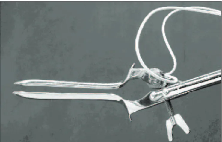
For the evaluation of the active and passive PFM forces and vaginal cavity aperture measurement, a stainless steel specular dynamometer was used. It captured the PFM strength in both anteroposterior (sagittal plane) and left-right directions ( fron-tal plane). he dynamometric device (Figure 1) was ixed to a mobile apparatus for a more efective adjustment. he device reliability was performed by the test-retest evaluation over three consecutive weeks and it was considered good and excel-lent. he standard error of measurement (SEM) for the mean anteroposterior strength and the mean left-right strength was 1.96 N and 1.86 N, respectively. he system was calibrated by a linear regression with weights ranging from 0 to 4 kg16.
he volunteers were evaluated in gynecological position. Before recording the data, general explanations about the pelvic loor was given and in order to check the correct PFM contrac-tion, a 2 second contraction was requested to the volunteers, considering this contraction valid only when an observable cranial movement of the perineum has occurred19.
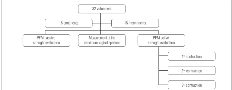
he PFM evaluation was divided into three steps, con-ducted in a single day for each woman from G1 and G2. In the irst step, the dynamometric speculum was positioned up to the maximum vaginal aperture that the volunteer could toler-ate, without discomfort or pain. he volunteers were instructed to relax their PFM and the passive force of these muscles was registered for 4 seconds. In the second step, with the specu-lum in the same position, the maximum vaginal aperture was measured (mm) with a digital paquimeter with a resolution of 0.01 mm (Digimess®
). After the registration of the passive force, with the speculum at the same position, the paquime-ter was placed in the speculum in order to measure vaginal aperture, always by the same researcher. In the third step, the
Figure 1. Side view of the equipment.
PFM active strength was evaluated with a 4.9 N of passive force (determined in a pilot study with 10 women, where they felt that the equipment touched the vaginal cavity without pain or discomfort), where three maximum isometric contractions of 4 seconds each were performed with a command (“squeeze, squeeze, squeeze”), instructed always by the same researcher, with a 2 minute interval, as shown in Figure 2.
he volunteers were requested to perform the PFM con-traction as isolated as possible, to minimize hip adductor, gluteus and abdominal muscles interferences. Abdominal and gluteus muscles were controlled by surface electromyography (EMG), to observe their activities during the PFM contractions, and when the contraction of these muscles was observed the PFM contraction was excluded.
he EMG was carried out by simple diferential electrodes made by two silver parallel bars, spaced 10 mm apart, with a pre-ampliier circuit with 20 times gain (±1%), a signal noise ratio lower than 3 μV RMS and a common mode rejection rate higher than 100 dB. he skin was cleaned with a solution of 70% alcohol before placing the EMG electrodes on the rectus-abdominis and gluteus muscles20 and the reference electrode was ixed on the
anterosuperior iliac crest with hydrosoluble gel. he EMG signal was collected by the signal acquisition module (SAM) EMG1000 (Lynx®
), with a sampling frequency of 2000 Hz, input range of
±5 V, a resolution of 16 bits, with a band-pass ilter from 20 to 1000 Hz (Butterworth ilter) and an acquisition data software Aqdados 7.2®
(Lynx®
). SAM was supplied by a battery and con-nected to a personal computer by an optical iber to isolate the equipment from the electric grid interferences21.
Before the evaluation the volunteers received instructions to empty their bladder and the data collection was not per-formed during the pre and menstrual periods to reduce hor-monal interference22.
he PFM active and passive forces were evaluated with dynamometric equipment. he equipment was covered with Olla®
condoms, lubricated with hydrosoluble gel.
he study can be considered as blinded because data was processed in speciic routines implemented on the MatLab 7.1®
software by a researcher who did not participate in the evalua-tion and data collecevalua-tion.
Data analysis
he sample size was calculated using the software GraphPad Statmate 2.0®
based on standard deviation data of active and passive forces of women with and without UI from Morin et al.13 study, with an alpha error of 0.05 and power of
80%, resulting in 16 participants in each group.
he variables age and BMI were compared between groups using Chi-square tests.
Variables such as the mean active strength, passive force and vaginal cavity aperture in continent and incontinent women were compared by using Student’s t test for
inde-pendent samples. Mann-Whitney test was applied for the independent samples for the data that were not normally distributed. Data was analyzed using SPSS 13.0®
. he signii-cance level of 5% was considered in all analysis.
Results
With regards to age and BMI variables, there was only dif-ference between the mean age of the groups (p=0.0012).
he anteroposterior active strength for continent women (mean ± standard deviation) (0.3±0.2 N) was greater compared to the values found in the evaluation of incontinent women
Measurement of the maximum vaginal aperture
32 volunteers
16 continents
PFM passive strenght evaluation
PFM active strenght evaluation
1st contraction
2nd contraction
3rd contraction
16 incontinents
Figure 2. Flowchart with the three steps of the study.
GROUP
FORCE (N) 95% CI APS
CONTINENT 0
1 2 3 4 5
INCONTINENT
Figure 3. Mean and 95% confidence intervals (CI) of the anteroposterior active strength (APS) of the pelvic floor muscles of the continent and incontinent groups (p<0.05).
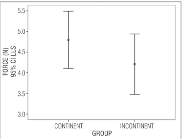
Figure 4. Mean and 95% confidence interval (CI) of the left-right active strength (LRS) of the pelvic floor muscles of the continent and incontinent groups (p>0.05).
FORCE (N) 95% CI LLS
3.0 3.5 4.0 4.5 5.0 5.5
GROUP
CONTINENT INCONTINENT
(0.1±0.1 N) and indicated a signiicant diference for the an-teroposterior active strength (p<0.01) - Figure 3; while left-right active strength (G1=0.43±0.1 N; G2=0.40±0.1 N) showed non-signiicant diference between groups (p=0.2) - Figure 4. he passive force (G1=1.1±0.2 N; G2=1.1±0.3 N) (p=0.89) and the vaginal cavity aperture (G1=21±3 mm; G2=24±4 mm) (p=0.06) did not difer between G1 and G2.
Discussion
The use of dynamometers to measure pelvic floor mus-cle function is recent and there are only few studies13,14,23
comparing the muscle strength in women with or without urinary incontinence.
he diference observed between the PFM anteroposterior active strength in continent and stress urinary incontinent women supports the hypothesis that the strength of these muscles is related to the continence function. hese results were not in agreement with the study of Morin et al.13 and
DeLancey et al.23,which evaluated the anteroposterior active
strength with a dynamometer and did not observe signiicant diferences in relation to the maximum strength of continent and incontinent groups. It can be assumed that these disagree-ments could be related to the evaluation methodology applied as well as the diference in equipment used in both studies. In the Morin et al.13 study, statistical analyses were adjusted
between group diferences for age and parity. In addition, they looked at many diferent variables speciic to force production, where they found diferences in endurance and rate of force development, but not peak strength.
Strength evaluation in diferent directions simultaneously was only performed in one study24, which had not compared
continent and urinary incontinent women. According to Nishiyama et al.25 pressure is directly proportional to strength
even thought they are diferent measures. Many studies11,26,27
with pressure manometers, also known as perineometers, compared the PFM pressure of continent and incontinent women and found a higher pressure in continent women corroborating with the results of the PFM anteroposterior active strength obtained in the present study.
Verelst and Leivseth28 tested dynamometric equipment that
measures strength in left-right direction. hey reported that the resulting force applied by the PFM runs in an anteropos-terior direction, therefore it would be the most appropriate di-rection to evaluate the PFM strength even though the strength applied in the left-right direction is important for a greater understanding of the PFM contraction behavior as the force applied by these muscles does not occur in one direction only. In 2007, the same authors14 compared the strength of women
with continence and SUI using this equipment and did not observe any signiicant diference in left-right active strength between the groups, in agreement with results obtained in the present research. Peng et al.15 used a vaginal probe with four
pressure sensors to measure the direction and amount of pres-sure applied in the vagina at rest, and during contractions of the PFM in 23 continents women and in 10 women with SUI. he authors observed that the pressure in the anteroposterior direction was signiicantly greater than that exerted on the left-right direction between the two groups, conirming the results obtained in this study.
he results found for the values of passive force be-tween the continent and stress urinary incontinent groups were not in agreement Morin et al.13 study, which had
evalu-ated the passive force and found lower values in incontinent
References
1. Keller SL. Urinary incontinence: occurrence, knowledge, and attitudes among women aged 55 and older in a rural Midwesten setting. J Wound Ostomy Continence Nurs. 1999;26(1):30-8.
2. Guarisi T, Pinto Neto AM, Osis MJ, Pedro AO, Paiva LHC, Faúndes A. Incontinência urinária entre mulheres climatéricas brasileiras: inquérito domiciliar. Rev Saúde Pública. 2001;35(5):428-35.
3. Kenton K, Mueller ER. The global burden of female pelvic floor disorders. BJU Int. 2006;98 Suppl 1:1-5.
4. Wagner TH, Hu TW. Economic costs of urinary incontinence in 1995. Urology. 1998;51(3):355-61.
5. DeLancey JO. The pathophysiology of stress urinary incontinence in women and its implications for surgical treatment. World J Urol. 1997;15(5):268-74.
6. Dumoulin C, Bourbonnais D, Lemieux MC. Development of a dynamometer for measuring the isometric force of the pelvic floor musculature. Neurourol Urodyn. 2003;22(7):648-53.
7. Bø K, Sherburn M. Evaluation of female pelvic-floor muscle function and strength. Phys Ther. 2005;85(3):269-82.
8. Balmforth JR, Mantle J, Bidmead J, Cardozo L. A prospective observational trial of pelvic floor muscle training for female stress urinary incontinence. BJU Int. 2006;98(4):811-7.
9. Peschers UM, Gingelmaier A, Jundt K, Leib B, Dimpfl T. Evaluation of pelvic floor muscle strength using four different techniques. Int Urogynecol J Pelvic Floor Dysfunct. 2001;12(1):27-30.
10. Bø K. Pelvic floor muscle strength and response to pelvic floor muscle training for stress urinary incontinence. Neurourol Urodyn. 2003;22(7):654-8.
11. Amaro JL, Moreira EC, Gameiro MOO, Padovani CR. Pelvic floor muscle evaluation in incontinent patients. Int Urogynecol J Pelvic Floor Dysfunct. 2005;16(5):352-4.
12. Rett MT, Simões JA, Herrmann V, Marques AA, Morais SS. Existe diferença na contratilidade da musculatura do assoalho pélvico feminino em diversas posições? Rev Bras Ginecol Obstet. 2005;27(1):12-9.
13. Morin M, Bourbonnais D, Gravel D, Dumoulin C, Lemieux MC. Pelvic floor muscle function in continent and stress urinary incontinent women using dynamometric measurements. Neurourol Urodyn. 2004;23(7):668-74.
14. Verelst M, Leivseth G. Force and stiffness of the pelvic floor as function of muscle length: a comparison between women with and without stress urinary incontinence. Neurourol Urodyn. 2007;26(6):852-7.
15. Peng Q, Jones R, Shishido K, Omata S, Constantinou CE. Spatial distribution of vaginal closure pressures of continent and stress urinary incontinent women. Physiol Meas. 2007;28(11): 1429-50.
16. Nunes FR, Martins CC, Guirro ECO, Guirro RRJ. Reliability of bidirectional and variable-opening equipment for the measurement of pelvic floor muscle strength. PM R. 2011;3(1): 21-6.
17. Dumoulin C, Gravel D, Bourbonnais D, Lemieux MC, Morin M. Reliability of dynamometric measurements of the pelvic floor musculature. Neurourol Urodyn. 2004;23(2):134-42.
18. Hundley AF, Wu JM, Visco AG. A comparison of perineometer to brink score for assessment of pelvic floor muscle strength. Am J Obstet Gynecol. 2005;192(5):1583-91.
19. Bø K. Pelvic floor muscle exercise for the treatment of female stress urinary incontinence: validity of vaginal pressure measurements of pelvic floor muscle strength and the necessity of supplementary methods for control of correct contraction. Neurourol Urodyn. 1990;9(5): 479-87.
women when compared with the continent sample. Several factors may have inluenced the values obtained from the pas-sive force evaluation such as vaginal aperture, instruments used and the population studied.
Verelst and Leivseth14 and DeLancey et al.23 had
quanti-fied the passive force in women with SUI and continence and had not observed any between-group differences, which corroborate with the results of passive force found in the present study.
Rahn et al.29 reported that the vagina has a property called
anisotropy, which means that the vagina is directionally de-pendent on the load applied to it, which may explain the dif-ferences found in strength in many studies as the instruments used were not the same. Besides, also based on that property, the way of the instrument was coupled to the vaginal cavity can also generate diferences in the results observed30.
he dynamometer used in this study measured the PFM strength in anteroposterior and left-right directions with a variable opening (in mm), while other studies with dynamom-eters used a predetermined opening of the equipment13,14,24.
his variable aperture in mm means that for each woman the opening (in mm) was one, and to maintain uniformity of the sample a value of the passive force was standardized (in N). herefore, the recording of the active force was performed after opening the equipment to 4.9 N for all women ensuring that the force was not inluenced by diferences in the equipment itted from one woman to another. As soon as determining the value of opening equal for all volunteers (mm), it probably
allows a better attachment in women who only need to apply a lesser contraction compared to women who the attachment was not very satisfactory30.
The between-group difference in age is also a difficulty found in other studies. Morin et al.13 in their study
compar-ing the strength of the PFM in the continent and inconti-nent groups reported that even with an identical procedure for both groups, significant differences were found in the variables age and parity. Verelst and Leivseth14 also found
significant difference in age between the continent and incontinent women. Hannestad et al.31 demonstrated an
increased prevalence of UI with advancing age.
One probably limitation of this study is the maximum PFM contraction that can include contraction of other muscles, especially rectus abdominis. In this study, we asked the volunteers to contract the PFM at the maximum contraction they could perform without the interference of other muscles, which could not stimulate the volunteers to perform a maximum contraction of the muscles studied as the purpose of the study was to evaluate the PFM itself without the influence of other muscle groups with the aim of achieving reliable data.
It would not be possible to evaluate women who have discomfort or pain while using the equipment to measure the PFM strength.
he current research highlights that there is a diferent be-havior of PFM in women who are continent and incontinent, in relation to anteroposterior active strength.
20. Cram JR, Kasman GS, Haltz J. Introduction to surface electromyography. Maryland: Aspen Publishers; 1998.
21. Guirro RRJ, Forti F, Bigaton DR. Proposal for electrical insulation of the electromyographic signal acquisition module. Electromyogr Clin Neurophysiol. 2006;46(6):355-63.
22. Sarwar R, Niclos BB, Rutherford OM. Changes in muscle strength, relaxation rate and fatiguability during the human menstrual cycle. J Physiol. 1996;493(Pt 1):267-72.
23. DeLancey JOL, Trowbridge ER, Miller JM, Morgan DM, Guire K, Feener DE, et al. Stress urinary incontence: relative importance of urethral support and urethral closure pressure. J Urol. 2008;179(6):2286-90.
24. Constantinou CE, Omata S. Direction sensitive sensor probe for the evaluation of voluntary and reflex pelvic floor contractions. Neurourol Urodyn. 2007;26(3):386-91.
25. Nishiyama M, Kimura Y, Nishiyama Y, Terazima M. Pressure-induced changes in the structure and function of the kinesin-microtubule complex. Biophys J. 2009;96(3):1142-50.
26. Mørkved S, Salvesen KA, Bø K, Eik-Nes S. Pelvic floor muscle strength and thickness in continent and incontinent nulliparous pregnant women. Int Urogynecol J Pelvic Floor Dysfunct. 2004;15(6):384-90.
27. Thompson JA, O’Sullivan PB, Briffa NK, Neumann P. Assessment of voluntary pelvic floor muscle contraction in continent and incontinent women using transperineal ultrasound, manual muscle testing and vaginal squeeze pressure measurements. Int Urogynecol J Pelvic Floor Dysfunct. 2006;17(6):624-30.
28. Verelst M, Leivseth G. Force-lenght relationship in the pelvic floor muscles under transverse vaginal distension: a method study in healthy women. Neurourol Urodyn. 2004;23(7): 662-7.
29. Rahn DD, Ruff MD, Brown SA, Tibbals HF, Word RA. Biomechanical properties of the vaginal wall: effect of pregnancy, elastic fiber deficiency, and pelvic organ prolapse. Am J Obstet Gynecol. 2008;198(5):590.e1-6.
30. Nunes FR, Guirro ECO, Martins CC, Guirro RRJ. Influence of visual feedback on pelvic floor muscle strength. Eur J Obstet Gynecol Reprod Biol. 2010;151(2): 217-20.
31. Hannestad YS, Rortveit G, Sandvik H, Hunskaar S; Norwegian EPINCONT study. Epidemiology of Incontinence in the County of Nord-Trøndelag. A community-based epidemiological survey of female urinary incontinence: The Norwegian EPINCONT Study. Epidemiology of Incontinence in the County of Nord-Trøndelag. J Clin Epidemiol. 2000;53(11): 1150-7.