Effect of supplementation of green
tea polyphenols on the developmental
competence of bovine oocytes
in vitro
Key Laboratory for Molecular Animal Nutrition, Ministry of Education, College of Animal Science, Zhejiang University, Hang Zhou, Zhejiang, China Z.G. Wang, S.D. Yu and
Z.R. Xu
Abstract
The objective of the present study was to examine the effect of green tea polyphenols (GTPs) supplementation during in vitro maturation, in vitro fertilization, and in vitro culture on the developmental
compe-tence of bovine oocytes. Cumulus-oocyte complexes aspirated from the ovaries were matured in vitro (38.5ºC for 24 h) and fertilized
(38.5ºC for 15-18 h) and embryos were cultured (38.5ºC for 192 h) in a defined conditioned medium with or without GTPs supplementa-tion. The GTPs used in the present study contained 99% catechin derivatives, with the major components being 50% (-)-epigallocate-chin gallate, 22% (-)-epicate(-)-epigallocate-chin gallate, 18% (-)-epigallocate(-)-epigallocate-chin, and 10% (-)-epicatechin. Four replicate trials were done for each type of experiment. GTPs supplementation (15 µM) of the maturation medium led to a significant increase in the rate of blastocyst formation (34.0 vs 21.4%, P < 0.05). However, the rate of blastocyst formation
was not improved when higher GTPs concentrations (20 or 25 µM) were added to the in vitro maturation medium. During in vitro
fertilization, supplementation with higher GTPs concentrations (20 or 25 µM) significantly reduced the rate of blastocyst formation (P < 0.05). Supplementation of the culture medium with 15 µM GTPs improved the rate of blastocyst formation, while higher GTPs concen-trations (25 µM) significantly reduced embryo development (P < 0.05). In conclusion, these results demonstrate that supplementation with GTPs at low concentration (15 µM) during in vitro maturation
and in vitro culture improved the developmental competence of
bovine oocytes.
Correspondence
S.D. Yu
College of Animal Science Zhejiang University Huajiachi Campus Hang Zhou 310029 China
Fax: +86-571-86994963 E-mail: [email protected] Research supported by the Zhejiang Science and Technology Committee (grant No. 2004C12037).
Received May 25, 2006 Accepted March 26, 2007
Key words
•Bovine oocytes •In vitro culture •Green tea polyphenols
•Embryo development
Introduction
The formation of reactive oxygen species (ROS) such as superoxide anions (O2-·), hy-droxyl radicals (OH·) and hydrogen peroxide (H2O2) is a normal process that occurs in the cell when there is a deviation of electrons to oxygen (O2) during electron transfer reactions (1). There is some evidence that ROS may be
beneficial at some steps of reproduction to permit successful gamete interaction (2). How-ever, an increasing number of in vitro studies
have demonstrated the detrimental effects of ROS on reproduction. The main detrimental effects include reduced sperm motility and axonemal protein phosphorylation (3), in vitro
In vitro oocyte or embryo culture results
in higher O2 concentrations than in in vivo environments, leading to increased ROS lev-els (7). ROS such as O2-· are able to diffuse and pass through cell membranes and alter most types of cellular molecules such as lipids, proteins and nucleic acids. This can affect the early development of mouse, ham-ster, and bovine embryos (8-10). Living or-ganisms possess natural protective equiva-lents known as ROS scavengers (antioxi-dants) that counteract the negative effects of ROS. These antioxidants include enzymes such as superoxide dismutase, which will eliminate O2-·, catalase and selenium-depend-ent glutathione peroxidase, which will trans-form H2O2 into H2O and O2, as well as lipid-and water-soluble antioxidants such as vita-mins C, E and uric acid (11). However, during in vitro oocyte and embryo culture
the levels of antioxidants are lower than in vivo because the oocytes or embryos are
divorced from the donor body and do not benefit from the maternal antioxidant pro-tection. The addition of an antioxidant to the medium, therefore, may be important for in vitro oocyte maturation and in vitro embryo
culture.
Tea (Camellia sinensis) is one of the
most popular beverages consumed world-wide. Green tea polyphenols (GTPs) are the major water-soluble components of green tea infusions. The major GTPs are (-)-epi-gallocatechin gallate (EGCG), (-)-epicate-chin gallate (ECG), (-)-epicate(-)-epicate-chin (EC), and (-)-epigallocatechin (EGC). These catechins have a strong antioxidant activity (12) and are potent scavengers of ROS superoxide, hydrogen peroxide, hydroxyl radicals, and nitric oxide produced by various chemicals (13).
There are no reports on the use of GTPs during in vitro maturation (IVM) of bovine oocytes and in vitro culture (IVC) of bovine
embryos. Thus, the objective of the present study was to examine the effects of GTPs supplementation during IVM, in vitro
fertili-zation (IVF) and IVC on the developmental competence of bovine oocytes.
Material and Methods
Reagents
All chemicals and media were purchased from Sigma (St. Louis, MO, USA) unless otherwise indicated. The green tea polyphe-nols (from the Tea Department of Zhejiang University) used in the present study con-tained 99% catechin derivatives, with the major components being 50% EGCG, 22% ECG, 18% EGC, and 10% EC. The average molecular weight of GTPs was estimated on the basis of the percent presence of major components.
In vitro maturation
Cumulus-oocyte complexes (COCs) were aspirated from 2- to 6-mm follicles of bovine ovaries obtained from a slaughterhouse. Oo-cytes with intact cumulus cells and evenly granulated cytoplasm were selected and ran-domly assigned to each treatment. Ten COCs were washed and incubated with droplets of IVM medium, which consisted of modified synthetic oviduct fluid (m-SOF) supplemented with minimum essential medium non-essen-tial amino acids (Gibco, Grand Island, NY, USA), minimum essential medium essential amino acids (Gibco), 1.5 mM glucose, and 1 mM glutamine. The SOF medium used in this study was based on the original formulation (14), with subsequent modifications (15). The IVM drops were covered with mineral oil and incubated at 38.5ºC in 5% CO2 in air at satu-rated humidity for 24 h.
In vitro fertilization
bicarbonate-buffered modified TALP) (17) and then placed in 50-µL droplets (10-12 COCs per droplet) of IVF medium containing 10 µg/ mL heparin. Frozen bull semen was thawed and prepared by the swim-up procedure (17). Sperm cells were added to the IVF drops at a final concentration of 2 x 10⁶/mL. Incuba-tion was carried out at 38.5ºC in 5% CO2 in air at saturated humidity for 15-18 h.
In vitro culture
Between 15 and 18 h after insemination, surrounding cumulus cells of presumptive zygotes were denuded by repeated pipetting in phosphate-buffered saline (PBS) and sub-sequently washed three times in PBS before being transferred to the IVC drops (20-30 embryos/50-µL drops). In all experiments, embryos were cultured in m-SOF medium containing 0.8% bovine serum albumin at 38.5ºC in 5% CO2 and 7% O2 with high humidity.
Cleavage and development of embryos to the blastocyst stage were assessed at 48 and 192 h (i.e., on 8th day) post-insemina-tion, respectively, under a stereomicroscope (60X). After removing the zona pellucida by immersing in acid Tyrode solution, pH 2.5, blastocysts were fixed in ethanol:acetic acid (3:1) and stained with 0.24% basic fuchsin. The number of cells in blastocysts was ex-amined by phase contrast microscopy.
Experimental design
Three separate experiments were per-formed to evaluate the effects of supplemen-tation with GTPs at different concentrations during different phases of the in vitro
pro-duction of bovine embryos. In Experiment 1, the GTPs at different concentrations (0, 10, 15, 20, or 25 µM) were added during IVM in defined condition medium under 20% O2 to reduce ROS production in the IVM medium. Oocytes cultured without GTPs supplementation were used as control.
In Experiment 2, bovine oocytes were matured in GTPs-free maturation medium. Oocytes were inseminated in the fertiliza-tion medium with different concentrafertiliza-tion of GTPs (0, 10, 15, 20, or 25 µM) under high (20%) O2 concentration and then compared with the control group (GTPs-free fertiliza-tion medium).
In Experiment 3, oocytes were matured and inseminated in GTPs-free medium. Vari-ous concentrations of GTPs (0, 10, 15, 20, or 25 µM) were added to the culture medium under low O2 (7%).
Statistical analysis
Data from four replicate trials were ana-lyzed statistically for comparison of each treat-ment by ANOVA and the Fisher protected least significant difference test using the STATVIEW program (Abacus Concepts, Inc., Berkeley, CA, USA). All percent values were subjected to arc sine transformation before statistical analysis. Data are reported as means ± SEM. A probability of P < 0.05 was consid-ered to be statistically significant.
Results
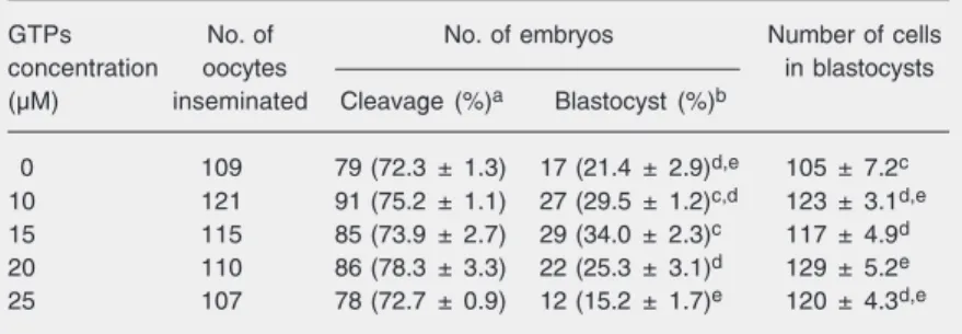
In the first experiment (Table 1), oocytes were cultured for 24 h in IVM medium
Table 1. Effect of green tea polyphenols (GTPs) concentration in the maturation medium on the development of bovine embryos in vitro.
GTPs No. of No. of embryos Number of cells
concentration oocytes in blastocysts
(µM) inseminated Cleavage (%)a Blastocyst (%)b
0 109 79 (72.3 ± 1.3) 17 (21.4 ± 2.9)d,e 105 ± 7.2c
10 121 91 (75.2 ± 1.1) 27 (29.5 ± 1.2)c,d 123 ± 3.1d,e
15 115 85 (73.9 ± 2.7) 29 (34.0 ± 2.3)c 117 ± 4.9d
20 110 86 (78.3 ± 3.3) 22 (25.3 ± 3.1)d 129 ± 5.2e
25 107 78 (72.7 ± 0.9) 12 (15.2 ± 1.7)e 120 ± 4.3d,e
aNumber of embryos is reported as median and percent of embryos cleaved as
percent of the number of inseminated oocytes (mean ± SEM), or bpercent of
blasto-cysts as percent of the number of cleaved embryos (mean ± SEM). c,d,eValues with
supplemented with GTPs at various concen-trations and then fertilized in vitro and
trans-ferred to IVC medium to assess the cleavage rates. There were no significant differences in cleavage rate among treatments. How-ever, on the 8th day after transfer in 7% O2, the presence of 15 µM GTPs during IVM significantly increased (P < 0.05) the rate of blastocyst formation and the cell number of blastocysts compared to control (34.0 vs
21.4% and 117 vs 105%). The cell numbers
of blastocysts were significantly higher in all four treatment groups than in the control group (P < 0.05). However, treatment with GTPs at a concentration of 25 µM showed a
significantly reduced number of blastocysts (P < 0.05).
In the second experiment (Table 2), oo-cytes were matured in GTPs-free maturation medium and fertilized in IVF medium sup-plemented with GTPs at various concentra-tions (0, 10, 15, 20, or 25 µM). No improve-ment in cleavage or blastocyst rate was found when 10 µM GTPs was added to the fertili-zation medium compared to control (78.3 vs
79.3% and 23.2 vs 25.1%). However,
sup-plementation with GTPs at 15, 20, and 25 µM concentrations significantly decreased the cleavage rates compared to control (P < 0.05). Furthermore, higher concentrations of GTPs (20 and 25 µM) significantly re-duced (P < 0.05) the rate of blastocyst for-mation. No improvement in the cell numbers of blastocysts was found when higher con-centrations of GTPs (20 and 25 µM) were added to fertilization medium compared to control.
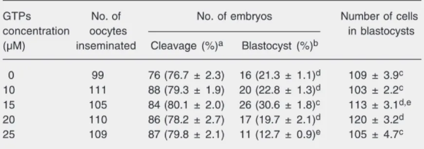
In the third experiment (Table 3), oo-cytes were matured in vitro and fertilized in
GTPs-free medium. Subsequently, the zy-gotes were cultured in IVC medium supple-mented with GTPs at various concentra-tions. There was no significant difference in cleavage rate between the treatment and con-trol groups (P > 0.05). The addition of 15 µM GTPs during IVC increased the rate of blas-tocyst formation and blasblas-tocyst cell number compared to control. However, the blasto-cyst rate and the cell numbers in blastoblasto-cysts were significantly reduced (P < 0.05) when 25 µM GTPs was added to the medium compared with 15 µM GTPs supplementa-tion.
Discussion
The present study assessed the effects of GTPs supplementation during IVM, IVF and IVC on the developmental competence of bovine oocytes. The principal finding is that supplementation with 15 µM GTPs during IVM and IVC improves the rate of
blasto-Table 2. Effect of green tea polyphenols (GTPs) concentration in the fertilization medium on the development of bovine embryos in vitro.
GTPs No. of No. of embryos Number of cells
concentration oocytes in blastocysts
(µM) inseminated Cleavage (%)a Blastocyst (%)b
0 106 84 (79.3 ± 2.4)c 21 (25.1 ± 1.8)c 98 ± 5.2c
10 110 86 (78.3 ± 3.1)c 20 (23.2 ± 1.3)cd 113 ± 2.1d
15 117 74 (63.5 ± 2.5)d 14 (19.0 ± 2.0)d 117 ± 1.9d
20 121 76 (62.7 ± 3.3)d 12 (15.8 ± 2.1)de 106 ± 3.2c
25 108 65 (60.4 ± 2.7)d 5 (7.7 ± 1.9)f 103 ± 2.7c
aNumber of embryos is reported as median and percent of embryos cleaved as
percent of the number of inseminated oocytes (mean ± SEM), or bpercent of
blasto-cysts as percent of the number of cleaved embryos (mean ± SEM). c,d,eValues with
different superscripts within a column are significantly different from each other (P < 0.05, ANOVA followed by the Fisher protected least significant difference test).
Table 3. Effect of green tea polyphenols (GTPs) concentration in the culture medium on the development of bovine embryos in vitro.
GTPs No. of No. of embryos Number of cells
concentration oocytes in blastocysts
(µM) inseminated Cleavage (%)a Blastocyst (%)b
0 99 76 (76.7 ± 2.3) 16 (21.3 ± 1.1)d 109 ± 3.9c
10 111 88 (79.3 ± 1.9) 20 (22.8 ± 1.3)d 103 ± 2.2c
15 105 84 (80.1 ± 2.0) 26 (30.6 ± 1.8)c 113 ± 3.1d,e
20 110 86 (78.2 ± 2.7) 17 (19.7 ± 2.1)d 120 ± 3.2d
25 109 87 (79.8 ± 2.1) 11 (12.7 ± 0.9)e 105 ± 4.7c
aNumber of embryos is reported as median and percent of embryos cleaved as
percent of the number of inseminated oocytes (mean ± SEM), or bpercent of
blasto-cysts as percent of the number of cleaved embryos (mean ± SEM). c,d,eValues with
cyst formation. However, the blastocyst rates were not improved by supplementation with 20 or 25 µM GTPs during IVM and IVC. Also, supplementation with GTPs at differ-ent concdiffer-entrations (0, 10, 15, 20, or 25 µM) during IVF did not improve the proportion of embryos reaching the blastocyst stage.
To our knowledge, the present study is the first to demonstrate the beneficial effect of GTPs supplementation of IVM or IVC medium on early bovine embryo develop-ment. Tea polyphenols, especially GTPs, have been shown to be useful as antidia-betic, antitumor, antiarthritic, and antioxi-dant agents (18-21). The antioxiantioxi-dant effects of tea polyphenols are thought to be associ-ated with their ability to stimulate the antiox-idant defense metabolism through redox-regulated transcription factors and mitogen-activated protein kinase-dependent cell cycle regulation (22,23).
The present results indicate that the addi-tion of 15 µM GTPs to the defined IVM medium (Experiment 1) significantly im-proved (P < 0.05) the rate of blastocyst formation. This enhancement could be at-tributed to the efficient GTPs-induced pro-tection of oocytes against oxidative stress during IVM. It has been reported that other antioxidants such as ß-mercaptoethanol, cys-teine and cystine added during IVM of bo-vine oocytes also improve the rate of em-bryo development to the blastocyst stage (24). These results suggest that supplemen-tation with appropriate GTPs concentrations during IVM could lead to subsequent im-provement of embryo development.
The results of Experiment 2 demonstrated that supplementation with low GTPs con-centrations (10 µM) during IVF had no ef-fect on the percentage of embryos produced, but higher concentrations of GTPs (15, 20, or 25 µM) significantly reduced the cleavage and blastocyst rates (P < 0.05). Ali et al. (24) and Luvoni et al. (7) reported that antioxi-dants such as superoxide dismutase and cys-teine present during IVF could significantly
reduce the percentage of embryos produced, suggesting that ROS might play a positive role during IVF. Furthermore, previous stud-ies implied that ROS were involved in the control of capacitation (25), acrosomal reac-tion (26) and fertilizareac-tion (22).
The results obtained here in Experiment 3 indicate that the proportion of oocytes developing to the blastocyst stage was sig-nificantly higher (P < 0.05) in the presence of a low GTPs concentration in a defined culture medium. This improvement in em-bryo development might be due to the anti-oxidant effect of GTPs, which scavenge ROS during in vitro embryo culture. Embryos are
inevitably exposed to high oxygen in vitro, a
fact that results in a higher production of ROS (7) than in vivo because of the
neces-sary manipulations with transient exposure to atmospheric oxygen. ROS seems to be responsible for numerous types of embryo damage. ROS such as superoxide anions are able to diffuse and pass through cell mem-branes and alter most types of cellular mol-ecules such as lipids, proteins and nucleic acids. The consequences are multiple, and include mitochondrial alterations, embryo cell-block, ATP depletion, and apoptosis (27). The detrimental effects of superoxide anion radicals on embryo development have been demonstrated in cattle (10,28). Fujitani et al. (29) reported that the development of bovine embryos to blastocysts was decreased in a dose-dependent manner when free radi-cals were generated in vitro by the addition
of 2,2'-azobis (2-amindinopropane) dihydro-chloride to the culture medium.
concentration ranges might have deleterious effects on the in vitro maturation events
occurring in both the nucleus and the cyto-plasm and on subsequent embryo develop-ment.
Our results demonstrate that 15 µM GTPs
supplementation during IVM or IVC im-proves the developmental competence of bovine oocytes possibly by protecting the embryos from oxidative stress. However, the GTPs supplementation during IVF may not improve the rate of blastocyst formation.
References
1. Ho YS, Dey MS, Crapo JD. Antioxidant enzyme expression in rat lungs during hyperoxia. Am J Physiol 1996; 270: L810-L818. 2. Miesel R, Drzejczak PJ, Kurpisz M. Oxidative stress during the
interaction of gametes. Biol Reprod 1993; 49: 918-923.
3. Aitken RJ, Buckingham D, Harkiss D. Use of a xanthine oxidase free radical generating system to investigate the cytotoxic effects of reactive oxygen species on human spermatozoa. J Reprod Fertil
1993; 97: 441-450.
4. Nasr-Esfahani MH, Johnson MH. Quantitative analysis of cellular glutathione in early preimplantation mouse embryos developing in vivo and in vitro. Hum Reprod 1992; 7: 1281-1290.
5. Watson AJ, Watson PH, Warnes D, Walker SK, Armstrong DT, Seamark RF. Preimplantation development of in vitro-matured and
in vitro-fertilized ovine zygotes: comparison between coculture on oviduct epithelial cell monolayers and culture under low oxygen atmosphere. Biol Reprod 1994; 50: 715-724.
6. Blondin P, Coenen K, Sirard MA. The impact of reactive oxygen species on bovine sperm fertilizing ability and oocyte maturation. J Androl 1997; 18: 454-460.
7. Luvoni GC, Keskintepe L, Brackett BG. Improvement in bovine embryo production in vitro by glutathione-containing culture media.
Mol Reprod Dev 1996; 43: 437-443.
8. Umaoka Y, Noda Y, Narimoto K, Mori T. Effects of oxygen toxicity on early development of mouse embryos. Mol Reprod Dev 1992; 31: 28-33.
9. McKiernan SH, Bavister BD. Environmental variables influencing in vitro development of hamster 2-cell embryos to the blastocyst stage.
Biol Reprod 1990; 43: 404-413.
10. Nagao Y, Saeki K, Hoshi M, Kainuma H. Effects of oxygen concen-tration and oviductal epithelial tissue on the development of in vitro
matured and fertilized bovine oocytes cultured in protein-free medi-um. Theriogenology 1994; 41: 681-687.
11. Knapen MF, Zusterzeel PL, Peters WH, Steegers EA. Glutathione and glutathione-related enzymes in reproduction. A review. Eur J Obstet Gynecol Reprod Biol 1999; 82: 171-184.
12. Dufresne CJ, Farnworth ER. A review of latest research findings on the health promotion properties of tea. J Nutr Biochem 2001; 12: 404-421.
13. Schroeder P, Klotz LO, Sies H. Amphiphilic properties of (-)-epicatechin and their significance for protection of cells against peroxynitrite. Biochem Biophys Res Commun 2003; 307: 69-73. 14. Tervit HR, Whittingham DG, Rowson LE. Successful culture in vitro
of sheep and cattle ova. J Reprod Fertil 1972; 30: 493-497. 15. Gardner DK, Lane M, Spitzer A, Batt PA. Enhanced rates of
cleav-age and development for sheep zygotes cultured to the blastocyst stage in vitro in the absence of serum and somatic cells: amino
acids, vitamins, and culturing embryos in groups stimulate develop-ment. Biol Reprod 1994; 50: 390-400.
16. Bavister BD, Leibfried ML, Lieberman G. Development of preimplan-tation embryos of the golden hamster in a defined culture medium.
Biol Reprod 1983; 28: 235-247.
17. Parrish JJ, Susko-Parrish JL, Leibfried-Rutledge ML, Critser ES, Eyestone WH, First NL. Bovine in vitro fertilization with frozen-thawed semen. Theriogenology 1986; 25: 591-600.
18. Ahmad N, Cheng P, Mukhtar H. Cell cycle dysregulation by green tea polyphenol epigallocatechin-3-gallate. Biochem Biophys Res Commun 2000; 275: 328-334.
19. Waltner-Law ME, Wang XL, Law BK, Hall RK, Nawano M, Granner DK. Epigallocatechin gallate, a constituent of green tea, represses hepatic glucose production. J Biol Chem 2002; 277: 34933-34940. 20. Lambert JD, Yang CS. Mechanisms of cancer prevention by tea
constituents. J Nutr 2003; 133: 3262S-3267S.
21. Raza H, John A. Green tea polyphenol epigallocatechin-3-gallate differentially modulates oxidative stress in PC12 cell compartments.
Toxicol Appl Pharmacol 2005; 207: 212-220.
22. Jiao HL, Ye P, Zhao BL. Protective effects of green tea polyphenols on human HepG2 cells against oxidative damage of fenofibrate.
Free Radic Biol Med 2003; 35: 1121-1128.
23. Williams RJ, Spencer JP, Rice-Evans C. Flavonoids: antioxidants or signalling molecules? Free Radic Biol Med 2004; 36: 838-849. 24. Ali AA, Bilodeau JF, Sirard MA. Antioxidant requirements for bovine
oocytes varies during in vitro maturation, fertilization and develop-ment. Theriogenology 2003; 59: 939-949.
25. Aitken RJ, Buckingham DW, West K, Brindle J. On the use of paramagnetic beads and ferrofluids to assess and eliminate the leukocytic contribution to oxygen radical generation by human sperm suspensions. Am J Reprod Immunol 1996; 35: 541-551.
26. de Lamirande E, Tsai C, Harakat A, Gagnon C. Involvement of reactive oxygen species in human sperm acrosome reaction in-duced by A23187, lysophosphatidylcholine, and biological fluid ultrafiltrates. J Androl 1998; 19: 585-594.
27. Guerin P, El Mouatassim S, Menezo Y. Oxidative stress and protec-tion against reactive oxygen species in the pre-implantaprotec-tion embryo and its surroundings. Hum Reprod Update 2001; 7: 175-189. 28. Shamsuddin M, Larsson B, Gustafsson H, Rodriguez-Martinez H. A
serum-free, cell-free culture system for development of bovine one-cell embryos up to blastocyst stage with improved viability. Therio-genology 1994; 41: 1033-1043.
30. Yang GY, Liao J, Li C, Chung J, Yurkow EJ, Ho CT, et al. Effect of black and green tea polyphenols on c-jun phosphorylation and H(2)O(2) production in transformed and non-transformed human bronchial cell lines: possible mechanisms of cell growth inhibition and apoptosis induction. Carcinogenesis 2000; 21: 2035-2039. 31. Sakagami H, Arakawa H, Maeda M, Satoh K, Kadofuku T, Fukuchi
K, et al. Production of hydrogen peroxide and methionine sulfoxide by epigallocatechin gallate and antioxidants. Anticancer Res 2001; 21: 2633-2641.