Po ssible invo lve me nt o f A
1
re ce pto rs
in the inhibitio n o f go nado tro pin
se cre tio n induce d by ade no sine
in rat he mipituitarie s
in vitro
1Departamento de Fisiologia, Centro de Ciências Biológicas, Universidade Federal do Pará, Belém, PA, Brasil
2Departamento de Neurologia e Psiquiatria, Centro de Ciências da Saúde, Universidade Federal de Pernambuco, Recife, PE, Brasil
3Departamento de Fisiologia, Faculdade de Medicina de Ribeirão Preto, Universidade de São Paulo, Ribeirão Preto, SP, Brasil
4Pennington Biomedical Research Center (LSU), Baton Rouge, LA, USA D.L.W. Picanço-Diniz1,
M.M. Valença2, A.L.V. Favaretto3, S.M. McCann4 and J. Antunes-Rodrigues3
Abstract
We investigated the participation of A1 or A2 receptors in the gonad-otrope and their role in the regulation of LH and FSH secretion in adult rat hemipituitary preparations, using adenosine analogues. A dose-dependent inhibition of LH and FSH secretion was observed after the administration of graded doses of the R-isomer of phenylisopropylad-enosine (R-PIA; 1 nM, 10 nM, 100 nM, 1 µM and 10 µM). The effect of R-PIA (10 nM) was blocked by the addition of 8-cyclopentyltheo-phylline (CPT), a selective A1 adenosine receptor antagonist, at the dose of 1 µM. The addition of an A2 receptor-specific agonist, 5-N-methylcarboxamidoadenosine (MECA), at the doses of 1 nM to 1 µM had no significant effect on LH or FSH secretion, suggesting the absence of this receptor subtype in the gonadotrope. However, a sharp inhibition of the basal secretion of these gonadotropins was observed after the administration of 10 µM MECA. This effect mimicked the inhibition induced by R-PIA, supporting the hypothesis of the pres-ence of A1 receptors in the gonadotrope. R-PIA (1 nM to 1 µM) also inhibited the secretion of LH and FSH induced by phospholipase C (0.5 IU/ml) in a dose-dependent manner. These results suggest the presence of A1 receptors and the absence of A2 receptors in the gonadotrope. It is possible that the inhibition of LH and FSH secretion resulting from the activation of A1 receptors may have occurred independently of the increase in membrane phosphoinositide synthe-sis.
Co rre spo nde nce
J. Antunes-Rodrigues Departamento de Fisiologia Faculdade de Medicina de Ribeirão Preto, USP Av. Bandeirantes, 3900
14049-900 Ribeirão Preto, SP Brasil
Research supported by FAPESP (Nos. 91/0567-0 and 94/3805-7), CNPq (Nos. 50167/91-7 and 521593/94-8) and PRO NEX (No. 76.97.1048-00).
Received March 19, 1999 Accepted July 14, 1999
Ke y words
·Adenosine ·LH ·FSH ·A1 receptor ·Anterior pituitary
Intro ductio n
The characterization of adenosine recep-tors is currently based on the selective bind-ing of analogues containbind-ing carboxamide groups in their structure to A2 receptors or of purine derivatives with modifications in the
K+ efflux), and A1B receptors (which may inhibit adenyl cyclase activity), with the ef-fects of their activation possibly being medi-ated by membrane GP (2). More recently, coupling of the A1 receptors to G0 and Gi has been characterized using the R-isomer of phenylisopropyladenosine (R-PIA) as the binding agonist in the preparation (3). Ca2+ efflux from the cell may also be mediated by GP after the activation of A1 receptors, a mechanism that utilizes calcium exchange with Na+ and is dependent on a pertussis toxin-sensitive pathway (4).
New adenosine receptor subtypes have been described on the basis of their affinity for 5’-N-ethylcarboxamideadenosine (NECA). A2a receptors have high affinity and A2b receptors have low affinity for the agonist. Variations in tissue distribution and differences in binding capacity indicate that these receptor subtypes may be different proteins (5,6).
The activation of purinergic receptors may also modify the capacity for inositol triphosphate synthesis in different types of experimental preparations (7,8). Studies on isolated sympathetic ganglia demonstrated that endogenously released adenosine may inhibit postsynaptic stimulation and [3 H]myo-inositol release (8). Other data have sug-gested that adenosine receptors that modu-late membrane phosphoinositide hydrolysis do not interfere with the generation of cyclic adenosine monophosphate (cAMP) in cere-bral cortex slices (9). However, experiments with GH3 cell lines showed that R-PIA ad-ministration inhibited the release of prolac-tin induced by thyrotropin releasing hor-mone (TRH) by blocking the synthesis of phosphatidylinositol and cAMP (10).
The participation of adenosine in pitu-itary gonadotropin secretion has not been fully clarified. In previous studies we dem-onstrated that adenosine causes a dose-de-pendent reduction of basal LH and FSH or LHRH-stimulated secretion by hemipitui-taries in vitro (11). The inhibitory effect of
adenosine was potentiated by the simulta-neous addition of dipyridamole, a blocker of adenosine reuptake by the cell, demonstrat-ing that the purinergic action may have re-sulted from the activation of outer mem-brane receptors (12). Although we did not observe any effect of dipyridamole alone, it is probable that adenosine may be released by pituitary cells. Other investigators have detected adenosine release (100 nM) into the incubation bath, associated with the release of the enzyme adenosine deaminase in prepa-rations of cells of the GH4C1 line, showing that the levels of released adenosine can be regulated within strict limits (13).
In the present study we report results suggesting the presence of subtype A1 aden-osine receptors in the gonadotrope and their involvement in the synthesis of membrane phosphoinositides.
Mate rial and Me thods
Male Wistar rats (200 to 220 g) housed in collective cages, at controlled temperature (22 to 24oC) and with 14 h of light and 10 h of dark, with free access to solid food and water, were used.
Drugs and solutions
Expe rime ntal proce dure s
After a period of adaptation to the labora-tory of approximately 1 h, the animals were sacrificed by decapitation at 10:00 a.m. in all experiments. The brain was removed and the anterior pituitary dissected in situ. The ante-rior pituitary was bisected longitudinally and immersed into refrigerated nutrient solution (4oC). Each hemipituitary was placed in an incubation flask containing 1 ml of nutrient solution (37oC). After 1 h of pre-incubation in a Dubnoff metabolic shaker (80 cycles/ min) for washing and stabilization of basal hormonal secretion levels, the medium was replaced with 1 ml of fresh solution contain-ing the test substances. After 60 min of incubation the samples were collected in chilled plastic tubes and kept at -20oC for later determination of LH and FSH by radio-immunoassay. The hemipituitaries were weighed and hormone concentrations in the nutrient solution were expressed as ng/mg tissue weight. At the end of each experiment, 56 mM KCl was added to evaluate the func-tional viability of cells in the preparation on the basis of LH and FSH release from intra-cellular stores. The cells maintained their secretory response for more than 135 min of incubation, thus guaranteeing the viability of the preparation (data not shown).
Radio im m uno assay
LH and FSH concentrations in the nutri-ent solution were determined by double-antibody radioimmunoassay (RIA) (14). The hormones for radioiodination and specific antibodies were obtained from the National Institute of Arthritis, Diabetes and Digestive Diseases (NADDK, Baltimore, MD, USA) Rat Pituitary Hormone Program.
Statistical analysis
Data are reported as means ± SEM and were analyzed using the GBSTAT computer
program. Statistical analysis was performed by analysis of variance (ANOVA), with the level of significance set at P<0.05 with the Newman-Keuls test.
Re sults
Inhibition of LH and FSH se cre tion induce d by the activation of A1 re ce ptors by R-PIA or MECA
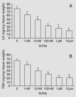
Increasing R-PIA concentrations (1 nM, 10 nM, 100 nM, 1 µM and 10 µM) caused a substantial and graded reduction of LH and FSH release. This effect was dose dependent and the maximum inhibition was reached with the 1 µM dose. The dose-response curves for the two hormones were similar in terms of the pattern of inhibition observed (Figure 1A,B).
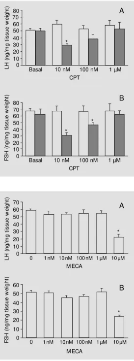
Previous incubation for 30 min with in-creasing doses of CPT (10 nM, 100 nM, 1 µM and 10 µM), an A1 receptor-specific antagonist, was performed to determine which dose would block the response in-duced by the addition of R-PIA to the nutri-ent solution. Treatmnutri-ent with the antagonist alone had no effect on basal LH or FSH
L
H
(
n
g
/m
g
t
is
s
u
e
w
e
ig
h
t) 90
70 60 50 40 30 20 10 0
0 1 nM 10 nM 100 nM 1 µM 10 µm
R-PIA
* *
* * *
80
F
S
H
(
n
g
/m
g
t
is
s
u
e
w
e
ig
h
t)
70 60 50 40 30 20 10 0 80
* *
*
* *
0 1 nM 10 nM 100 nM 1 µM 10 µm
R-PIA
A
B
Figure 1 - Effects of A1 receptor
activation by different concen-trations of R-PIA on basal LH (A) and FSH (B) secretion. Wistar rat hemipituitaries w ere pre-in-cubated for 60 min for stabiliza-tion of preparastabiliza-tion and incu-bated in new Earle salt solution supplemented w ith 0.1% BSA and 15 mM HEPES at 37oC, pH
Administration of MECA at concentra-tions of 1 µM or less did not induce any changes in basal LH and FSH secretion. A significant inhibition of approximately 50% of LH and FSH secretion occurred when MECA was added at the dose of 10 µM (Figure 3A,B).
Effe cts of R-PIA administration on LH and FSH se cre tion stimulate d by phospholipase C
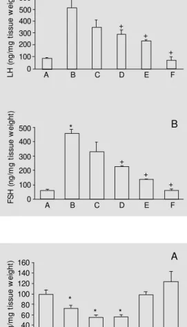
In this experiment we used phospholipase C to determine whether the inhibitory effect of R-PIA on LH and FSH secretion continued after the activation of inositol triphosphate and diacylglycerol promoted by this enzyme. The addition of phospholipase C (0.5 IU/ml) to the incubation medium induced a substantial in-crease in basal LH and FSH levels in the nutrient solution. When phospholipase C was added in combination with different doses of R-PIA (1 nM, 10 nM, 100 nM, 1 µM and 10 µM) there was a dose-dependent decrease in LH and FSH secretion stimulated by phospho-lipase C (Figure 4A,B).
Effe cts of ade nosine administration on LH and FSH se cre tion inhibite d by LiCl
To determine whether functional impair-ment of the cycle of membrane phosphoino-sitide synthesis interferes with basal or aden-osine-inhibited LH and FSH secretion, we added 5 mM LiCl alone or in combination with 10 nM adenosine to the incubation medium. LiCl induced a significant decrease in basal LH and FSH secretion but had no effect on the secretion of these hormones when added in combination with 1 mM myo-inositol. This substrate, in turn, had no effect on LH and FSH secretion when added alone to the preparation. The administration of 10 nM adenosine alone or in combination with 5 mM LiCl elicited a significant decrease in basal LH and FSH secretion, with no signifi-cant differences between these effects (Fig-ure 5A,B).
Figure 3 - Effects of administra-tion of different M ECA concen-trations on basal LH (A) and FSH (B) secretion. The preparation conditions are the same as de-scribed in the legend to Figure 1. Data are reported as means ± SEM (N = 5). * P<0.05 compared to control (0) (New man-Keuls test). L H ( n g /m g t is s u e w e ig h t) 70 60 50 40 30 20 10 0 80 F S H ( n g /m g t is s u e w e ig h t) 70 60 50 40 30 20 10 0 80
Basal 10 nM 100 nM 1 µM
CPT
*
*
*
Basal 10 nM 100 nM 1 µM
CPT Figure 2 - Effects of previous
in-cubation (30 min) w ith CPT, an A1 antagonist, on LH (A) and FSH
(B) secretion inhibited by 10 nM R-PIA (closed bars). The prepara-tion condiprepara-tions are the same as described in the legend to Fig-ure 1. Dat a are report ed as means ± SEM (N = 5). * P<0.05 com pared t o cont rol (basal) (New man-Keuls test).
A
B
levels at any of the doses tested. Under con-ditions of equimolality, the antagonist did not block the inhibitory effects of R-PIA on hormonal secretion. Partial blockade occurred after administration of 100 nM CPT at 10 times higher concentration. Only at a 100 times higher concentration (1 µM) did we observe total blockade of the agonist effects on LH and FSH secretion (Figure 2A,B).
L H ( n g /m g t is s u e w e ig h t) 70 60 50 40 30 20 10 0
0 1 nM 100 nM 1 µM
M ECA
10 nM 10 µM
* A B F S H ( n g /m g t is s u e w e ig h t) 60 50 40 30 20 10 0
0 1 nM 100 nM 1 µM
M ECA
10 nM 10 µM
D iscussio n
The effects of R-PIA were similar for LH and FSH secretion, suggesting the existence of a single purinergic regulatory mechanism for both hormones (Figure 1A,B). This hy-pothesis is supported by the similar behavior resulting from the blockade of these effects induced by previous administration of CPT, a specific A1 receptor antagonist (Figure 2A,B). This blockade demonstrates the ex-istence of A1 receptors in the gonadotropes. The lack of effect of CPT on basal LH and FSH secretion suggests the absence of sig-nificant actions of endogenous adenosine in this type of experiment. On the other hand, studies with cultures of GH4C1 cell lines demonstrated adenosine release accompa-nied by adenosine deaminase release under basal experimental conditions. Besides, an increase in prolactin secretion was found when the enzyme was added alone, suggest-ing that released adenosine may have a tonic-inhibitory autocrine action (13). The pres-ence of an inhibitory effect of the A2 recep-tor agonist MECA on LH and FSH secretion only with the use of supramicromolar con-centrations may be by an effect on A1 recep-tors. The lack of effect of MECA at lower concentrations, where it would selectively activate A2 receptors, suggests the probable absence of A2 receptors in the gonadotrope (Figure 3A,B).
The increased LH and FSH basal secre-tion observed after the addisecre-tion of phospho-lipase C (Figure 4A,B) or the inhibition in-duced by the addition of LiCl (Figure 5A,B) to the medium supports other findings indi-cating the contribution of membrane phos-phoinositides as cell messengers to the regu-lation of the basal secretion of these hor-mones (15). The progressive and dose-re-lated inhibition of phospholipase C-stimu-lated LH and FSH secretion by R-PIA sug-gests that the activation of A1 receptors may inhibit the increase in LH and FSH secretion by a mechanism not dependent on the
secre-L H ( n g /m g t is s u e w e ig h t) 160 140 120 100 80 60 40 20 0 * * * E D
B C F
A F S H ( n g /m g t is s u e w e ig h t) 80 70 60 50 40 30 20 10 0 E D
B C F
A
*
* *
A
B
Figure 5 - Effects of administra-tion of 0.01 µM adenosine on LH (A) and FSH (B) secretion in-hibited by 5 mM LiCl. The prepa-ration conditions are the same as described in the legend to Figure 1. A = Basal, B = 10 nM adenosine, C = 5 mM LiCl + 10nM adenosine, D = 5 mM LiCl, E = 5 mM LiCl + 1 mM inositol, F = 1 mM myo-inositol. Data are reported as means ± SEM (N = 5). * P<0.05 com pared t o cont rol (A) (New man-Keuls test).
L H ( n g /m g t is s u e w e ig h t) 600 500 400 300 200 100 0 E D
B C F
+ A + + * A F S H ( n g /m g t is s u e w e ig h t) 500 400 300 200 100 0 + + + * E D
B C F
A
Figure 4 - Effects of administra-tion of different R-PIA doses on LH (A) and FSH (B) secretion stimulated by phospholipase C (0.5 IU/ml). The preparation con-ditions are the sam e as de-scribed in the legend to Figure 1. Data are reported as means ± SEM (N = 5). * P<0.01, +P<0.05
com pared t o cont rol (A) (New man-Keuls test).
B
combination of adenosine + LiCl and those obtained by the separate addition of each substance impairs the interpretation of the results.
Among the possible mechanisms that may mediate the inhibition of gonadotropin in-duced by the activation of A1 receptors, the one based on membrane GiP (guanine nucle-otide-binding membrane proteins) inhibition may best fit the present case. GiP inhibition may result in a decrease of both adenyl cy-clase and phospholipase C activity.
Recent studies have demonstrated that adenosine inhibits prolactin secretion by act-ing on a purinergic receptor that modulates the activity of the enzymes adenyl cyclase and phospholipase C by a mechanism de-pending on membrane GiP activation (16). Other studies have shown that administra-tion of R-PIA inhibits the release of PRL induced by TRH by inhibiting the decrease in phosphatidylinositol and cAMP synthesis in GH3 cell lines (10).
An alternative hypothesis is the blockade of Ca2+ influx or the increased Ca2+ efflux from the cell directly mediated at the mem-brane GP level. It has been demonstrated that the activation of A1 receptors reduced the concentration of basal cytoplasmic Ca2+ and of Ca2+ stimulated by protein kinase C in GH3B6 cell lines (17), suggesting that the inhibition may depend on the firing of a mechanism preceding the activation of this kinase. Ca2+ efflux from the cell may also be mediated by GP after A1 receptor activation, a mechanism based on Ca2+ exchange with Na+ and depending on a pathway sensitive to pertussis toxin (4). On the other hand, it has been recently demonstrated that A1
recep-tors decrease intracellular free calcium. This would induce a decrease in nitric oxide (NO) synthase activation in the gonadotropes, re-sulting in decreased NO synthesis. NO stimu-lates LH and FSH release by activating gua-nylate cyclase that synthesizes cGMP from GTP. cGMP concentrations increase in the gonadotropes inducing activation of LH and FSH release. Adenosine decreases NO re-lease and concentration in the gonadotrope cells, as well as cGMP formation followed by a consequent decrease of LH and FSH release (18).
In conclusion, the present results show that the activation of A1 receptors by R-PIA induced a dose-dependent inhibition of pitu-itary LH and FSH secretion by a mechanism not depending on increased membrane phos-phoinositide synthesis induced by phospho-lipase C, leading us to formulate the hypoth-eses that this inhibition may have occurred by mediation of a membrane GiP which, once activated by A1 receptors, may act at different levels, inhibiting cAMP and phos-phoinositide synthesis, and Ca2+ influx, or stimulating Ca2+ efflux from the cell. Aside from these preliminary speculations, the mechanisms involved in signal transduction by the activation of A1 receptors remain obscure and further experiments are needed to clarify the mechanisms.
Ackno wle dgm e nts
We thank Rubens Fernando de Melo, Leonardo Fidelis Filho, Gilberto Lopes, Manoel Corrêa de Lima, Marina Holanda and Maria Valci Aparecida dos Santos Silva for skillful technical assistance.
Re fe re nce s
1. Stone TW (1985). Summary of a sympo-sium discussion on purine receptors no-menclature. In: Stone TW (Editor), Phar-macology and Physiological Roles. VHC Publishers, Deerfield Beach, FL. 2. Fredholm BB & Dunw iddie TV (1988).
How does adenosine inhibit transmitter release? Trends in Pharmacological Sci-ences, 9: 130-134.
3. Freissmuth M , Selzer E & Schütz W (1991). Interactions of purified bovine brain A1-adenosine receptors w ith
G-pro-teins. Reciprocal modulation of agonist and antagonist binding. Biochemical Jour-nal, 275: 651-656.
heart sarcolemma is mediated by a per-tussis toxin-sensitive G protein. Journal of Biological Chem ist ry, 265: 16851-16855.
5. Lupica CR, Cass WA, Zahniser NR & Dunw iddie TV (1990). Effects of the se-lective adenosine A2 receptor agonist
CGS21680 on in vitro electrophysiology, cAM P formation and dopamine release in rat hippocampus and striatum. Journal of Pharmacology and Experimental Thera-peutics, 252: 1134-1141.
6. Hutchison KA & Fox IH (1989). Purifica-tion and characterizaPurifica-tion of the adenosine A2-like binding site from human placental membranes. Journal of Biological Chem-istry, 264: 19898-19903.
7. Arend LJ, Handler JS, Rhim JS, Gusovsky F & Spielman WS (1989). Adenosine-sen-sit ive phosphoinoAdenosine-sen-sit ide t urnover in a new ly established renal cell line. Ameri-can Journal of Physiology, 256: 1067-1074.
8. Rubio R, Bencherif M & Berne RM (1989). Inositol phospholipid metabolism during and follow ing synaptic activation: role of adenosine. Journal of Neurochemistry, 52: 797-806.
9. Alexander SPH, Kendall DA & Hill SJ (1989). Differences in the adenosine
re-ceptors modulating inositol phosphates and cyclic AM P accumulation in mamma-lian cerebral cortex. British Journal of Pharmacology, 98: 1241-1248.
10. Delahunty TM , Cronin M J & Linden J (1988). Regulation of GH3-cell function via
adenosine A1 receptors. Inhibition of
pro-lactin release, cyclic AM P production and inositol phosphate generation. Biochemi-cal Journal, 255: 69-77.
11. Picanço-Diniz DLW, López-Jiménez M , Valença M M , Favaretto ALV & Antunes-Rodrigues J (1989). Effect of adenosine on gonadotropin and prolactin secretion by hemipituitaries in vitro. Brazilian Jour-nal of M edical and Biological Research, 22: 783-785.
12. Picanço-Diniz DLW , Valença M M , Favaretto ALV & Antunes-Rodrigues J (1992). Dipyridamole amplifies the effects of adenosine on gonadotropin and prolac-tin release from the rat anterior pituitary gland. M edical Science Research, 20: 783-785.
13. Dorflinger LJ & Schonbrunn A (1985). A-denosine inhibits prolactin and grow th hormone secretion in a clonal pituitary cell line. Endocrinology, 117: 2330-2338. 14. Nisw ender GD, Chen CL, M igdley Jr AR,
M eites J & Ellis S (1969).
Radioimmu-noassay for rat prolactin. Proceedings of the Society for Experimental Biology and M edicine, 130: 793-797.
15. Naor Z (1990). Signal transduction mecha-nisms of Ca2+ mobilizing hormones: The
case of gonadotropin-releasing hormone. Endocrine Review s, 11: 326-353. 16. Scorziello A, Landolfi E, Grim aldi M ,
M eucci O, Vent ra C, Avallone A, Postiglione A & Schettini G (1993). Direct effect of adenosine on prolactin secretion at the level of the single rat lactotroph: involvement of pertussis toxin-sensitive and insensitive transducing mechanisms. Journal of M olecular Endocrinology, 11: 325-334.
17. M ollard P, Guérineau N, Chiavaroli C, Schlegel W & Cooper DM F (1991). Aden-osine A1 receptor-induced inhibition of
Ca2+ transients linked to action potentials
in clonal pituitary cells. European Journal of Pharmacology, 206: 271-277. 18. Yu WH, Kimura M , Walczew ska A, Porter