Potential mechanisms linking probiotics to diabetes:
a narrative review of the literature
Mecanismos potenciais ligando probióticos a diabetes: uma revisão
narrativa da literatura
Maryam Miraghajani
I, Somayeh Shahraki Dehsoukhteh
II, Nahid Raie
III, Sahar Golpour Hamedani
III, Sima Sabihi
III, Reza Ghiasvand
IVIsfahan University of Medical Sciences, Isfahan, Iran
ABSTRACT
CONTEXT AND OBJECTIVE: Some studies have suggested a wide range of possible mechanisms through which probiotics may play a role in diabetes prevention and treatment. However, the underlying mecha-nisms are not fully understood. We conducted this study to review the potential mechamecha-nisms suggested for the efect of probiotics in diabetes.
DESIGN AND SETTING: Narrative review conducted at the Food Security Research Center of Isfahan. METHODS: A search in the electronic databases MEDLINE (PubMed), Cochrane Library, Web of Science and Google scholar was performed up to October 2016.
RESULTS: The initial search yielded 1214 reports. After removing duplicates, 704 titles and abstracts were screened. Finally, out of 83 full-text articles that were reviewed for eligibility, 30 articles were included in the inal analysis. The anti-diabetic mechanisms for probiotics reported encompass intraluminal and direct efects on the intestinal mucosa and microbiota (n = 13), anti-inlammatory and immunomodulatory ef-fects (n = 10), antioxidative eef-fects (n = 5), eef-fects on endoplasmic reticulum (ER) stress and expression of genes involved in glucose homeostasis and insulin resistance (n = 6), with some studies pointing to more than one mechanism.
CONCLUSION: The results may throw some light on the capacity of probiotics as a novel approach to-wards controlling diabetes. However, further human studies are warranted to elucidate and conirm the potential role of probiotics in diabetes prevention and treatment. Also, it needs to be ascertained whether the efectiveness of probiotics in diabetes prevention and treatment is dependent on the strain of the microorganisms.
RESUMO
CONTEXTO E OBJETIVO: Alguns estudos têm sugerido ampla gama de possíveis mecanismos, pelos quais os probióticos podem desempenhar um papel na prevenção e tratamento do diabetes. No entanto, os mecanismos subjacentes não são totalmente compreendidos. Realizamos este estudo para revisar os possíveis mecanismos sugeridos para o efeito dos probióticos na diabetes.
TIPO DE ESTUDO E LOCAL: Revisão narrativa conduzida no Food Security Research Centro de Isfahan. MÉTODOS: Busca sistemática nas bases de dados eletrônicas MEDLINE (PubMed), Cochrane Library, Web of Science e Google scholar até outubro de 2016.
RESULTADOS: A busca inicial resultou em 1.214 artigos. Após a remoção de duplicatas, foram pesquisados 704 títulos e resumos. Finalmente, de 83 artigos completos revisados para elegibilidade, 30 foram incluídos na análise inal. Os mecanismos antidiabéticos relatados dos probióticos abrangem efeitos intraluminais e diretos na mucosa e microbiota intestinal (n = 13), efeitos anti-inlamatórios e imunomoduladores (n = 10), efeitos antioxidativos (n = 5), efeitos sobre o estresse de retículo endoplasmático (RE) e expressão de genes envolvidos na homeostase da glicose e resistência à insulina (n = 6), com alguns estudos apontando para mais de um mecanismo.
CONCLUSÃO: Os resultados podem lançar alguma luz sobre os probióticos como uma nova abordagem no controle do diabetes, no entanto, mais estudos em humanos são justiicados para elucidar e conirmar o papel potencial dos probióticos na prevenção e tratamento do diabetes. Além disso, deverá ser deter-minado se a eicácia dos probióticos na prevenção e tratamento do diabetes é dependente da cepa dos microrganismos.
IPhD. Doctoral Student, Cancer Research Center,
Shahid Beheshti University of Medical Sciences, Tehran, Iran.
IIMSc. Coach, Department of Statistics, Faculty of
Sciences, Zabol University, Zabol, Iran.
IIIMSc. Master’s Student, Food Security Research
Center, Department of Community Nutrition, School of Nutrition and Food Science, Isfahan University of Medical Sciences, Isfahan, Iran.
IVPhD. Professor, Food Security Research Center,
Department of Community Nutrition, School of Nutrition and Food Science, Isfahan University of Medical Sciences, Isfahan, Iran.
KEY WORDS:
Molecular mechanisms of pharmacological action. Probiotics.
Diabetes mellitus. Review. Microbiota.
PALAVRAS-CHAVE:
Mecanismos moleculares de ação farmacológica. Probióticos.
INTRODUCTION
Probiotics are live microorganisms that may exert beneicial efects regarding the suiciency of consumption via their impact on the microbial balance of the gut.1 he most commonly used
probiotics are Lactobacillus, Biidobacterium and Saccharomyces boulardii, which have diferent efects depending on the dosage, length of therapy and administration route.2
Given the influence of the gut microbiota on metabolic conditions including diabetes and on improving host metabo-lism, the concept of manipulating the gut microbiota has gained considerable interest over recent years. Use of probiotics has been suggested as one of the approaches towards modifying the clonal flora.3
Diabetes mellitus is a chronic metabolic disease with major complications largely inluenced by glycemic measures.1 he Global
Burden of Disease 2015 study (GBD 2015) showed that diabetes was among the leading causes of years of life lost (YLLs) in most regions.2 Also, diabetes was shown to be a leading cause of
dis-ability-adjusted life years (DALYs), for which the observed burden exceeded expected levels in many localities.3 he rise in diabetes
prevalence is set to pose one of the most important challenges to healthcare systems over the coming years.4
A growing body of evidence suggests that favorable associa-tions exist between probiotic consumption and metabolic proile among diabetes subjects.5 However, the potential mechanisms
underlying the efects of probiotics on glycemia-related param-eters are not fully understood. One of the main mechanisms pos-tulated may involve increased glucagon-like peptide 1 (GLP-1) secretion from enteroendocrine L-cells to improve carbohydrate metabolism, decrease glucotoxicity and increase insulin sensitivity of target cells.6 Other proposed mechanisms to explain the action
of probiotics on diabetes relate to anti-inlammatory, antioxidant and immunomodulatory efects and alteration of the expression of some genes involved in diabetes.7-10
Moreover, probiotic intake afects the structure of the gut lora, which might improve the integrity of the intestinal epithelium, weaken the immune responses and diminish the toll-like recep-tor 4 pathway, which in turn reduces pro-inlammarecep-tory signaling and enhances insulin sensitivity.11,12
Given the various statements regarding the efects of probiot-ics on diabetes that have been made, the aim of the present study was to focus on possible mechanisms for probiotics that might explain some of their beneicial efects in relation to diabetes, in the form of a review.
OBJECTIVE
he aim of the present study was to focus on possible mecha-nisms for probiotics that might explain some of their beneicial efects in relation to diabetes, in the form of a narrative review.
METHODS
Search strategy
A search of the electronic databases MEDLINE (via PubMed) and Cochrane Library (via Wiley) and the electronic reposi-tories Web of Science and Google Scholar was performed. he search was last performed in October 2016, using combinations of search terms including “probiotics” OR “probiotic” OR “lactic acid bacteria” OR “lactobacillus” OR “lactobacilli” OR “biido-bacterium” OR “biidobacteria” AND “diabetes mellitus”, without any restrictions, in order to ind studies focusing on the mecha-nisms linking probiotics with diabetes.
Eligibility criteria
Studies were included if they assessed the efect of a single or combination of live probiotics on diabetes. On the other hand, studies presented only as abstracts with no full-text available, non-English literature, studies involving patients with other met-abolic diseases such as obesity or hypercholesterolemia, studies with no probiotic genus/strains reported, studies using synbiotics (i.e. probiotics combined with prebiotics), study protocols, pilot studies, letters, editorials, obviously irrelevant studies and studies that included non-diabetic patients or animals were all excluded.
Selection strategy
he eligibility of all potential studies identiied for inclusion was inde-pendently assessed by two reviewers. Discrepancies regarding study inclusion were resolved through discussion with a third reviewer. Initially, titles and abstracts were veriied and then an assessment of full texts was conducted. he reference lists of eligible articles or rel-evant review papers were screened for other eligible papers.
Data extraction
Study characteristics from eligible articles such as the irst author’s name, year of publication, study design, subjects or ani-mal models, probiotic strain and suggested mechanisms for pro-biotics on diabetes were extracted by two authors. he details of all eligible articles are outlined in Table 1.10-39
RESULTS
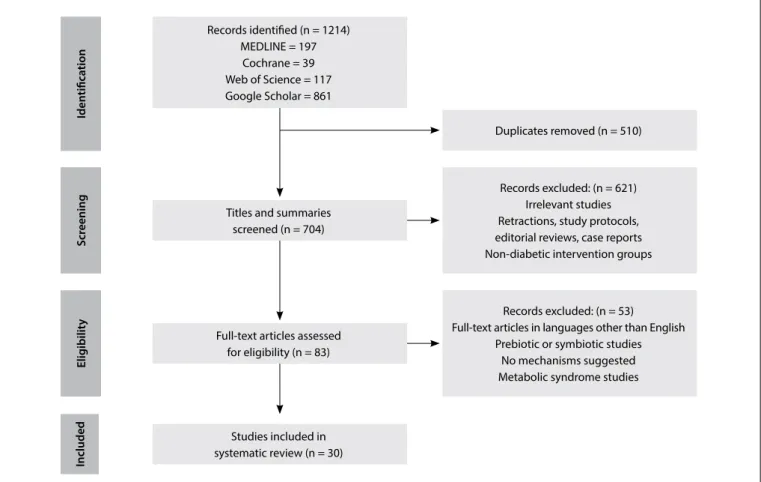
Our initial search retrieved 1,214 articles. Ater removing dupli-cates, 704 titles and abstracts were screened. hen, from among these articles, 83 full texts were assessed for eligibility. Finally, 30 studies were included in this review. A lowchart of the study selection process is illustrated in Figure 1.
Local efects of probiotics in the intestine
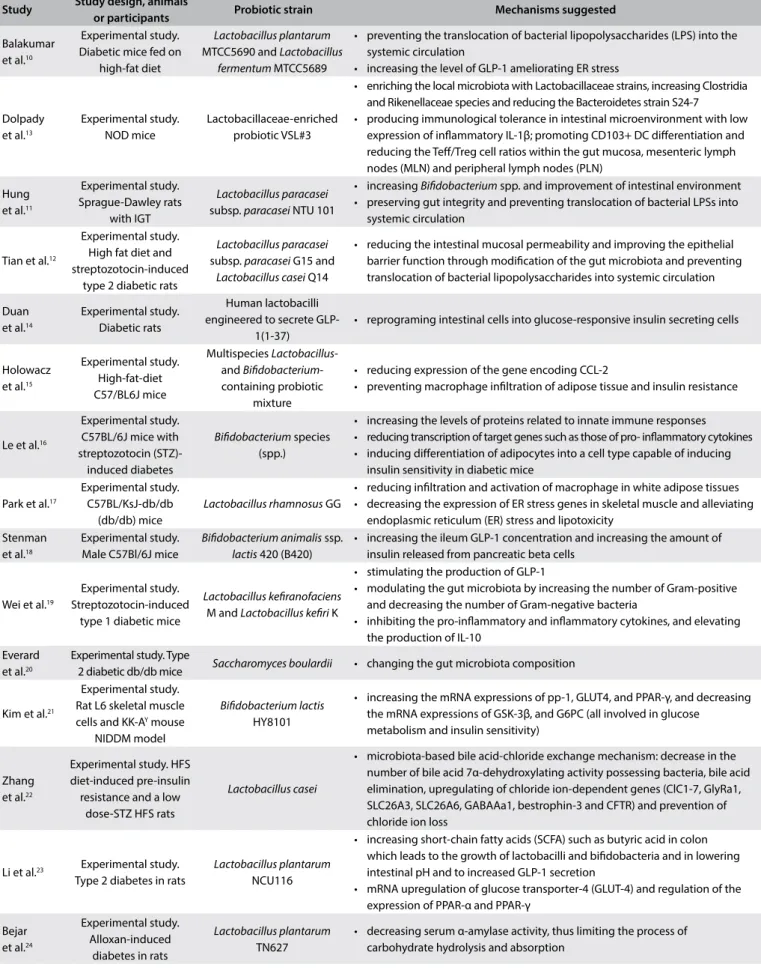
Study Study design, animals
or participants Probiotic strain Mechanisms suggested
Balakumar et al.10
Experimental study. Diabetic mice fed on
high-fat diet
Lactobacillus plantarum
MTCC5690 and Lactobacillus fermentum MTCC5689
• preventing the translocation of bacterial lipopolysaccharides (LPS) into the systemic circulation
• increasing the level of GLP-1 ameliorating ER stress
Dolpady et al.13 Experimental study. NOD mice Lactobacillaceae-enriched probiotic VSL#3
• enriching the local microbiota with Lactobacillaceae strains, increasing Clostridia and Rikenellaceae species and reducing the Bacteroidetes strain S24-7
• producing immunological tolerance in intestinal microenvironment with low expression of inlammatory IL-1β;promoting CD103+ DC diferentiation and reducing the Tef/Treg cell ratios within the gut mucosa, mesenteric lymph nodes (MLN) and peripheral lymph nodes (PLN)
Hung et al.11 Experimental study. Sprague-Dawley rats with IGT Lactobacillus paracasei
subsp. paracasei NTU 101
• increasing Biidobacterium spp. and improvement of intestinal environment • preserving gut integrity and preventing translocation of bacterial LPSs into
systemic circulation
Tian et al.12
Experimental study. High fat diet and streptozotocin-induced
type 2 diabetic rats
Lactobacillus paracasei
subsp. paracasei G15 and
Lactobacillus casei Q14
• reducing the intestinal mucosal permeability and improving the epithelial barrier function through modiication of the gut microbiota and preventing translocation of bacterial lipopolysaccharides into systemic circulation
Duan et al.14
Experimental study. Diabetic rats
Human lactobacilli engineered to secrete
GLP-1(1-37)
• reprograming intestinal cells into glucose-responsive insulin secreting cells
Holowacz et al.15
Experimental study. High-fat-diet C57/BL6J mice
Multispecies Lactobacillus -and Biidobacterium -containing probiotic
mixture
• reducing expression of the gene encoding CCL-2
• preventing macrophage iniltration of adipose tissue and insulin resistance
Le et al.16
Experimental study. C57BL/6J mice with streptozotocin (STZ)-induced diabetes
Biidobacterium species (spp.)
• increasing the levels of proteins related to innate immune responses • reducing transcription of target genes such as those of pro- inlammatory cytokines • inducing diferentiation of adipocytes into a cell type capable of inducing
insulin sensitivity in diabetic mice
Park et al.17
Experimental study. C57BL/KsJ-db/db
(db/db) mice
Lactobacillus rhamnosus GG
• reducing iniltration and activation of macrophage in white adipose tissues • decreasing the expression of ER stress genes in skeletal muscle and alleviating
endoplasmic reticulum (ER) stress and lipotoxicity Stenman
et al.18
Experimental study. Male C57Bl/6J mice
Biidobacterium animalis ssp.
lactis 420 (B420)
• increasing the ileum GLP-1 concentration and increasing the amount of insulin released from pancreatic beta cells
Wei et al.19
Experimental study. Streptozotocin-induced
type 1 diabetic mice
Lactobacillus keiranofaciens
M and Lactobacillus keiri K
• stimulating the production of GLP-1
• modulating the gut microbiota by increasing the number of Gram-positive and decreasing the number of Gram-negative bacteria
• inhibiting the pro-inlammatory and inlammatory cytokines, and elevating the production of IL-10
Everard et al.20
Experimental study. Type
2 diabetic db/db mice Saccharomyces boulardii • changing the gut microbiota composition
Kim et al.21
Experimental study. Rat L6 skeletal muscle cells and KK-AY mouse
NIDDM model
Biidobacterium lactis
HY8101
• increasing the mRNA expressions of pp-1, GLUT4, and PPAR-γ, and decreasing the mRNA expressions of GSK-3β, and G6PC (all involved in glucose
metabolism and insulin sensitivity)
Zhang et al.22
Experimental study. HFS diet-induced pre-insulin resistance and a low
dose-STZ HFS rats
Lactobacillus casei
• microbiota-based bile acid-chloride exchange mechanism: decrease in the number of bile acid 7α-dehydroxylating activity possessing bacteria, bile acid elimination, upregulating of chloride ion-dependent genes (ClC1-7, GlyRa1, SLC26A3, SLC26A6, GABAAa1, bestrophin-3 and CFTR) and prevention of chloride ion loss
Li et al.23 Experimental study.
Type 2 diabetes in rats
Lactobacillus plantarum
NCU116
• increasing short-chain fatty acids (SCFA) such as butyric acid in colon which leads to the growth of lactobacilli and biidobacteria and in lowering intestinal pH and to increased GLP-1 secretion
• mRNA upregulation of glucose transporter-4 (GLUT-4) and regulation of the expression of PPAR-α and PPAR-γ
Bejar et al.24
Experimental study. Alloxan-induced
diabetes in rats
Lactobacillus plantarum
TN627
• decreasing serum α-amylase activity, thus limiting the process of carbohydrate hydrolysis and absorption
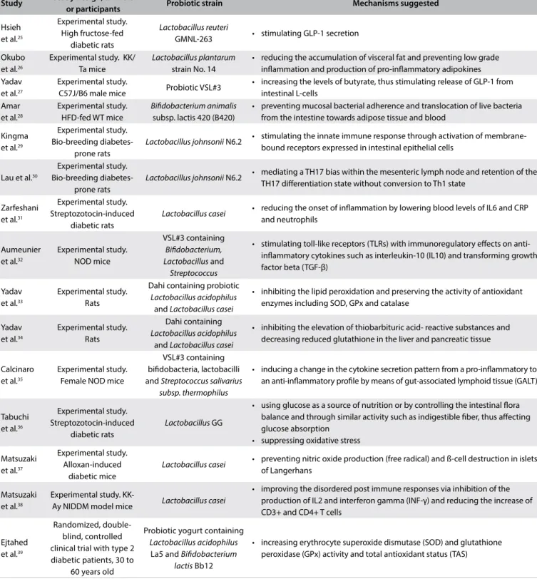
Study Study design, animals
or participants Probiotic strain Mechanisms suggested
Hsieh et al.25
Experimental study. High fructose-fed
diabetic rats
Lactobacillus reuteri
GMNL-263 • stimulating GLP-1 secretion
Okubo et al.26
Experimental study. KK/ Ta mice
Lactobacillus plantarum
strain No. 14
• reducing the accumulation of visceral fat and preventing low grade inlammation and production of pro-inlammatory adipokines Yadav
et al.27
Experimental study.
C57J/B6 male mice Probiotic VSL#3
• increasing the levels of butyrate, thus stimulating release of GLP-1 from intestinal L-cells
Amar et al.28
Experimental study. HFD-fed WT mice
Biidobacterium animalis
subsp. lactis 420 (B420)
• preventing mucosal bacterial adherence and translocation of live bacteria from the intestine towards adipose tissue and blood
Kingma et al.29
Experimental study. Bio-breeding
diabetes-prone rats
Lactobacillus johnsonii N6.2 • stimulating the innate immune response through activation of membrane-bound receptors expressed in intestinal epithelial cells
Lau et al.30
Experimental study. Bio-breeding
diabetes-prone rats
Lactobacillus johnsonii N6.2 • mediating a TH17 bias within the mesenteric lymph node and retention of the TH17 diferentiation state without conversion to Th1 state
Zarfeshani et al.31
Experimental study. Streptozotocin-induced
diabetic rats
Lactobacillus casei • reducing the onset of inlammation by lowering blood levels of IL6 and CRP
and neutrophils
Aumeunier et al.32
Experimental study. NOD mice
VSL#3 containing
Biidobacterium, Lactobacillus and
Streptococcus
• stimulating toll-like receptors (TLRs) with immunoregulatory efects on anti-inlammatory cytokines such as interleukin-10 (IL10) and transforming growth factor beta (TGF-β)
Yadav et al.33
Experimental study. Rats
Dahi containing probiotic
Lactobacillus acidophilus
and Lactobacillus casei
• inhibiting the lipid peroxidation and preserving the activity of antioxidant enzymes including SOD, GPx and catalase
Yadav et al.34
Experimental study. Rats
Dahi containing
Lactobacillus acidophilus
and Lactobacillus casei
• inhibiting the elevation of thiobarbituric acid- reactive substances and decreasing reduced glutathione in the liver and pancreatic tissue
Calcinaro et al.35
Experimental study. Female NOD mice
VSL#3 containing biidobacteria, lactobacilli and Streptococcus salivarius
subsp. thermophilus
• inducing a change in the cytokine secretion pattern from a pro-inlammatory to an anti-inlammatory proile by means of gut-associated lymphoid tissue (GALT)
Tabuchi et al.36
Experimental study. Streptozotocin-induced
diabetic rats
Lactobacillus GG
• using glucose as a source of nutrition or by controlling the intestinal lora balance and through similar activity such as indigestible iber, thus afecting glucose absorption
• suppressing oxidative stress
Matsuzaki et al.37
Experimental study. Alloxan-induced
diabetic mice
Lactobacillus casei • preventing nitric oxide production (free radical) and ß-cell destruction in islets
of Langerhans
Matsuzaki et al.38
Experimental study.
KK-Ay NIDDM model mice Lactobacillus casei
• improving the disordered post immune responses via inhibition of the production of IL2 and interferon gamma (INF-γ) and reducing the increase of CD3+ and CD4+ T cells
Ejtahed et al.39
Randomized, double-blind, controlled clinical trial with type 2 diabetic patients, 30 to
60 years old
Probiotic yogurt containing
Lactobacillus acidophilus
La5 and Biidobacterium lactis Bb12
• increasing erythrocyte superoxide dismutase (SOD) and glutathione peroxidase (GPx) activity and total antioxidant status (TAS)
Table 1. Continues...
resistance in mice and suppression of endotoxemia by probiotic supplementation is considered to be a protective mechanism.40 In
this regard, Balakumar et al.10 stated that probiotic interventions
increased the gene expression proile of the intestinal tight junc-tion markers and gut integrity, thereby preventing translocajunc-tion of bacterial lipopolysaccharides (LPS) into the systemic circulation.
Iden
tiica
tion
Elig
ibilit
y
S
cr
eening
Included
Records identiied (n = 1214) MEDLINE = 197
Cochrane = 39 Web of Science = 117 Google Scholar = 861
Titles and summaries screened (n = 704)
Duplicates removed (n = 510)
Full-text articles assessed for eligibility (n = 83)
Records excluded: (n = 53)
Full-text articles in languages other than English Prebiotic or symbiotic studies
No mechanisms suggested Metabolic syndrome studies
Records excluded: (n = 621) Irrelevant studies Retractions, study protocols, editorial reviews, case reports Non-diabetic intervention groups
Studies included in systematic review (n = 30)
Figure 1. Flow chart of study selection process.
Similarly, presence of Lactobacillus paracasei subsp. paracasei G15 and Lactobacillus casei Q14 in the gut has shown a clear corre-lation with reduced intestinal mucosal permeability and improved epithelial barrier function, through modiication of the gut micro-biota. In turn, this has been shown to lower the circulating levels of LPS and inlammatory cytokines, including interleukin (IL)-1β and IL-8, and possibly to alleviate the inlammatory status and islet β-cell dysfunction.19
Treatment with the probiotic Biidobacterium animalis subsp. lactis 420 (B420) in another study14 led to protection against
diabe-tes through prevention of mucosal bacterial adherence and trans-location of live bacteria from the intestine towards adipose tissue and blood, which caused inlammation and insulin resistance.
Incretins, especially glucagon-like peptide 1 (GLP-1) secreted by intestinal L-cells, are a group of metabolic hormones that inhibit postprandial hyperglycemia by increasing the amount of insulin released from pancreatic beta cells.40 Several studies10,18,25 have
shown that the beneicial efects of probiotic interventions on glucose tolerance and insulin sensitivity were related to increased levels of GLP-1.
Also, administration of Lactobacillus keiranofaciens M and Lactobacillus keiri K was found to stimulate GLP-1 production,
with a concomitant decrease in the numbers of Gram-negative bacteria, which could trigger inlammation.19
he inluence of human lactobacilli engineered to secrete GLP-1 on hyperglycemia has been investigated by Duanet al.14
hey showed that these lactobacilli reprogram intestinal cells into glucose-responsive insulin-secreting cells and that they therefore had the ability to ameliorate hyperglycemia and diabetes.
Moreover, in some studies, the effect of probiotics on dia-betes has been linked to increases in the levels of short-chain fatty acids (SCFAs), especially butyrate in the colon.23,27 SCFAs
are probably key components in the growth of lactobacilli and bifidobacteria and in lowering intestinal pH. All of these are expected to have beneficial effects on diabetes. In addition, SCFAs have been linked to increased GLP-1 secretion in both animal and human models.
Dolpady et al.13 also demonstrated prevention of type 1 diabetes
(T1D) through enriching the local microbiota with Lactobacillaceae strains and through inducing substantial modiications in the micro-biota composition, with increased levels of species of Clostridia and Rikenellaceae and decreased levels of the Bacteroidetes strain S24-7, when a Lactobacillaceae-enriched VSL#3 probiotic was administered. In addition, these modiications generated a pro-tolerogenic intestinal microenvironment with low expression of inlammatory IL-1β. he VSL#3-induced protolerogenic micro-environment promotes CD103+ dendritic cell diferentiation and reduces T efectors/T regulatory cell (Tef/Treg) ratios within the gut mucosa, mesenteric lymph nodes (MLN) and peripheral lymph nodes (PLN), which results in autoimmune diabetes prevention. Pancreatic inlammation caused by type 1 diabetes results in leakage of a-amylase into the bloodstream, thus eliciting higher levels of serum pancreatic a-amylase, a key enzyme involved in carbohydrate digestion. Administration of L. plantarum TN627 to diabetic rats was found to signiicantly decrease serum α-amylase activity, thus limiting the process of carbohydrate hydrolysis and absorption. Consequently, beneicial efects were observed on the glycemic index.24
The efects of probiotics on the inlammatory and immune response pathways
Altered production or function of circulating innate immune pro-teins, cellular pattern-recognition receptors and inlammatory cytokines have been linked to insulin resistance and diabetes.41
Lactobacillus keiranofaciens M and lactobacillus keiri K were reported to mitigate progression of type 1 diabetes through inhib-iting pro-inlammatory and inlammatory cytokines and elevating the production of IL-10. IL-10 inhibits the levels of pro-inlamma-tory cytokines (tumor necrosis factor-alpha) and h1 cytokines (IL-1β, IL-2, IL-6) and prevents β cell destruction.19
Moreover, administration of Biidobacterium spp. increased the levels of innate immune response proteins, including IκB kinase alpha (IKKα), nuclear factor-kappa B inhibitor alpha (IκBα), extra-cellular-signal-regulated kinase 2 (ERK2) and protein kinase B (Akt). Akt may afect IKKα and even result in activation of IκBα, which may in turn inhibit the efects of NF-κBand, thus leading to reduced transcription of target genes such as those of pro-inlam-matory cytokines. On the other hand, ERK, a widely-expressed protein kinase, is an intracellular signaling molecule involved in functions relating to regulation of cell proliferation, diferentiation and survival. Increased ERK2 levels may induce diferentiation of adipocytes into a cell type capable of inducing insulin sensitivity in diabetic mice fed with Biidobacterium spp.16
Furthermore, Lactobacillus rhamnosus GG (LGG) treat-ment was shown17 to reduce iniltration and activation of
mac-rophages, which is critical for initiation and ampliication of
chronic inlammation in white adipose tissues. Hence, the insu-lin-sensitizing efect of LGG may occur through alleviating this inlammatory pathway.
Another study26 indicated that administration of Lactobacillus plantarum No. 14 prevents development of insulin resistance, mainly through reducing accumulations of visceral fat, which prevents pro-duction of pro-inlammatory adipokines. Pro-inlammatory adi-pokines interfere with the insulin-signaling pathway of peripheral tissues and facilitate development of insulin resistance.
In addition, there is evidence that oral treatment with VSL#3, a probiotic compound containing biidobacteria, lactobacilli and Streptococcus salivarius subsp. thermophilus, induces a change in the cytokine secretion pattern from a pro-inlammatory to an anti-inlammatory proile in the gut-associated lymphoid tissue (GALT), which is associated with qualitative modiication of islet-speciic destructive autoimmunity and, possibly, diabetes prevention.35
Consistent with the abovementioned data, protective action by Lactobacillus casei in relation to diabetes was correlated with less frequent onset of inlammation, through lowered levels of IL6, CRP and neutrophils in blood.31Lactobacillus casei also has the
potential to decrease blood glucose levels through improvement of disordered post-immune responses via inhibition of produc-tion of IL2 and interferon gamma (INF-γ) and reducproduc-tion of the increases in CD3+ and CD4+ T cell counts.38
Kingma et al.29 showed that Lactobacillus johnsonii (Ljo) N6.2
stimulates the innate immune response through activation of the membrane-bound receptors expressed in intestinal epithelial cells. hese receptors activate type 1 interferon (INF), which are key players in innate immunity. herefore, a higher state of immuno-logical activation would be achieved, thereby preventing diabe-tes. Moreover, this strain inhibits type 1 diabetes through medi-ating T-helper 17 (h17) bias within the mesenteric lymph nodes. Retention of the h17 diferentiation state, without conversion to a h1 state, which is critical to diabetogenesis, prevents or delays the onset of type 1 diabetes.30
Stimulation of toll-like receptors (TLRs), which have immuno-regulatory efects on anti-inlammatory cytokines, can prevent the onset of autoimmune diseases. TLR-mediated efects of probiotics involve immune-regulatory cytokines such as interleukin IL-10 and transforming growth factor (TGF)-β and some regulatory T cells, under the experimental conditions that result in protection from spontaneous diabetes.32
The efects of probiotics on oxidative stress
Tabuchi et al.36 showed that Lactobacillus GG lowered the level of
MDA per gram of liver weight, which conferred suppression of oxidative stress and improved glucose tolerance.
Other authors concluded that the inhibitory effect of Lactobacillus casei on the incidence of diabetes was partially depen-dent on prevention of nitric oxide production, given that this is a free radical that is involved in the ß-cell destruction process in islets of Langerhans.37,44
On the other hand, foods containing probiotics have been shown to protect against indices relating to diabetes. In one study, probiotic yogurt consumption increased the activity levels of eryth-rocyte superoxide dismutase (SOD) and glutathione peroxidase (GPx), which scavenge free radicals, and improved the total anti-oxidant status (TAS).39
Another mechanism that was proposed to explain the action of fermented milk products containing probiotic bacteria on diabe-tes was through diminishing the elevation of thiobarbituric acid-reactive substances and increasing glutathione levels in the liver and pancreatic tissues of diabetic rats. hese indings indicated that this drink had good antioxidant properties.33
Probiotic milk has consistently been found to exert antioxi-dant efects through inhibiting lipid peroxidation and preserv-ing the activity of antioxidant enzymes, includpreserv-ing SOD, GPx and catalase (CAT).34
The efects of probiotics on gene expression
Some studies on interactions between probiotics and gene expres-sion have suggested that type 2 diabetes in rats is ameliorated through mRNA upregulation of glucose transporter-4 (GLUT-4) through Lactobacillus plantarum NCU116 treatment.23 his has a
critical role in glucose uptake.45 Moreover, NCU can regulate
glu-cose homeostasis and insulin sensitivity in diabetic rats via regu-lating PPAR-α and PPAR-γ gene expression. hese genes play key roles in inlammation and glucose homeostasis.46
Biidobacterium spp. also has an impact on enhanced expres-sion of proteins involved in the insulin-signaling pathway, includ-ing IR-β, IRS-1 and Akt. his results in improved glucose uptake and blood glucose reduction.16
Zhang et al.22 postulated that prevention of the onset of type
2 diabetes through using L. casei Zhang may occur via a microbi-ota-based bile acid-chloride exchange mechanism. Hyperglycemia relates to high levels of plasma bile acids and urine chloride ion loss. High intracellular chloride ion levels in β-cells of the pan-creas are essential for the electrical activity of the β-cell mem-brane and for insulin release. L. casei Zhang administration was found to cause a decrease in the quantity of bacteria with bile acid 7α-dehydroxylating activity and, therefore, bile acid elimi-nation was enhanced. In turn, chloride ion loss was signiicantly prevented by L. casei via upregulation of chloride ion-dependent
genes (ClC1-7, GlyRa1, SLC26A3, SLC26A6, GABAAa1, bestro-phin-3 and CFTR).
In addition, discovery of the antidiabetic activity of Biidobacterium lactis HY 8101 has shed new light on the mecha-nisms for probiotics and their importance in diabetes.21 Its
antidia-betic activity occurs through increasing the mRNA expression of pp-1 (glycogen synthesis-related enzymes), GLUT4 (glucose uptake-related genes) and PPAR-γ (insulin sensitivity-uptake-related genes) and decreasing the mRNA expression of GSK-3β (glycogen synthesis-related enzymes) and G6PC (gluconeogenesis-synthesis-related enzymes), which are all involved in glucose metabolism and insulin sensitivity. Another investigation15 also provided evidence that a
mul-tispecies mixture of probiotics containing Lactobacillus and Biidobacterium reduced expression of the gene encoding CCL-2. he latter is an important chemokine for macrophage iniltra-tion of adipose tissue and contributes towards insulin resistance.47
Finally, endoplasmic reticulum (ER) stress has been men-tioned as one of the main causes of development of inlammation and insulin resistance. ER stress appears to act directly as a nega-tive modulator of the insulin signaling pathway, but also indirectly by promoting lipid accumulation.48 Two studies10,17 showed that
probiotic interventions alleviated lipotoxicity and ER stress gene expression in skeletal muscle, which resulted in improvement of glucose tolerance.
DISCUSSION
One signiicant question regarding clinical use of probiotics is the mechanism underlying the wide range of actions. However, the increasing number of studies that are being conducted with the aim of establishing probiotic mechanisms relating to diabetes conditions indicate that there is a promising future for probiot-ics in treating this disease. To the best of our knowledge, this is the irst review on the mechanisms of probiotic function relating to diabetes. It is hoped that gaining a mechanistic understanding of probiotic action will provide the rationale to support develop-ment of new hypothesis-driven studies to deine the clinical ei-cacy of preventive, adjunctive or alternative treatments for dia-betes. Also, such eforts could suitably help in selecting strains for speciic investigation and applications under these conditions and may uncover novel probiotic functions.
efects of probiotics in relation to diabetes (ive studies). Finally, six studies suggested that probiotics might have efects through altering the expression of genes involved in ER stress and glucose homeostasis and insulin resistance.
he strengths of this review include its use of an outcome clas-siication for diferent possible mechanisms of probiotics in rela-tion to diabetes. However, several limitarela-tions need to be taken into account in interpreting our indings. It should be mentioned that, except for one study, all of these mechanisms have been veriied in animal studies. Moreover, it seems that such efects depend on the type of bacteria, dose and duration of consumption, manner and frequency of administration, environmental factors and complex interactions between probiotics, cells and metabolic pathways that are rarely mediated by a single mechanism.49
In addition, it is important to take into consideration the risk of bias across diferent studies, such as publication, perfor-mance and reporting bias, along with potential conlicts of inter-est. Such factors might limit the ability to draw robust conclu-sions from these studies. Given that we only had limited access to some databases such as Embase, and that studies not reported in English were excluded, it is possible that more rigorous report-ing of study results would improve the quality of the evidence in further studies.
Nonetheless, elucidation of the mechanisms linking the microbiome to diabetes can provide a rational basis for dietary consumption of probiotic microorganisms in relation to dia-betes. In addition, evaluation of the mechanism of action for probiotics both in healthy subjects and in diabetic patients, so as to address the influence of these microorganisms on gene expression for different pathways, is needed in order to better understand the role that probiotics might have in prevention and treatment of diabetes.
CONCLUSIONS
In conclusion, there is some evidence suggesting various poten-tial mechanisms of action for probiotics in relation to diabetes prevention and treatment. Further studies are needed to conirm the underlying pathways involved in the beneicial efects from each strain, along with assessment of other confounding factors.
REFERENCES
1. Hemarajata P, Versalovic J. Efects of probiotics on gut microbiota:
mechanisms of intestinal immunomodulation and neuromodulation.
Therap Adv Gastroenterol. 2013;6(1):39-51.
2. Ozdemir O. Various efects of diferent probiotic strains in allergic
disorders: an update from laboratory and clinical data. Clin Exp Immunol.
2010;160(3):295-304.
3. Idzior Waluś B, Waluś-Miarka M. Is now the time for probiotics in diabetes
management? Pol Arch Med Wewn. 2015;125(11):797-8.
4. Tonucci LB, Olbrich Dos Santos KM, Licursi de Oliveira L, Rocha Ribeiro
SM, Duarte Martino HS. Clinical application of probiotics in type 2
diabetes mellitus: A randomized, double-blind, placebo-controlled
study. Clin Nutr. 2015;pii: S0261-5614(15)00331-3.
5. Kasińska MA, Drzewoski J. Efectiveness of probiotics in type 2 diabetes:
a meta-analysis. Pol Arch Med Wewn. 2015;125(11):803-13.
6. Tremaroli V, Bäckhed F. Functional interactions between the gut
microbiota and host metabolism. Nature. 2012;489(7415):242-9.
7. Kim JJ, Sears DD. TLR4 and Insulin Resistance. Gastroenterol Res Pract.
2010;2010. pii: 212563.
8. Moher D, Liberati A, Tetzlaf J, Altman DG; PRISMA Group. Preferred
reporting items for systematic reviews and meta-analyses: the PRISMA
statement. Ann Intern Med. 2009;151(4):264-9, W64.
9. Everard A, Cani PD. Diabetes, obesity and gut microbiota. Best Pract
Res Clin Gastroenterol. 2013;27(1):73-83.
10. Balakumar M, Prabhu D, Sathishkumar C, et al. Improvement in glucose
tolerance and insulin sensitivity by probiotic strains of Indian gut origin
in high-fat diet-fed C57BL/6J mice. Eur J Nutr. 2016. [Epub ahead of
print].
11. Hung S-C, Tseng W-T, Pan T-M. Lactobacillus paracasei subsp. paracasei
NTU 101 ameliorates impaired glucose tolerance induced by a
high-fat, high-fructose diet in Sprague-Dawley rats. Journal of Functional
Foods. 2016;24:472-81. Available from: http://www.sciencedirect.com/
science/article/pii/S1756464616301086. Accessed in 2017 (Feb 2).
12. Tian P, Li B, He C, et al. Antidiabetic (type 2) efects of Lactobacillus
G15 and Q14 in rats through regulation of intestinal permeability and
microbiota. Food Funct. 2016;7(9):3789-97.
13. Dolpady J, Sorini C, Di Pietro C, et al. Oral Probiotic VSL#3 Prevents
Autoimmune Diabetes by Modulating Microbiota and Promoting
Indoleamine 2,3-Dioxygenase-Enriched Tolerogenic Intestinal
Environment. J Diabetes Res. 2016; 2016:7569431.
14. Duan FF, Liu JH, March JC. Engineered commensal bacteria reprogram
intestinal cells into glucose-responsive insulin-secreting cells for the
treatment of diabetes. Diabetes. 2015;64(5):1794-803.
15. Holowacz S, Guigné C, Chêne G, et al. A multispecies Lactobacillus- and
Biidobacterium-containing probiotic mixture attenuates body weight
gain and insulin resistance after a short-term challenge with a high-fat
diet in C57/BL6J mice. PharmaNutrition.2015; 3(3): 1017. Available from:
http://www.sciencedirect.com/science/article/pii/S2213434415000158.
Accessed in 2017 (Feb 2).
16. Le TK, Hosaka T, Nguyen TT, et al. Biidobacterium species lower serum
glucose, increase expressions of insulin signaling proteins, and improve
adipokine proile in diabetic mice. Biomed Res. 2015;36(1):63-70.
17. Park KY, Kim B, Hyun CK. Lactobacillus rhamnosus GG improves glucose
tolerance through alleviating ER stress and suppressing macrophage
activation in db/db mice. J Clin Biochem Nutr. 2015;56(3):240-6.
18. Stenman LK, Waget A, Garret C, et al. Probiotic B420 and prebiotic
polydextrose improve eicacy of antidiabetic drugs in mice. Diabetol
19. Wei S-H, Chen Y-P, Chen M-J. Selecting probiotics with the abilities of
enhancing GLP-1 to mitigate the progression of type 1 diabetes in
vitro and in vivo. Journal of Functional Foods. 2015;18 (Part A):473-86.
Available from: http://www.sciencedirect.com/science/article/pii/
S1756464615004004. Accessed in 2017 (Feb 2).
20. Everard A, Matamoros S, Geurts L, Delzenne NM, Cani PD. Saccharomyces
boulardii administration changes gut microbiota and reduces hepatic
steatosis, low-grade inlammation, and fat mass in obese and type 2
diabetic db/db mice. MBio. 2014;5(3):e01011-14.
21. Kim SH, Huh CS, Choi ID, et al. The anti-diabetic activity of
Biidobacterium lactis HY8101 in vitro and in vivo. J Appl Microbiol.
2014;117(3):834-45.
22. Zhang Y, Guo X, Guo J, et al. Lactobacillus casei reduces susceptibility
to type 2 diabetes via microbiota-mediated body chloride ion inlux.
Sci Rep. 2014; 4:5654.
23. Li C, Ding Q, Nie SP, et al. Carrot juice fermented with Lactobacillus
plantarum NCU116 ameliorates type 2 diabetes in rats. J Agric Food
Chem. 2014;62(49):11884-91.
24. Bejar W, Hamden K, Ben Salah R, Chouayekh H. Lactobacillus plantarum
TN627 signiicantly reduces complications of alloxan-induced diabetes
in rats. Anaerobe. 2013; 24:4-11.
25. Hsieh FC, Lee CL, Chai CY, et al. Oral administration of Lactobacillus
reuteri GMNL-263 improves insulin resistance and ameliorates
hepatic steatosis in high fructose-fed rats. Nutr Metab (Lond).
2013;10(1):35.
26. Okubo T, Takemura N, Yoshida A, Sonoyama K. KK/Ta Mice Administered
Lactobacillus plantarum Strain No. 14 Have Lower Adiposity and Higher
Insulin Sensitivity. Biosci Microbiota Food Health. 2013;32(3):93-100.
27. Yadav H, Lee JH, Lloyd J, Walter P, Rane SG. Beneicial metabolic efects
of a probiotic via butyrate-induced GLP-1 hormone secretion. J Biol
Chem. 2013;288(35):25088-97.
28. Amar J, Chabo C, Waget A, et al. Intestinal mucosal adherence and
translocation of commensal bacteria at the early onset of type 2
diabetes: molecular mechanisms and probiotic treatment. EMBO
Mol Med. 2011;3(9):559-72.
29. Kingma SD, Li N, Sun F, et al. Lactobacillus johnsonii N6.2 stimulates
the innate immune response through Toll-like receptor 9 in Caco-2
cells and increases intestinal crypt Paneth cell number in biobreeding
diabetes-prone rats. J Nutr. 2011;141(6):1023-8.
30. Lau K, Benitez P, Ardissone A, et al. Inhibition of type 1 diabetes correlated
to a Lactobacillus johnsonii N6.2-mediated Th17 bias. J Immunol.
2011;186(6):3538-46.
31. Zarfeshani A, Khaza’ai H, Mohd Ali R, et al. Efect of Lactobacillus casei on
the Production of Pro-Inlammatory Markers in Streptozotocin-Induced
Diabetic Rats. Probiotics Antimicrob Proteins. 2011;3(3-4):168-74.
32. Aumeunier A, Grela F, Ramadan A, et al. Systemic Toll-like receptor
stimulation suppresses experimental allergic asthma and autoimmune
diabetes in NOD mice. PloS One. 2010;5(7): e11484.
33. Yadav H, Jain S, Sinha PR. Antidiabetic efect of probiotic dahi containing
Lactobacillus acidophilus and Lactobacillus casei in high fructose fed
rats. Nutrition. 2007;23(1):62-8.
34. Yadav H, Jain S, Sinha PR. Oral administration of dahi containing
probiotic Lactobacillus acidophilus and Lactobacillus casei delayed
the progression of streptozotocin-induced diabetes in rats. J Dairy
Res. 2008;75(2):189-95.
35. Calcinaro F, Dionisi S, Marinaro M, et al. Oral probiotic administration
induces interleukin-10 production and prevents spontaneous
autoimmune diabetes in the non-obese diabetic mouse. Diabetologia.
2005;48(8):1565-75.
36. Tabuchi M, Ozaki M, Tamura A, et al. Antidiabetic efect of Lactobacillus
GG in streptozotocin-induced diabetic rats. Biosci Biotechnol Biochem.
2003;67(6):1421-4.
37. Matsuzaki T, Nagata Y, Kado S, et al. Efect of oral administration of
Lactobacillus casei on alloxan-induced diabetes in mice. APMIS.
1997;105(8):637-42.
38. Matsuzaki T, Yamazaki R, Hashimoto S, Yokokura T. Antidiabetic efects
of an oral administration of Lactobacillus casei in a
non-insulin-dependent diabetes mellitus (NIDDM) model using KK-Ay mice. Endocr
J. 1997;44(3):357-65.
39. Ejtahed HS, Mohtadi-Nia J, Homayouni-Rad A, et al. Probiotic yogurt
improves antioxidant status in type 2 diabetic patients. Nutrition.
2012;28(5):539-43.
40. Holst JJ. The physiology of glucagon-like peptide 1. Physiol Rev.
2007;87(4):1409-39.
41. Fernández-Real JM, Pickup JC. Innate immunity, insulin resistance and
type 2 diabetes. Diabetologia. 2012; 55(2):273-8.
42. Maritim AC, Sanders RA, Watkins JB 3rd. Diabetes, oxidative stress, and
antioxidants: a review. J Biochem Mol Toxicol. 2003;17(1):24-38.
43. Rahbani-Nobar M, Rahimi-Pour A, Rahbani-Nobar M, Adi-Beig F,
Mirhashemi SM. Total antioxidant capacity, superoxide dismutase
and glutathione peroxidase in diabetic patients. Medical Journal of
Islamic Academy Sciences. 1999;12(4):109-14. Available from: http://
www.journalagent.com/ias/pdfs/IAS_12_4_109_114.pdf. Accessed in
2017 (Feb 2).
44. Kasuga A, Nakaki T, Takei I, et al. Nitric oxide is important for mouse
beta-cell line killing by peritoneal exudate cells obtained from
cyclophosphamide treated non-obese diabetic mice. Endrocr J.
1995;42(2):259-63.
45. Kim SW, Park KY, Kim B, Kim E, Hyun CK. Lactobacillus rhamnosus GG
improves insulin sensitivity and reduces adiposity in high-fat diet-fed
mice through enhancement of adiponectin production. Biochem
Biophys Res Commun. 2013;431(2):258-63.
46. Soares FL, de Oliveira Matoso R, Teixeira LG, et al. Gluten-free diet
reduces adiposity, inlammation and insulin resistance associated
with the induction of PPAR-alpha and PPAR-gamma expression. J Nutr
47. Kanda H, Tateya S, Tamori Y, et al. MCP-1 contributes to macrophage
iniltration into adipose tissue, insulin resistance, and hepatic steatosis
in obesity. J Clin Invest. 2006;116(6):1494-505.
48. Flamment M, Hajduch E, Ferré P, Foufelle F. New insights into ER
stress-induced insulin resistance. Trends Endocrinol Metab. 2012;23(8):381-90.
49. Boyle RJ, Robins-Browne RM, Tang ML. Probiotic use in clinical practice:
what are the risks? Am J Clin Nutr. 2006;83(6):1256-64; quiz 1446-7.
Acknowledgements: This study was supported by the Isfahan University of Medical Sciences, Isfahan, Iran
Authors’ contributions: NR, SGH and SS searched databases and
selected articles. MM, NR, SGH, SS and RGH wrote the manuscript. All
authors read and approved the content of the manuscript
Sources of funding: None Conlict of interest: None
Address for correspondence:
Reza Ghiasvand
Department of Community Nutrition
School of Nutrition and Food Science
Isfahan University of Medical Sciences
Isfahan — Iran
Tel. 0098 313792-3153
Fax. 0098 313668-2509