Retrospective cohort of trisomy 18 (Edwards syndrome)
in southern Brazil
Coorte retrospectiva de trissomia do cromossomo 18 (síndrome de Edwards)
no sul do Brasil
Daniela Denardin
I, Fabíola Elizabete Savaris
I, André Campos da Cunha
II, Rosilene da Silveira Betat
II, Jorge Alberto Bianchi Telles
III,
Luciano Vieira Targa
IV, Aline Weiss
V, Paulo Ricardo Gazzola Zen
VI, Rafael Fabiano Machado Rosa
VIIHospital Materno Infantil Presidente Vargas (HMIPV) and Universidade Federal de Ciências da Saúde de Porto Alegre (UFCSPA),
Porto Alegre, Rio Grande do Sul, Brazil
ABSTRACT
CONTEXT AND OBJECTIVE: Trisomy 18 (T18), or Edwards syndrome, is a chromosomal disease character-ized by a broad clinical picture and a poor prognosis. Our aim was to describe clinical, radiological and survival data of a cohort of patients prenatally diagnosed with T18.
DESIGN AND SETTING: Retrospective single cohort in the Fetal Medicine Service of Hospital Materno Infantil Presidente Vargas (HMIPV).
METHODS: All sequential patients with T18 registered at the Fetal Medicine Service of HMIPV between January 2005 and September 2013 were considered. We gathered their clinical, radiological and survival data and used the Kaplan-Meier test for survival analysis.
RESULTS: Ten patients were diagnosed with T18, of whom seven (70%) were female. The majority (90%) were referred due to malformations seen on ultrasound. The mean gestational age at the irst evaluation was 25.5 weeks. At karyotyping, the defects were considered multiple in only four patients (40%). All the fetuses presented full trisomy of chromosome 18. The main abnormality observed was congenital heart disease (n = 7). Intrauterine death occurred in half of the patients (50%). All live patients (n = 5) were born through cesarean section presenting low weight and low Apgar scores. The median length of survival after birth was 18 days.
CONCLUSIONS: T18 is associated with a high risk of fetal and neonatal death. The majority of the patients present major malformations identiied through ultrasound, such as congenital heart defects, which could help in identifying such cases prenatally.
RESUMO
CONTEXTO E OBJETIVO: A trissomia do cromossomo 18 (T18), ou síndrome de Edwards, é uma doença cromossômica caracterizada por um quadro clínico amplo e prognóstico pobre. Nosso objetivo foi descrever os dados clínicos, radiológicos e de sobrevida de uma coorte de pacientes com diagnóstico pré-natal de T18.
TIPO DE ESTUDO E LOCAL: Coorte única retrospectiva no Serviço de Medicina Fetal do Hospital Materno Infantil Presidente Vargas (HMIPV).
MÉTODOS: Foram considerados todos os pacientes consecutivos com T18 registrados no Serviço de Me-dicina Fetal do HMIPV entre janeiro de 2005 e setembro de 2013. Foram coletados os seus dados clínicos, radiológicos e de sobrevida. Foi utilizado o teste de Kaplan-Meier para análise de sobrevida.
RESULTADOS: 10 pacientes foram diagnosticados com T18, 7 (70%) do sexo feminino. A maioria (90%) foi encaminhada devido a malformações detectadas no ultrassom. A média da idade gestacional na primeira avaliação foi de 25,5 semanas. Ao cariótipo, os defeitos foram considerados múltiplos em apenas 4 pacien-tes (40%). Todos apresentaram trissomia livre do cromossomo 18. A principal anormalidade observada foi a cardiopatia congênita (n = 7). Morte intraútero ocorreu em metade dos pacientes (50%). Todos os pacientes vivos (n = 5) nasceram através de parto cesáreo, apresentando baixo peso e baixos escores de Apgar. A mediana de sobrevida após o nascimento foi de 18 dias.
CONCLUSÕES: A T18 associa-se a risco elevado de morte fetal e neonatal. A maioria dos pacientes apre-senta malformações identiicadas através do ultrassom, como cardiopatias congênitas, que poderiam au-xiliar na sua identiicação pré-natal.
IMD. Physician, Residency Program on Obstetrics
and Gynecology, Hospital Materno Infantil Presidente Vargas (HMIPV), Porto Alegre, Rio Grande do Sul, Brazil.
IIMD. Obstetrician, Fetal Medicine, Hospital
Materno Infantil Presidente Vargas (HMIPV), Porto Alegre, Rio Grande do Sul, Brazil.
IIIMD. Fetologist, Fetal Medicine, Hospital
Materno Infantil Presidente Vargas (HMIPV), Porto Alegre, Rio Grande do Sul, Brazil.
IVMD. Pediatric Radiologist, Hospital Materno
Infantil Presidente Vargas (HMIPV), Porto Alegre, Rio Grande do Sul, Brazil.
VMD. Neonatologist, Hospital Materno Infantil
Presidente Vargas (HMIPV), Porto Alegre, Rio Grande do Sul, Brazil.
VIPhD. Adjunct Professor of Clinical Genetics
and of the Postgraduate Program on Pathology, Universidade Federal de Ciências da Saúde de Porto Alegre (UFCSPA), and Clinical Geneticist, Universidade Federal de Ciências da Saúde de Porto Alegre (UFCSPA) and Complexo Hospitalar Santa Casa de Porto Alegre (CHSCPA), Porto Alegre, Rio Grande do Sul, Brazil.
VIIPhD. Clinical Geneticist, Universidade Federal
de Ciências da Saúde de Porto Alegre (UFCSPA), Complexo Hospitalar Santa Casa de Porto Alegre (CHSCPA) and Hospital Materno Infantil Presidente Vargas (HMIPV), Porto Alegre, Rio Grande do Sul, Brazil.
KEY WORDS: Trisomy.
Chromosomes, human, pair 18. Karyotype.
Prenatal diagnosis. Survival analysis.
PALAVRAS-CHAVE: Trissomia.
Cromossomos humanos par 18. Cariótipo.
INTRODUCTION
Trisomy 18 (T18) was one of the irst chromosomal abnormali-ties to be described. It was irst reported by Edwards et al. in 1960 and, therefore, is also known as Edwards syndrome. Nowadays, it is considered to be the second most common chromosomal abnormality involving the autosomes, only behind trisomy 21 (Down syndrome).1,2 It has an estimated prevalence of approxi-mately 1:3,600-8,500 live births.3 T18 is clinically characterized by a broad clinical picture, with more than 130 diferent ind-ings already described, and a prognosis that is considered poor.2,4 Most fetuses diagnosed during gestation are spontaneously aborted and, among those that are born alive, most die within the irst six months.1,2,4
In Brazil, prenatal identiication of patients with T18 is important in determining issues relating to their evolution and prognosis, as well as their birth and clinical management. Many countries have also adopted a more interventionist stance and have made more investment in cases that come to birth, thereby increasingly respecting family autonomy in decision-making.1,2 However, in Brazil, there is a paucity of studies evaluating both the prenatal diagnosis and the natural history of patients with T18, especially from the beginning of pregnancy.4,5
OBJECTIVE
Our aim was to describe clinical, radiological and survival data from a cohort of patients prenatally diagnosed with T18.
METHODS
All sequential patients with T18 registered at the Fetal Medicine Service of Hospital Materno Infantil Presidente Vargas between January 2005 and September 2013 were considered. We gath-ered clinical, radiological and survival data on the patients from their medical records. his project was approved by the hospital’s Research Ethics Committee.
he information retrieved from medical records consisted of the reason for referral; gestational age at irst assessment; maternal and paternal ages; maternal pregnancy history; presence of diseases and threatened abortion in the current pregnancy; results from irst-trimester sonographic screening, echocardiography and karyotyping; delivery and perinatal features; and postnatal evaluation, survival and autopsy results.
Gestational age was determined according to the earlier ultrasound. he main reasons for fetal karyotyping were catego-rized as described by Kessler et al.6 he patients were classiied according to the number of major and minor sonographic mark-ers observed before and ater puncturing for fetal karyotyping in accordance with Raniga et al.7 Abnormalities identiied through imaging studies performed during prenatal care were also clas-siied as single or multiple defects, in accordance with Staebler
et al.,8 before and ater performing fetal karyotyping. To deter-mine the congenital heart defect observed, we used the classiica-tion suggested by Botto et al.9
he Kaplan-Meier test was used to construct the survival curve, by means of the BioEstat 5.0 sotware.
RESULTS
Over this period of about nine years, ten patients were diagnosed with T18. he majority of them (90%) had been referred due to presence of malformations in an ultrasound evaluation (only one patient presented increased nuchal translucency). he mean gesta-tional age at the irst evaluation was 25.5 weeks. he maternal age ranged from 20 to 45 years (mean of 34.4 years) and the paternal age ranged from 30 to 40 years (mean of 33.9 years). Advanced maternal age (≥ 35 years) was observed in six cases (60%) (Table 1). All the cases in the sample were singleton pregnancies. Regarding the maternal history of pregnancy, two mothers were primiparous (20%). One of them (10%) presented gestational diabetes. here were no cases of preeclampsia or even threatened abortion. hree mothers (30%) had family histories of malforma-tions, and two of them had had malformed fetuses in a previous pregnancy. Half of the patients underwent irst-trimester sono-graphic screening. hree of these screenings (60%) were consid-ered normal. Two patients (40%) presented a cystic hygroma. In one of them, the nasal bone was not identiied and there was tricuspid regurgitation. Abnormalities were seen in all cases in evaluations through obstetric and morphological ultrasound. Six patients (70%) underwent echocardiography and additional heart abnormalities were observed in three cases (all of these were ventricular septal defects) (Table 1 and 2).
Out of all the patients, 7 (70%) were female. All the live patients (n = 5) were born through cesarean section. Pelvic presen-tation was observed in two cases (40%). Only one was premature. Low birth weight (< 2,500 grams) was observed in all cases: the weight ranged from 1,460 to 2,475 grams, with a mean of 2,030 grams. he length ranged from 36 to 44.5 cm (mean of 41.3 cm) and the head circumference from 30 to 34.5 cm (mean of 32.7 cm) (Table 1). All patients (n = 5) presented Apgar score < 7 at irst minute, and 3 at the ith minute. Additional ind-ings observed during the postnatal period and potentially iden-tiied through ultrasound during the prenatal period consisted
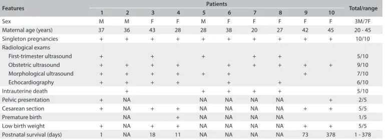
Table 1. Gestational, perinatal and survival indings observed among the patients of the sample
Features Patients Total/range
1 2 3 4 5 6 7 8 9 10
Sex M M F F M F F F F F 3M/7F
Maternal age (years) 37 36 43 28 28 38 20 27 42 45 20 - 45
Singleton pregnancies + + + + + + + + + + 10/10
Radiological exams
First-trimester ultrasound + + + + + 5/10
Obstetric ultrasound + + + + + + + + + 9/10
Morphological ultrasound + + + + + + + 7/10
Echocardiography + + + + + + 6/10
Intrauterine death + + + + + 5/10
Pelvic presentation + NA NA NA NA NA + 2/5
Cesarean section + NA + + NA NA NA NA + + 5/5
Premature birth NA + NA NA NA NA 1/5
Low birth weight + NA + + NA NA NA NA + + 5/5
Postnatal survival (days) 1 NA 18 11 NA NA NA NA 73 378 1 - 378
M = male; F = female; NA = not applicable.
Table 2. Prenatal indings among the patients of the sample by the end of the pregnancy, divided into minor and major markers as
described by Raniga et al.7
Features Patients Total
1 2 3 4 5 6 7 8 9 10
Minor markers (n) 1 0 0 1 0 0 0 1 1 1 0-1
Single umbilical artery + + + 3/10
Short long bones + + 2/10
Major markers (n) 2 4 3 2 3 2 3 1 3 2 1-4
Congenital heart defect + + + + + + + 7/10
Cystic hygroma + + 2/10
Micrognathia + + 2/10
Omphalocele + + 2/10
Corpus callosum agenesis + 1/10
Ventriculomegaly + 1/10
Microcephaly + 1/10
Cleft lip/palate + 1/10
Esophageal atresia + 1/10
Diaphragmatic hernia + 1/10
Radial anomaly + 1/10
Clenched ists with overlapping ingers + 1/10
Club foot + 1/10
Myelomeningocele + 1/10
Ambiguous genitalia + 1/10
Intrauterine growth restriction + 1/10
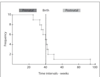
of ambiguous genitalia (n = 2), clenched ists with overlapping ingers (n = 2) and micrognathia (n = 1). he survival of these patients ranged from 1 to 378 days (median of 18 days). hree patients (60%) died within the irst month. Only one completed the irst year of life. In none of the cases did the family authorize an autopsy (Figure 1 and Table 1).
DISCUSSION
some type of malformation, who have been attended within the Brazilian national health system (Sistema Único de Saúde, SUS). As seen from the results, the number of cases with prena-tal diagnosis of T18 at this service is about one per year, which corresponds to about 10% of the demand from live births in the state (taking the year 2005 as the reference point, which had notiications of 11 cases). We believe that many cases of T18 were diagnosed only during the postnatal period. his is in accordance with the results obtained by Rosa et al. in the other study conducted in Brazil.4
here was a high rate of maternal age ≥ 35 years in our study, seen in 60% of the cases. he average maternal age observed (34.4 years) was similar to that described by Rosa et al.4 (33.9 years). he maternal age was similar to the paternal age (mean of 33.9 years). Diseases of pregnancy have been described among patients with T18.4 Rosa et al.4 observed a higher prev-alence of preeclampsia. Sugayama et al.5 also described a high rate of unspeciied hypertension in pregnancies of individuals with T18. A threat of abortion was also reported in 20% of the cases evaluated by Rosa et al.4 However, there were no cases of preeclampsia or threatened abortion in our sample. We cannot rule out the possibility that these indings may be related to our small sample size.
The first cases of prenatal diagnosis of T18 date from the 1970s.2 Today, diagnostic suspicion of T18 may be raised dur-ing the prenatal period through fetal ultrasonography, with measurement of nuchal translucency in the first trimester. This can be confirmed by fetal chromosome analysis through
procedures such as puncturing the chorionic villi and amnio-centesis.2 The finding of increased nuchal translucency is con-sidered to be the most sensitive finding for diagnosing T18 up to the 16th week of gestation.10 However, it was notewor-thy in our sample that only one of the five patients (20%) who underwent this evaluation presented increased nuchal trans-lucency. Moreover, this was the only case referred to our ser-vice due to this finding. This feature may have been related to our small sample size. On the other hand, it is interesting to note that only half of the sample was assessed by means of ultrasound during first trimester, thus showing that many patients are coming in late for evaluation.
Fetal echocardiography can also detect a congenital heart defect that may suggest the presence of T18. he sonographic inding of congenital heart disease is considered to be the most sensitive feature for diagnosing T18 ater the 16th week of preg-nancy.10 In our sample, congenital heart disease was the main anomaly found during pregnancy (it was identiied in 70% of the patients), and use of fetal echocardiography allowed detection of additional defects that had not been identiied through morpho-logical and obstetric ultrasound. his highlights the importance of performing echocardiography in these cases. he main defects, as observed in our sample, are of septal type, especially ventric-ular septal defects. Conotruncal defects, such as a double outlet of the right ventricle and tetralogy of Fallot, and let obstructive defects, such as hypoplastic let heart, are considered less com-mon acom-mong patients with T18.11
Other common manifestations reported during pregnancy include intrauterine growth restriction and polyhydramnios. he latter has been described in 9-52% of pregnancies,5,12,13 and it seems related to abnormalities of sucking and swallowing presented by the fetus. However, despite its frequency, intra-uterine growth restriction and polyhydramnios were uncom-mon in our sample. Polyhydramnios was observed only in the case with esophageal atresia (patient 4) (Table 1). We believe that in this case, the polyhydramnios was secondary to the digestive tract malformation presented by the fetus, which pre-vented adequate swallowing of amniotic luid and hence led to accumulation around it.
It is important to highlight that the association between intra-uterine growth restriction and major malformation consistent with the phenotype of T18 oten leads to prenatally diagnosing it ater the 20th week of gestation. According to Viora et al.,14 modern ultrasound examinations clearly present high sensitivity (greater than 90%) for detecting fetuses with T18. his inding is also in agreement with the observations made by Yeo et al.,15 who found that multiple abnormalities were usually observed in fetal sonog-raphy, typically involving the brain, heart and upper limbs.16 In the study by Yeo et al.,15 all the fetuses had four or more abnormalities.
Figure 1. Kaplan-Meier curve showing the survival presented by the patients during the prenatal and postnatal periods. Note especially that half of them presented intrauterine death and that out of those who were born alive, three died within the irst month of life and only one patient lived for longer than one year.
10
8
6
4
2
Fr
equenc
y
20 40 40 80 100
In our sample, using the method of Raniga et al.,7 the number of major markers was 1.8 per case and the number of minor markers was 0.3 per case, at the time of karyotyping. Another important aspect of our sample was that at the time of fetal karyotyping, only four patients had multiple defects. hus, it is important to have a high degree of suspicion in cases with malformations presenting a greater association with T18, such as omphalocele, diaphragmatic hernia, myelomeningocele and esophageal atresia.1,2
he most common chromosomal abnormality observed in these patients is full trisomy of chromosome 18,1,2 and all the patients of our sample presented this inding. Full trisomy of chromosome 18 has a relationship with advanced maternal age, due to the phenomenon of non-disjunction of chromosomes.2 In our sample, as pointed out earlier, we found a high rate of mothers aged over 35 years (60%). However, it is important to be aware that T18 can be secondary to other chromosomal abnormalities such as translocations, which may have impor-tant implications regarding genetic counseling for the patients and their families.2
We found that females predominated in our sample (70%). his inding is in accordance with the literature, which shows that the proportion of females has ranged from 56 to 78%.4,5,17-19 However, it is noteworthy that some authors have found equal frequencies of the sexes in evaluations performed during the pre-natal period,20 especially before the 18th week of gestation.21 hese features may perhaps be related to the fact that female patients have been associated with a greater chance of being born alive and surviving for longer periods than boys.2
In our series, all the live patients were born through cesarean section. hese high rates of cesarean section have also been fre-quently described in the literature (50 to 90%).4,5,13,17,19,20 It is note-worthy that there are some studies speciically drawing attention to this inding.17 In our sample, we believe that this feature was related to prenatal detection of major malformations, which thus inluenced the choice of cesarean section as the delivery route.
Several studies have drawn attention to the high rate of pre-maturity described among patients with T18 (48%).4,5,13,19,20 However, in our series only one patient (20%) was premature. Regarding low birth weight, our frequency of 100% was similar to that described in the literature,13,19,20 including studies devel-oped in Brazil.4,5 Regarding Apgar scores, the high rate of patients presenting scores below 7 (suggestive of some degree of anoxia) at the irst and ith minute that we observed in our study was similar to what was described by Lin et al.19 and Rosa et al.4
A signiicant proportion of the fetuses with T18 die while still in utero, as observed in our sample (50%). According to Morris and Savva,22 it is estimated that 72% of pregnancies with fetuses with T18 end in miscarriage or stillbirth between the 12th week and full term. he median survival ater birth among patients
with T18 that has been reported in the literature has usually ranged from 2.5 to 14.5 days,3,13,18-20,23,24 and we obtained a simi-lar value (18 days). It is noteworthy that the value described in the other study developed in Brazil, by Rosa et al.,4 was higher (31 days). hose authors associated this inding with possi-ble postnatal selection, since most of the patients in their study had been referred by other medical units within the state and had not been born in the hospital. hey did not rule out the possi-bility that patients with greater severity of disease may not have survived to the point of being referred to their hospital for eval-uation and diagnosis. Interestingly, one of our patients (10%) presented survival longer than one year, and some other stud-ies have reported that about 5-10% of the patients live longer this age.1,2 Some authors have also reported, as pointed out earlier, that female patients were more likely to be born alive and survive for a longer period of time than males.3,19,24 Moreover, the only patient who lived longer than one year was a female.
he birth of a child with T18 may represent a great chal-lenge, with complex ethical implications. Even though termina-tion of pregnancy in cases of fetuses with T18 is not permitted by Brazilian law, prenatal identiication of such cases is of great importance to the family and the medical team, since it provides important information regarding prognosis and management for these patients. A multidisciplinary approach is usually nec-essary not only during pregnancy but also ater birth. Moreover, diagnosing T18 is of critical importance for appropriate genetic counseling for families, so that correct risk calculation for future pregnancies can be made. Recurrence in cases of full trisomy of chromosome 18 is considered rare.
CONCLUSIONS
T18 is a chromosomal disease associated with a high risk of fetal and neonatal death. he majority of the patients present major malformations identiied through ultrasound, such as congen-ital heart defects, and this could help in prenatally identifying this condition. Among the live births, most have low birth weight and low Apgar scores. We believe that further studies, especially involving a larger number of individuals and diferent regions of the country, are required in order to better delineate the current setting of the prenatal diagnosis and natural history of patients with T18 in Brazil.
REFERENCES
1. Cereda A, Carey JC. The trisomy 18 syndrome. Orphanet J Rare Dis.
2012;7:81.
2. Rosa RF, Rosa RC, Zen PR, Graziadio C, Paskulin GA. Trissomia 18:
revisão dos aspectos clínicos, etiológicos, prognóstico e éticos
[Trisomy 18: review of the clinical, etiologic, prognostic, and ethical
3. Rasmussen SA, Wong LY, Yang Q, May KM, Friedman JM.
Population-based analyses of mortality in trisomy 13 and trisomy 18. Pediatrics.
2003;111(4 Pt 1):777-84.
4. Rosa RF, Rosa RC, Lorenzen MB, et al. Trisomy 18: experience of a
reference hospital from the south of Brazil. Am J Med Genet A.
2011;155A(7):1529-35.
5. Sugayama SMM, Kim CA, Leone CR, et al. História natural de 24
pacientes com trissomia 18 (síndrome de Edwards) e de 20 pacientes
com trissomia 13 (síndrome de Patau) [Natural history of 24 patients
with trisomy 18 (Edwards’ syndrome) and 20 patients with trisomy 13
(Patau’s syndrome)]. Pediatria (São Paulo). 1999;21(1):69-77.
6. Kessler RG, Sanseverino MTV, Leistner-Segal S, Magalhães JAA,
Giugliani R. Prenatal diagnosis of fetal chromosomal abnormalities:
Report of an 18-year experience in a Brazilian public hospital. Genet
Mol Biol. 2008;31(4):829-33.
7. Raniga S, Desai PD, Parikh H. Ultrasonographic soft markers of
aneuploidy in second trimester: are we lost? MedGenMed. 2006;8(1):9.
8. Staebler M, Donner C, Van Regemorter N, et al. Should determination
of the karyotype be systematic for all malformations detected by
obstetrical ultrasound? Prenat Diagn. 2005;25(7):567-73.
9. Botto LD, Correa A, Erickson JD. Racial and temporal variations in the
prevalence of heart defects. Pediatrics. 2001;107(3):E32.
10. Yang JH, Chung JH, Shin JS, et al. Prenatal diagnosis of trisomy 18:
report of 30 cases. Prenat Diagn. 2005;25(2):119-22.
11. Rosa RF, Rosa RC, Lorenzen MB, et al. Trisomy 18: frequency, types, and
prognosis of congenital heart defects in a Brazilian cohort. Am J Med
Genet A. 2012;158A(9):2358-61.
12. Moerman P, Fryns JP, Goddeeris P, Lauweryns JM. Spectrum of
clinical and autopsy indings in trisomy 18 syndrome. J Genet Hum.
1982;30(1):17-38.
13. Young ID, Cook JP, Mehta L. Changing demography of trisomy 18.
Arch Dis Child. 1986;61(10):1035-6.
14. Viora E, Zamboni C, Mortara G, et al. Trisomy 18: Fetal ultrasound
indings at diferent gestational ages. Am J Med Genet A.
2007;143(6):553-7.
15. Yeo L, Guzman ER, Day-Salvatore D, et al. Prenatal detection of fetal
trisomy 18 through abnormal sonographic features. J Ultrasound
Med. 2003;22(6):581-90; quiz 591-2.
16. Bronsteen R, Lee W, Vettraino IM, Huang R, Comstock CH.
Second-trimester sonography and trisomy 18. J Ultrasound Med.
2004;23(2):233-40.
17. David TJ, Glew S. Morbidity of trisomy 18 includes delivery by
caesarean section. Lancet. 1980;2(8207):1295.
18. Goldstein H, Nielsen KG. Rates and survival of individuals with trisomy
13 and 18. Data from a 10-year period in Denmark. Clin Genet.
1988;34(6):366-72.
19. Lin HY, Lin SP, Chen YJ, et al. Clinical characteristics and survival of
trisomy 18 in a medical center in Taipei, 1988-2004. Am J Med Genet
A. 2006;140(9):945-51.
20. Embleton ND, Wyllie JP, Wright MJ, Burn J, Hunter S. Natural history of
trisomy 18. Arch Dis Child Fetal Neonatal Ed. 1996;75(1):F38-41.
21. Niedrist D, Riegel M, Achermann J, Rousson V, Schinzel A. Trisomy
18: changes in sex ratio during intrauterine life. Am J Med Genet A.
2006;140(21):2365-7.
22. Morris JK, Savva GM. The risk of fetal loss following a prenatal diagnosis
of trisomy 13 or trisomy 18. Am J Med Genet A. 2008;146(7):827-32.
23. Brewer CM, Holloway SH, Stone DH, Carothers AD, FitzPatrick
DR. Survival in trisomy 13 and trisomy 18 cases ascertained from
population based registers. J Med Genet. 2002;39(9):e54.
24. Niedrist D, Riegel M, Achermann J, Schinzel A. Survival with trisomy
18--data from Switzerland. Am J Med Genet A. 2006;140(9):952-9.
Sources of funding: None
Conlict of interest: None
Date of irst submission: November 20, 2013
Last received: April 22, 2014
Accepted: July 11, 2014
Address for correspondence: Rafael Fabiano Machado Rosa
Genética Clínica, Universidade Federal de Ciências da Saúde de Porto
Alegre (UFCSPA) - Complexo Hospitalar Santa Casa de Porto Alegre
(CHSCPA)
Rua Sarmento Leite, 245/403
Centro — Porto Alegre (RS) — Brasil
CEP 90050-170
Tel. (+55 51) 3303-8771