Whole-body difusion-weighted magnetic resonance
imaging versus FDG-PET/CT for initial lymphoma staging:
systematic review on diagnostic test accuracy studies
Ressonância magnética de corpo total com difusão comparada à FDG-PET/CT no
estadiamento inicial do linfoma: revisão sistemática de estudos de acurácia diagnóstica
Rodrigo Regacini
I, Andrea Puchnick
II, David Carlos Shigueoka
III, Wagner Iared
IV, Henrique Manoel Lederman
VDepartment of Diagnostic Imaging, Universidade Federal de São Paulo-Escola Paulista de Medicina (Unifesp-EPM), São Paulo, Brazil
ABSTRACT
CONTEXT AND OBJECTIVE: Positron emission tomography with [18]F-luoro-2-deoxyglucose (FDG-PET/CT) has been advocated as the method of choice for lymphoma staging, since it enables whole-body analysis with high sensitivity for detection of afected areas and because it combines capacities for anatomical and functional assessment. With technological advances, magnetic resonance imaging (MRI) has emerged as an alternative to FDG-PET/CT. This systematic review with meta-analysis aimed to compare whole-body difusion-weighted MRI (WB-MRI) with FDG-PET/CT for lymphoma staging.
DESIGN AND SETTING: Systematic review on diagnostic test accuracy studies conducted at a public university.
METHODS: The Medline, Scopus, Embase and Lilacs databases were searched for studies published up to September 2013 that compared WB-MRI and FDG-PET/CT for lymphoma staging. The reference lists of included studies were checked for any relevant additional citations.
RESULTS: Six studies that evaluated the initial lymphoma staging in 116 patients were included. WB-MRI and FDG-PET/CT agreed in 90.5% of the cases (κ = 0.871; P < 0.0001). In most of the studies, when there was disagreement between the methods, WB-MRI overstaged in relation to FDG-PET/CT. The sensitivity of WB-MRI and FDG-PET/CT, in comparison with the clinical-radiological standard, ranged from 59 to 100% and from 63 to 100% respectively.
CONCLUSION: WB-MRI is a highly sensitive method for initial lymphoma staging. It has excellent agreement with FDG-PET/CT and is a great alternative for managing lymphoma patients, without using ionizing radiation or an intravenous contrast agent.
RESUMO
CONTEXTO E OBJETIVO: A tomograia por emissão de pósitrons com 2-[18F]-luoro-2-deoxi-D-glicose (FDG-PET/CT) tem sido defendida como método de escolha para o estadiamento do linfoma por realizar o estudo do corpo inteiro com boa sensibilidade para detecção das áreas acometidas e por combinar as capacidades de avaliação anatômica e funcional. Com os avanços tecnológicos, a ressonância magnética tem se apresentando como alternativa à FDG-PET/CT. Esta revisão sistemática com metanálise visa comparar a ressonância magnética de corpo inteiro (WB-MRI) com difusão com a FDG-PET/CT no estadiamento do linfoma.
TIPO DE ESTUDO E LOCAL: Revisão sistemática de estudos de acurácia diagnóstica conduzida em universidade pública.
MÉTODOS: Foi conduzida uma busca nos bancos de dados Medline, Embase, Scopus e Lilacs por estudos publicados até setembro de 2013 comparando a WB-MRI com a FDG-PET/CT no estadiamento do linfoma. As referências bibliográicas dos estudos incluídos foram checadas com a inalidade de encontrar citações adicionais relevantes.
RESULTADOS: Foram incluídos seis estudos que avaliaram o estadiamento inicial do linfoma de 116 pacientes. A WB-MRI e a FDG-PET/CT concordaram em 90,5% dos casos (κ = 0,871; P < 0,0001). Na maioria dos estudos, quando houve discordância, a WB-MRI estabeleceu estadiamento superior à FDG-PET/CT. A sensibilidade da WB-MRI e da FDG-PET/CT, em relação ao padrão clínico-radiológico, variou de 59% a 100% e de 63% a 100%, respectivamente.
CONCLUSÃO: A WB-MRI apresenta alta sensibilidade no estadiamento inicial do linfoma, excelente concordância com a FDG-PET/CT e representa uma ótima alternativa no manejo de pacientes com linfoma, sem utilizar radiação ionizante ou meio de contraste intravenoso.
IMD, MSc. Radiologist, Discipline of Pediatric
Radiology, Department of Diagnostic Imaging, Universidade Federal de São Paulo-Escola Paulista de Medicina (Unifesp-EPM), São Paulo, Brazil.
IIBSc. Professor and Coordinator of Educational
and Research Support, Department of Diagnostic Imaging, Universidade Federal de São Paulo-Escola Paulista de Medicina (Unifesp-EPM), São Paulo, Brazil.
IIIMD, PhD. Adjunct Professor, Department of
Diagnostic Imaging, Universidade Federal de São Paulo-Escola Paulista de Medicina (Unifesp-EPM), São Paulo, Brazil.
IVMD, PhD. Assistant Research Radiologist,
Department of Diagnostic Imaging, Universidade Federal de São Paulo-Escola Paulista de Medicina (Unifesp-EPM), São Paulo, Brazil
VMD, PhD. Full Professor and Head of the
Discipline of Pediatric Radiology, Department of Diagnostic Imaging, Universidade Federal de São Paulo-Escola Paulista de Medicina (Unifesp-EPM), São Paulo, Brazil.
KEY WORDS:
Magnetic resonance imaging. Difusion magnetic resonance imaging. Whole body imaging.
Lymphoma.
Positron-emission tomography.
PALAVRAS-CHAVE:
Imagem por ressonância magnética. Imagem de difusão por ressonância magnética. Imagem corporal total.
Linfoma.
INTRODUCTION
Lymphomas account for approximately 5-6% of all
malig-nancies.1 Over two-thirds of these cases are non-Hodgkin
lymphomas (NHL), and Hodgkin’s lymphoma (HL) makes up
the rest.1 Ater a histopathological diagnosis has been
estab-lished, the imaging-based initial staging will inluence the choice of therapy and prognosis, aid in radiation therapy plan-ning for localized disease and provide a baseline for treatment
response monitoring.2,3 HL and NHL staging is currently based
on the Cotswolds modiication of the Ann Arbor classiication system.4 his system uses the number of tumor sites, the extent
of involvement (nodal or extranodal) and its distribution as staging factors, whereas the Cotswolds modiication also takes tumor burden into account.
Several imaging methods have been used for this purpose and, of these, computed tomography (CT) is currently the most
popular.2,3 Over recent years, [18]F-luoro-2-deoxyglucose
pos-itron emission tomography/computed tomography (FDG-PET/ CT) has emerged as the most accurate method of all. It is based on the principle that malignant tissues exhibit higher glucose metabolism than that of healthy tissue5 and enables whole-body
scanning with high sensitivity for detection of afected areas while combining the anatomical and functional assessment capa-bilities of CT and PET.6,7 However, its sensitivity and speciicity
vary according to histological subtype,8,9 and use of PET/CT has
been correlated with substantial radiation exposure, particularly because scans must oten be obtained repeatedly over the treat-ment course. Recent studies have shown that radiation exposure secondary to diagnostic imaging leads to increased lifetime risk of malignant tumors, especially in children.10-12
Magnetic resonance imaging (MRI) has emerged as a safer alternative for lymphoma staging, since progress in MRI tech-niques now enables rapid whole-body scanning13 while potentially
providing the same information as FDG-PET/CT.14,15 he
func-tional assessment in whole-body MRI (WB-MRI) is based on difusion-weighted imaging (DWI), a method that maps water molecule movement in tissue (within cells, in the extracellular medium and across cell membranes). In the presence of lym-phomas, the Brownian motion of water molecules is restricted due to increased tissue cellularity and elevated nucleus-to-cyto-plasm ratio, which will produce relatively high signal intensity on DWI, compared with normal tissues.16 Using this principle,
dif-fusion MRI can detect tumor-related changes that are not limited
to anatomical information.17 Furthermore, apparent difusion
coeicient (ADC) quantiication on DWI can provide useful information on treatment response and help distinguish benign
from malignant lymph nodes.18
Over the last decade, a growing number of studies have compared WB-MRI and FDG-PET/CT in patients with
lymphoma, using a variety of approaches. In studies focusing solely on initial lymphoma staging, the two methods are usu-ally compared in two ways: taking into account the accuracy of each method for detection of individual lesions (on the basis of the number of lesions detected); or taking into account the inal staging score, regardless of the number of lesions detected through each method.
Comparative analysis on these studies can be quite chal-lenging when this attempts to focus on the ability of each method to detect individual lesions. The difficulty is mostly due to the wide range of WB-MRI protocols used, which precludes proper comparison. However, since the ultimate objective of initial lymphoma imaging is to define the dis-ease stage at baseline, studies can be compared on the basis of the staging scores indicated by each method, regardless of the number of lesions detected.
OBJECTIVES
Within this context, this study aimed to compare whole-body difusion-weighted MRI (WB-MRI) with PET/CT for lymphoma staging by means of a meta-analysis, in order to identify whether the data available in the literature are suicient to establish that WB-MRI is a safe alternative for lymphoma staging.
METHODS
Type of study and participants
his was a systematic review of diagnostic test accuracy studies, with meta-analysis. he spectrum of patients included HL and NHL cases.
he present study was approved by the local Research Ethics Committee, under number 0135/12HE.
Inclusion criteria
All diagnostic test accuracy studies, comparing WB-MRI versus FDG-PET/CT for initial lymphoma staging, with the added util-ity of DWI in WB-MRI, which were published up to September 2013, were assessed.
Exclusion criteria
Search strategy
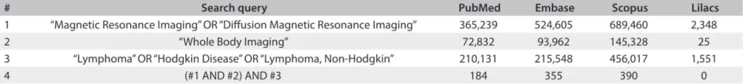
he Medline (via PubMed), Embase, Lilacs and Scopus data-bases were searched for relevant studies on the performance of WB-MRI versus other imaging methods for lymphoma evalu-ation. he references of each study included were checked for potentially relevant additional citations. he results from our search strategy are shown in Table 1. he search was last updated on September 27, 2013.
Article selection and quality assessment
For the irst stage of the selection, two investigators (RR, AP) conducted independent assessments of the titles and abstracts of articles identiied by the abovementioned search strategy. Studies on the diagnostic performance of WB-MRI for lym-phoma staging or follow-up were included. Animal studies, reviews, meta-analyses, abstracts, editorials, letters to the editor, case reports, tutorials and practice guidelines were excluded. All clearly ineligible articles were also excluded.
For the second stage, all potentially eligible studies were set aside for full-text reading, critical appraisal and data extraction, conducted independently by the same investigators (RR, AP). Any disagreements arising between them at either stage were resolved through discussion and reaching a consensus.
Study quality was assessed using the Quality Assessment of
Diagnostic Accuracy Studies (QUADAS-2).19 he QUADAS-2
tool enables more transparent ratings for bias and for the applica-bility of diagnostic accuracy studies. hree responses to questions regarding the risk of bias and applicability concerns were possi-ble: “low”, “high” or “unclear”.
Reference standard
Lymphoma staging provided by a set of clinical and radiological data was used to compare each technique in accordance with the Ann Arbor staging system. he inal staging established needed to take into account all the clinical information available at the time of diagnosis, such as physical examination, laboratory and histological results, and bone marrow biopsy, and also the infor-mation available during the clinical and imaging follow-up (CT, FDG-PET/CT, WB-MRI or other methods). his follow-up was used to determine the status of lesions ater treatment. For example, if they became larger during the follow-up period or
decreased in size ater treatment, they were considered positive for the presence of lymphoma.
Statistical analysis
he Review Manager (RevMan) version 5.1 (Cochrane Collaboration, Oxford, England) was used to calculate sensi-tivity and speciicity, with 95% conidence intervals (CIs), for WB-MRI and FDG-PET/CT, in comparison with a clinical-radiological standard reference. he results from each individual study were presented in forest plots.
Statistical analyses were performed using the SPSS 16.0 sot-ware package. he unweighted kappa (κ) statistic was used to test agreement between WB-MRI and FDG-PET/CT in the initial lymphoma staging. Agreement was considered poor at a κ value of 0, weak at 0.01-0.20, fair at 0.21-0.40, moderate at 0.41-0.60, good at 0.61-0.80 and excellent at 0.81-1.0.20 P values < 0.05 were
considered indicative of signiicant diferences.
RESULTS
he search strategy chosen yielded 929 citations. Ater careful reading of the titles and abstracts and exclusion of duplicates, 19 articles were selected for full-text analysis and critical appraisal. hirteen failed to meet the inclusion criteria (or met the exclusion criteria) and were excluded from further analysis: one study compared WB-MRI versus FDG-PET/CT for assessment of treatment response across a patient spectrum previously used in another study included in this systematic review;21 one study
used FDG-PET/CT as a single reference standard for lymphoma
staging;22 two studies assessed the performance of WB-MRI in
lymphoma, but only for detection of bone involvement;23,24 two
studies only compared WB-MRI with conventional CT;25,26 four
studies had samples that included patients with diseases other
than lymphoma;27-30 one study only compared WB-MRI with a
reference standard;31 and two studies only compared WB-MRI
with conventional CT and bone scintigraphy.32,33
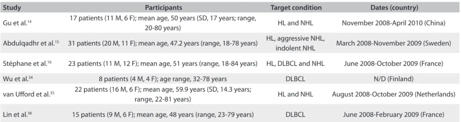
On completion of the search and retrieval strategy, six pro-spective cohort studies were included for meta-analysis.14-16,34-36
Most of them either failed to conduct separate analyses on HL and NHL or conducted pooled analyses on diferent histological subtypes of NHL. he study by Wu et al.34 limited its analysis only
to a single histological subtype of NHL. Table 2 provides a sum-mary of the key features of these studies.
# Search query PubMed Embase Scopus Lilacs
1 “Magnetic Resonance Imaging” OR “Difusion Magnetic Resonance Imaging” 365,239 524,605 689,460 2,348 2 “Whole Body Imaging” 72,832 93,962 145,328 25 3 “Lymphoma” OR “Hodgkin Disease” OR “Lymphoma, Non-Hodgkin” 210,131 215,548 456,017 1,551
4 (#1 AND #2) AND #3 184 355 390 0
Table 1. Search strategy results
Quality assessment on studies included
Figure 1 summarizes the risk of bias and applicability judgments on the six studies included. he methodological quality graph presents the percentage of included studies for which the item was rated “low”, “high” or “unclear”, for each quality assessment domain. he graph shows that the potential area of concern was the description of the reference standard.
Patient selection criteria were clearly described in all the studies included. Regarding the reference standard, van Uford et al.35 and Lin et al.36 did not describe it clearly. All the other
quality assessment parameters were considered satisfactory across all six studies.
he QUADAS-2 score, expressed as a percentage of the max-imum score, was 90% on average (range, 71-100%) in the six studies included. In the quality assessment, all of the studies were considered to present low risk of bias and low concerns about applicability.
Summary assessment of the sensitivity of WB-MRI for lymphoma staging
he sensitivity of WB-MRI and FDG-PET/CT for initial lym-phoma staging versus that of the reference standard ranged from
59% to 100% and from 63% to 100% respectively (Figure 2).
Gu et al.,14 Abdulqadhr et al.,15 Stéphane et al.16 and Lin et al.36
Study Participants Target condition Dates (country)
Gu et al.14 17 patients (11 M, 6 F); mean age, 50 years (SD, 17 years; range,
20-80 years) HL and NHL November 2008-April 2010 (China) Abdulqadhr et al.15 31 patients (20 M, 11 F); mean age, 47.2 years (range, 18-78 years) HL, aggressive NHL,
indolent NHL March 2008-November 2009 (Sweden) Stéphane et al.16 23 patients (11 M, 12 F); mean age, 51 years (range, 18-84 years) HL, DLBCL and NHL June 2008-October 2009 (France)
Wu et al.34 8 patients (4 M, 4 F); age range, 32-78 years DLBCL N/D (Finland)
van Uford et al.35 22 patients (16 M, 6 F); mean age, 59.9 years (SD, 14.3 years;
range, 22-81 years) HL and NHL August 2008-October 2009 (Netherlands) Lin et al.36 15 patients (9 M, 6 F); mean age, 48 years (range, 23-79 years) DLBCL June 2008-February 2009 (France) Table 2. Key features of the studies included in this meta-analysis (all of them were prospective cohort studies)
M = male; F = female; SD = standard deviation; HL = Hodgkin’s lymphoma; NHL = non-Hodgkin lymphoma; DLBCL = difuse large B-cell lymphoma; N/D = no data.
Figure 1. Graphical representation of study quality assessment.
Flow and timing References standard Index tests Patient selection
Proportion of studies with low, high, or unclear risk bias
Proportion of studies with low, high, or unclear concerns regarding applicability
References standard Index tests Patient selection
QU
AD
AS
-2 Domain
QU
AD
AS
-2 Domain
0% 20% 40% 60% 80% 100%
0% 20% 40% 60% 80% 100%
reported high sensitivity for both methods, whereas Wu et al.34
and van Uford et al.35 found lower sensitivity values.
Agreement between WB-MRI and FDG-PET/CT for lymphoma staging
In the study by Gu et al.,14 there was agreement between
WB-MRI and FDG-PET/CT staging in 15 of their 17 patients. In the remaining two patients, WB-MRI overstaged one and understaged the other. In the latter patient, the staging with both methods was considered inadequate in relation to the reference standard because both of them failed to detect bone marrow inil-tration, which was later conirmed by means of bone marrow biopsy. In the study by Abdulqadhr et al.,15 there was agreement
between WB-MRI and FDG-PET/CT staging in 28 of their 31 patients. In the remaining three patients, low-grade lymphoma had higher staging through WB-MRI than through FDG-PET/ CT, which was later validated by means of clinical staging. In the studies by Stéphane et al.16 and Wu et al.34 WB-MRI and
FDG-PET/CT yielded the same staging in all patients, although three were incorrectly staged with both methods in the study by Wu et al.34 In the sample of van Uford et al.,35 WB-MRI and
FDG-PET/CT agreed regarding the staging of 17 of their 22 patients. WB-MRI overstaged ive patients in relation to FDG-PET/CT, and only one of these patients, who had bone marrow iniltra-tion later conirmed by biopsy, was correctly staged by means of the imaging method. Finally, in the study by Lin et al.,36 WB-MRI
and FDG-PET/CT yielded similar staging for 14 patients. In the sole case in which the staging was diferent between the methods, it was higher with WB-MRI than with FDG-PET/CT, although
Specificity Sensitivity
Specificity Sensitivity
0 0.2 0.4 0.6 0.8 1
WB-MRI
FDG-PET/CT
Not estimable
Specificity Sensitivity
TP FP FN TN Study Not estimable 0 0 0 0 0 0 0 0 0 0 0 0
1.00 [0.89, 1.00] 0.88 [0.64, 0.99] 0.93 [0.68, 1.00] 1.00 [0.85, 1.00] 0.59 [0.36, 0.79] 0.63 [0.24, 0.91] Wu et al.34 2011
van Uford et al.35 2011
Stéphane et al.16 2013
Lin et al.36 2010
Gu et al.14 2011
Abdulqadhr et al.15 2011 0
2 1 0 9 3 31 15 14 23 13 5 Not estimable Not estimable Not estimable Not estimable Not estimable Specificity Sensitivity
TP FP FN TN Study Not estimable 0 0 0 0 0 0 0 0 0 0 0 0
0.90 [0.74, 0.98] 0.94 [0.71, 1.00] 0.93 [0.68, 1.00] 1.00 [0.85, 1.00] 0.73 [0.50, 0.89] 0.63 [0.24, 0.91] Wu et al.34 2011
van Uford et al.35 2011
Stéphane et al.16 2013
Lin et al.36 2010
Gu et al.14 2011
Abdulqadhr et al.15 2011 3
1 1 0 6 3 28 16 14 23 16 5 Not estimable Not estimable Not estimable Not estimable
0 0.2 0.4 0.6 0.8 1
0 0.2 0.4 0.6 0.8 1 0 0.2 0.4 0.6 0.8 1
Figure 2. Forest plots of the sensitivity of imaging methods for lymphoma staging versus a comparison reference standard. TP = true positive, FP = false positive, FN = false negative, TN = true negative. Brackets show 95% conidence intervals. The igure shows the sensitivity for each study (squares) and 95% conidence intervals (horizontal lines). Speciicity was not calculable, since all patients had lymphoma.
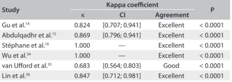
Study Kappa coeicient P
κ CI Agreement
Gu et al.14 0.824 [0.707; 0.941] Excellent < 0.0001
Abdulqadhr et al.15 0.869 [0.796; 0.941] Excellent < 0.0001
Stéphane et al.16 1.000 — Excellent < 0.0001
Wu et al.34 1.000 — Excellent < 0.0001
van Uford et al.35 0.683 [0.564; 0.803] Good < 0.0001
Lin et al.36 0.847 [0.712; 0.981] Excellent < 0.0001 Table 3. Agreement between WB-MRI and FDG-PET/CT for lymphoma staging in each of the studies included
WB-MRI = whole-body difusion-weighted magnetic resonance imaging; FDG-PET/CT = [18]F-luoro-2-deoxyglucose positron emission tomography; CI = conidence interval.
both methods staged the patient incorrectly, compared with the reference standard. he kappa statistic was indicative of excel-lent overall agreement between WB-MRI and FDG-PET/CT (κ = 0.871 [0.782; 0.960]; P < 0.0001). Table 3 summarizes the agreement between the two methods in each study.
DISCUSSION
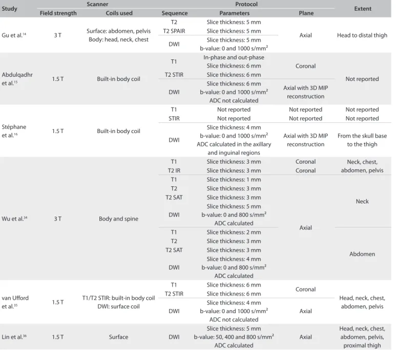
thicknesses and body areas (Table 4) are a particular cause for concern. Lymph node size cutofs and the criteria used to classify an organ or extranodal lesion as “involved” also difered across studies. In view of this heterogeneity, lesions that were consid-ered positive with one imaging method may have been classiied as negative with the other.
However, all the studies also conducted staging in accordance with the Ann Arbor classiication, which is based on a clinical and radiological reference standard, to which both WB-MRI and FDG-PET/CT staging were compared. Lymphoma staging in the
Ann Arbor system is dependent on disease distribution (above or below the diaphragm) and on the afected lymph node sites. Detection of one abnormal lymph node or extranodal lesion suf-ices for a region to be classiied as involved. Hence, for staging purposes, detection of additional lesions in a region or organ classiied as positive is of no utility. his enables comparison between studies, despite methodological diferences, in terms of the ability of each imaging method to establish staging.
Overall, the six studies included in this meta-analysis assessed 116 patients. here was agreement in staging between
Study Scanner Protocol Extent
Field strength Coils used Sequence Parameters Plane
Gu et al.14 3 T Surface: abdomen, pelvis
Body: head, neck, chest
T2 Slice thickness: 5 mm
Axial Head to distal thigh T2 SPAIR Slice thickness: 5 mm
DWI Slice thickness: 5 mm b-value: 0 and 1000 s/mm²
Abdulqadhr
et al.15 1.5 T Built-in body coil
T1 In-phase and out-phase
Slice thickness: 6 mm Coronal
Not reported T2 STIR Slice thickness: 6 mm
DWI
Slice thickness: 6 mm b-value: 0 and 1000 s/mm²
ADC not calculated
Axial with 3D MIP reconstruction
Stéphane
et al.16 1.5 T Built-in body coil
T1 Not reported Not reported Not reported STIR Not reported Not reported Not reported
DWI
Slice thickness: 4 mm b-value: 0 and 1000 s/mm² ADC calculated in the axillary
and inguinal regions
Axial with 3D MIP reconstruction
From the skull base to the thigh
Wu et al.34 3 T Body and spine
T1 Slice thickness: 3 mm Coronal Neck, chest, abdomen, pelvis T2 IR Slice thickness: 3 mm Coronal
T1 Slice thickness: 1 mm
Axial
Neck T2 Slice thickness: 3 mm
T2 SAT Slice thickness: 3 mm DWI
Slice thickness: 5 mm b-value: 0 and 800 s/mm²
ADC calculated T1 Slice thickness: 2 mm
Abdomen T2 Slice thickness: 3 mm
T2 SAT Slice thickness: 3 mm DWI
Slice thickness: 4 mm b-value: 0 and 800 s/mm²
ADC calculated
van Uford
et al.35 1.5 T
T1/T2 STIR: built-in body coil DWI: surface coil
T1 Slice thickness: 6 mm
Coronal
Head, neck, chest, abdomen, pelvis T2 STIR Slice thickness: 6 mm
DWI
Slice thickness: 4 mm b-value: 0 and 1000 s/mm²
ADC not calculated
Axial
Lin et al.36 1.5 T Surface DWI
Slice thickness: 5 mm b-value: 50, 400 and 800 s/mm²
ADC calculated
Axial
Head, neck, chest, abdomen, pelvis, proximal thigh
SPAIR = spectral presaturation attenuated inversion recovery; STIR = short tau inversion recovery; DWI = difusion-weighted imaging; SAT = fat saturation; ADC = apparent difusion coeicient; MIP = maximum intensity projection; WB-MRI = whole-body difusion-weighted magnetic resonance imaging.
WB-MRI and FDG-PET/CT in 105 cases (90.5%). In nearly all cases of diferences in staging, WB-MRI yielded a higher grade than FDG-PET/CT. Overall, there was excellent agreement between the two methods (κ = 0.871; P < 0.0001).
Some characteristics inherent to WB-MRI may lead to false-positive results, such as its limited ability to distinguish malignant from benign causes of lymph node enlargement, particularly in inguinal and axillary nodes, and its extreme sensitivity for small
lymph nodes, even on DWI sequences,14 as well as the T2
shine-through efect, which refers to an area of high signal on DWI mimicking restricted difusion due to very prolonged spin-spin relaxation time.34 In such cases, ADC quantitative analysis can
be helpful for reducing the number of false positives in these cases. he causes of false-negative indings inherent to WB-MRI include diaphragmatic motion artifacts37,38 and artifacts in the
hilar region due to respiratory and cardiac motion,35 as well as
falsely elevated ADC values in these areas.25 Stéphane et al.16 also
reported diiculties in analyzing hilar regions.
Several intrinsic factors may hinder interpretation of FDG-PET/CT results. Non-pathological variability in FDG uptake by healthy tissues, FDG uptake attributable to inlammation, altered biodistribution of FDG due to hyperglycemia or hyper-insulinemia and, particularly, the bone marrow activation commonly found in cancer patients ater treatment may lead to false positives.34
Addition of DWI to WB-MRI protocols provides improved lymph node viewing, compared with conventional sequences, thus increasing the accuracy of the method for detection of lesions,14 whereas ADC value analysis improves speciicity. In the
study by Lin et al.,36 DWI using the lesion size criterion yielded
sensitivity and speciicity of 90% and 94% respectively, in com-parison with FDG-PET/CT. Addition of visual ADC analysis reduced the sensitivity to 81% and increased the speciicity to approximately 100%.
ADC quantiication can also provide useful information on treatment response.18 In the study by Lin et al.,36 the mean ADC
(×10−3 mm2/s) in regions with restricted difusion was 0.75,
ver-sus 1.6 in regions with no restriction. In the study by Wu et al.,34
the ADC correlated inversely with the maximum standardized
uptake value (SUVmax) of FDG-PET/CT, thus suggesting that
these parameters were comparable.
A diferent WB-MRI protocol was used in each of the six studies included, and this lack of standardization hindered comparison of WB-MRI with other, better established methods. herefore, development of standardized protocols is critical to establishing the role of WB-MRI for staging and monitoring of lymphoma and other malignant conditions. Moreover, it may be of interest to compare diferent strength ields, such as 1.5 T versus 3.0 T. Despite this heterogeneity, ive of the six studies
included showed excellent agreement between WB-MRI and
FDG-PET/CT, and one of them showed good agreement.35
Abdulqadhr et al.15 obtained T1-weighted and T2-weighted
cor-onal images with SPAIR and axial DWI images, with 3D MIP reconstruction with a total scan time of roughly 50 minutes. Stéphane et al.16 used a similar protocol, except for the use of
T2-weighted images with STIR, acquired for a total scan time
of 40 minutes. Gu et al.14 used axial T2 and T2 with SPAIR
sequences and axial DWI with 3D reconstruction for a total scan time of 44 to 52 minutes. Lin et al.36 also obtained
excel-lent results with axial DWI alone and a total scan time of 30 to 45 minutes. his study also included ADC calculation, thus improving the speciicity of the method, which is essential for
treatment response assessment. Wu et al.34 also measured ADC
and obtained results that corroborate its importance in patient follow-up, but the test protocol was complex and no informa-tion on total scan time was provided. van Uford et al.35 used a
protocol consisting of coronal T1-weighted, T2-weighted and STIR images (total scan time, 25 to 30 minutes) and axial DWI (total scan time, 20 to 25 minutes). his was the only study in which WB-MRI showed good agreement with FDG-PET/CT, and this was due to lack of experience in WB-MRI interpreta-tion by the examining radiologists. In our opinion, a WB-MRI protocol can be built only with DWI, which has shown excellent results for lymphoma staging in comparison with FDG-PET/ CT. ADC analysis, visual or otherwise, should also be provided for, since assessment of the functional evolution of residual lesions plays an important role in treatment monitoring.39
Diagnostic accuracy studies usually assess the accuracy of a test method under evaluation (index test) in relation to that of a gold-standard, well-established comparison method (ref-erence standard), for detection of the presence or absence of a target condition. Conversely, the present review did not set out to assess the ability of WB-MRI or FDG-PET/CT to detect the pres-ence or abspres-ence of lymphoma, but the ability of either method to yield a correct disease stage, in comparison with a reference stan-dard. he clinical and radiological reference standard to which the results of WB-MRI and FDG-PET/CT were independently compared was based on a set of parameters assessed over time. Since this reference standard establishes the deinitive baseline staging that will be used for patient management and treatment planning, it may be considered to be the true measurement. herefore, we were able to calculate the sensitivity of the index methods as used in each of the studies included and compare them with the reference standard, i.e. to ascertain the ability of each method to stage the target condition correctly in relation to
a true measurement.40 We found that both WB-MRI and
FDG-PET/CT exhibited high sensitivity in the studies by Gu et al.,14
WB-MRI and 90 to 94% for FDG-PET/CT. he highest sensitiv-ity (100% for both methods) was found in the study by Stéphane et al.16 In the study by Wu et al.,34 because of a poorly
representa-tive patient spectrum and because both methods staged three out of the eight patients incorrectly, the overall sensitivity was 63%. In the study by van Uford et al.,35 the sensitivity of WB-MRI was
59%, and that of FDG-PET/CT, 73%.
One limitation of the present review derives from the use of a clinical and radiological reference standard. Stéphane et al.16
used FDG-PET/CT as the gold standard method, although they also explicitly used clinical and imaging follow-up data to set up
the diferences between WB-MRI and FDG-PET/CT. Gu et al.14
also referred FDG-PET/CT as the reference standard for assess-ment of lesions on an individual basis and established the Ann Arbor staging using data such as physical examination, inte-grated FDG-PET/CT images at baseline and follow-up, and bone
marrow biopsy results. Both Abdulqadhr et al.15 and van Uford
et al.35 separately staged the patients using WB-MRI and
FDG-PET/CT. For the former, diferences in staging between the two methods were resolved using biopsy results and clinical and CT follow-ups; for the latter, these diferences were resolved using the contrast-enhanced full-dose component of the FDG-PET/
CT examination and bone marrow biopsy. Lin et al.36 staged
patients by means of physical examination, contrast-enhanced CT, FDG-PET/CT and bone marrow biopsy. Neither van Uford et al.35 nor Lin et al.36 made it clear whether follow-up
examinations were also included in determining the inal stag-ing. Wu et al.34 established lymphoma staging through detailed
medical history, physical examination, standard laboratory tests, CT scans of the chest, abdomen and pelvis and bone marrow
biopsy. All authors except Wu et al.34 included WB-MRI and/or
FDG-PET/CT as part of the reference standard and, because of this, incorporation bias may have occurred, which would prob-ably increase the level of agreement between the two index tests and the reference standard, and hence overestimate the mea-surements of diagnostic accuracy.19
Proven presence or absence of viable tumor tissue in anatom-ical pathology specimens is the most accurate reference standard in the ield of oncology. However, since lymphomas oten pres-ent as a difuse disease, surgical exploration of all potpres-ential sites of involvement for histological analysis is ethically and practi-cally unfeasible and may not afect treatment planning; therefore clinical and radiological staging is widely accepted as the refer-ence standard.
WB-MRI provides several advantages over FDG-PET/CT. It does not emit ionizing radiation, which is particularly useful in
children and young adults41,42 and when patients must undergo
repeated imaging for follow-ups, as in lymphoma cases. FDG-PET/CT exposes patients to substantial radiation doses and,
consequently, is associated with increased risk of later malig-nancies.42 Furthermore, thorough patient preparation is required
before FDG-PET/CT, and because a cyclotron is required to pro-duce FDG, it is not widely available.43
CONCLUSION
WB-MRI is a highly sensitive method for initial lymphoma stag-ing. It has excellent agreement with FDG-PET/CT and is a great alternative for managing lymphoma patients, without using ionizing radiation or an intravenous contrast agent. However, in order to deine the role of WB-MRI in clinical practice, fur-ther studies are needed to assess the performance of WB-MRI in comparison with FDG-PET/CT, with regard to early and late response evaluation.
REFERENCES
1. Siegel R, Naishadham D, Jemal A. Cancer statistics, 2012. CA Cancer J
Clin. 2012;62(1):10-29.
2. Armitage JO. Staging non-Hodgkin lymphoma. CA Cancer J Clin.
2005;55(6):368-76.
3. Connors JM. State-of-the-art therapeutics: Hodgkin’s lymphoma.
J Clin Oncol. 2005;23(26):6400-8.
4. Lister TA, Crowther D, Sutclife SB, et al. Report of a committee
convened to discuss the evaluation and staging of patients
with Hodgkin’s disease: Cotswolds meeting. J Clin Oncol.
1989;7(11):1630-6.
5. Lapela M, Leskinen S, Minn HR, et al. Increased glucose metabolism
in untreated non-Hodgkin’s lymphoma: a study with positron
emission tomography and luorine-18-luorodeoxyglucose. Blood.
1995;86(9):3522-7.
6. Schöder H, Noy A, Gönen M, et al. Intensity of 18luorodeoxyglucose
uptake in positron emission tomography distinguishes between
indolent and aggressive non-Hodgkin’s lymphoma. J Clin Oncol.
2005;23(21):4643-51.
7. Juweid ME, Cheson BD. Role of positron emission tomography in
lymphoma. J Clin Oncol. 2005;23(21):4577-80.
8. Tsushima Y, Takano A, Taketomi-Takahashi A, Endo K. Body
difusion-weighted MR imaging using high b-value for malignant tumor
screening: usefulness and necessity of referring to T2-weighted
images and creating fusion images. Acad Radiol. 2007;14(6):643-50.
9. Jerusalem G, Beguin Y, Najjar F, et al. Positron emission tomography
(PET) with 18F-luorodeoxyglucose (18F-FDG) for the staging
of low-grade non-Hodgkin’s lymphoma (NHL). Ann Oncol.
2001;12(6):825-30.
10. Brenner D, Elliston C, Hall E, Berdon W. Estimated risks of
radiation-induced fatal cancer from pediatric CT. AJR Am J Roentgenol.
2001;176(2):289-96.
11. Kleinerman RA. Cancer risks following diagnostic and therapeutic
12. Mathews JD, Forsythe AV, Brady Z, et al. Cancer risk in 680,000 people
exposed to computed tomography scans in childhood or adolescence:
data linkage study of 11 million Australians. BMJ. 2013;346:f2360.
13. Walker RE, Eustace SJ. Whole-body magnetic resonance imaging:
techniques, clinical indications, and future applications. Semin
Musculoskelet Radiol. 2001;5(1):5-20.
14. Gu J, Chan T, Zhang J, et al. Whole-body difusion-weighted imaging:
the added value to whole-body MRI at initial diagnosis of lymphoma.
AJR Am J Roentgenol. 2011;197(3):W384-91.
15. Abdulqadhr G, Molin D, Aström G, et al. Whole-body
difusion-weighted imaging compared with FDG-PET/CT in staging of
lymphoma patients. Acta Radiol. 2011;52(2):173-80.
16. Stéphane V, Samuel B, Vincent D, et al. Comparison of PET-CT
and magnetic resonance difusion weighted imaging with body
suppression (DWIBS) for initial staging of malignant lymphomas. Eur
J Radiol. 2013;82(11):2011-7.
17. de Bazelaire C, de Kerviler E. From multislice CT to whole-body
biomarker imaging in lymphoma patients. Eur Radiol. 2011;21(3):555-8.
18. Dudeck O, Zeile M, Pink D, et al. Difusion-weighted magnetic resonance
imaging allows monitoring of anticancer treatment efects in patients
with soft-tissue sarcomas. J Magn Reson Imaging. 2008;27(5):1109-13.
19. Whiting PF, Rutjes AW, Westwood ME, et al. QUADAS-2: a revised tool
for the quality assessment of diagnostic accuracy studies. Ann Intern
Med. 2011;155(8):529-36.
20. Landis JR, Koch GG. The measurement of observer agreement for
categorical data. Biometrics. 1977;33(1):159-74.
21. Lin C, Itti E, Luciani A, et al. Whole-body difusion-weighted imaging
with apparent difusion coeicient mapping for treatment response
assessment in patients with difuse large B-cell lymphoma: pilot
study. Invest Radiol. 2011;46(5):341-9.
22. Punwani S, Taylor SA, Bainbridge A, et al. Pediatric and adolescent
lymphoma: comparison of whole-body STIR half-Fourier RARE MR
imaging with an enhanced PET/CT reference for initial staging.
Radiology. 2010;255(1):182-90.
23. Ribrag V, Vanel D, Leboulleux S, et al. Prospective study of bone
marrow iniltration in aggressive lymphoma by three independent
methods: whole-body MRI, PET/CT and bone marrow biopsy. Eur J
Radiol. 2008;66(2):325-31.
24. Kwee TC, Fijnheer R, Ludwig I, et al. Whole-body magnetic resonance
imaging, including difusion-weighted imaging, for diagnosing
bone marrow involvement in malignant lymphoma. Br J Haematol.
2010;149(4):628-30.
25. Kwee TC, van Uford HM, Beek FJ, et al. Whole-body MRI, including
difusion-weighted imaging, for the initial staging of malignant
lymphoma: comparison to computed tomography. Invest Radiol.
2009;44(10):683-90.
26. Brennan DD, Gleeson T, Coate LE, et al. A comparison of whole-body
MRI and CT for the staging of lymphoma. AJR Am J Roentgenol.
2005;185(3):711-6.
27. Krohmer S, Sorge I, Krausse A, et al. Whole-body MRI for primary
evaluation of malignant disease in children. Eur J Radiol.
2010;74(1):256-61.
28. Stecco A, Romano G, Negru M, et al. Whole-body difusion-weighted
magnetic resonance imaging in the staging of oncological patients:
comparison with positron emission tomography computed
tomography (PET-CT) in a pilot study. Radiol Med. 2009;114(1):1-17.
29. Goo HW, Choi SH, Ghim T, Moon HN, Seo JJ. Whole-body MRI of
paediatric malignant tumours: comparison with conventional
oncological imaging methods. Pediatr Radiol. 2005;35(8):766-73.
30. Daldrup-Link HE, Franzius C, Link TM, et al. Whole-body MR imaging
for detection of bone metastases in children and young adults:
comparison with skeletal scintigraphy and FDG PET. AJR Am J
Roentgenol. 2001;177(1):229-36.
31. Li S, Xue HD, Li J, et al. Application of whole body difusion weighted
MR imaging for diagnosis and staging of malignant lymphoma. Chin
Med Sci J. 2008;23(3):138-44.
32. Kellenberger CJ, Miller SF, Khan M, et al. Initial experience with FSE
STIR whole-body MR imaging for staging lymphoma in children. Eur
Radiol. 2004;14(10):1829-41.
33. Iizuka-Mikami M, Nagai K, Yoshida K, et al. Detection of bone marrow
and extramedullary involvement in patients with non-Hodgkin’s
lymphoma by whole-body MRI: comparison with bone and 67Ga
scintigraphies. Eur Radiol. 2004;14(6):1074-81.
34. Wu X, Kellokumpu-Lehtinen PL, Pertovaara H, et al.
Difusion-weighted MRI in early chemotherapy response evaluation of patients
with difuse large B-cell lymphoma--a pilot study: comparison with
2-deoxy-2-luoro- D-glucose-positron emission tomography/
computed tomography. NMR Biomed. 2011;24(10):1181-90.
35. van Uford HM, Kwee TC, Beek FJ, et al. Newly diagnosed lymphoma:
initial results with whole-body T1-weighted, STIR, and
difusion-weighted MRI compared with 18F-FDG PET/CT. AJR Am J Roentgenol.
2011;196(3):662-9.
36. Lin C, Luciani A, Itti E, et al. Whole-body difusion-weighted magnetic
resonance imaging with apparent difusion coeicient mapping
for staging patients with difuse large B-cell lymphoma. Eur Radiol.
2010;20(8):2027-38.
37. Kwee TC, Takahara T, Ochiai R, Nievelstein RA, Luijten PR.
Difusion-weighted whole-body imaging with background body signal
suppression (DWIBS): features and potential applications in oncology.
Eur Radiol. 2008;18(9):1937-52.
38. Kwee TC, Takahara T, Ochiai R, et al. Whole-body difusion-weighted
magnetic resonance imaging. Eur J Radiol. 2009;70(3):409-17.
39. Cheson BD, Pistner B, Juweid ME, et al. Revised response criteria for
malignant lymphoma. J Clin Oncol. 2007;25(5):579-86.
40. Cooper KL, Harnan S, Meng Y, et al. Positron emission tomography
(PET) for assessment of axillary lymph node status in early breast
cancer: A systematic review and meta-analysis. Eur J Surg Oncol.
41. Brenner DJ, Elliston CD. Estimated radiation risks potentially
associated with full-body CT screening. Radiology. 2004;232(3):735-8.
42. Huang B, Law MW, Khong PL. Whole-body PET/CT scanning:
estimation of radiation dose and cancer risk. Radiology.
2009;251(1):166-74.
43. Kwee TC, Kwee RM, Nievelstein RA. Imaging in staging of malignant
lymphoma: a systematic review. Blood. 2008;111(2):504-16.
This article was presented in the form of a dissertation by the author
Rodrigo Regacini on October 18, 2012, to Universidade Federal de São
Paulo-Escola Paulista de Medicina (Unifesp-EPM), São Paulo, Brazil
Sources of funding: None
Conlict of interest: None
Date of irst submission: January 31, 2014
Last received: September 26, 2014
Accepted: October 28, 2014
Address for correspondence: Rodrigo Regacini
Rua Napoleão de Barros, 800
Vila Clementino — São Paulo (SP) — Brasil
CEP 04024-002
Tel. (+55 11) 5908-7900