Characteristics of fetuses evaluated due to suspected
anencephaly: a population-based cohort study
in southern Brazil
Características de fetos avaliados por suspeita de anencefalia: um estudo de coorte
de base populacional do Sul do Brasil
Emanuele Pelizzari
I, Carolina Melendez Valdez
I, Jamile dos Santos Picetti
II, André Campos da Cunha
III, Cristine Dietrich
III,
Paulo Renato Krahl Fell
III, Luciano Vieira Targa
IV, Paulo Ricardo Gazzola Zen
V, Rafael Fabiano Machado Rosa
VIHospital Materno Infantil Presidente Vargas (HMIPV) and Universidade Federal de Ciências da Saúde de Porto Alegre (UFCSPA),
Porto Alegre, Rio Grande do Sul, Brazil
ABSTRACT
CONTEXT AND OBJECTIVE: Anencephaly is considered to be the most common type of neural tube defect. Our aim was to assess the clinical and gestational features of a cohort of fetuses with suspected anencephaly.
DESIGN AND SETTING: Population-based retrospective cohort study in a referral hospital in southern Brazil.
METHODS: The sample consisted of fetuses referred due to suspected anencephaly, to the Fetal Medicine Service of Hospital Materno Infantil Presidente Vargas, between January 2005 and September 2013. Clini-cal, radiologiClini-cal, pathological and survival data were gathered.
RESULTS: Our sample was composed of 29 fetuses. The diagnosis of suspected anencephaly was made on average at 21.3 weeks of gestation. Seven fetuses had malformations that afected other organs, and these included oral clefts (n = 4) and congenital heart defects (n = 2). In 16 cases, there was termination of pregnancy (n = 12) or intrauterine death (n = 4). Regarding those who were born alive (n = 13), all of them died in the irst week of life. After postnatal evaluation, the diagnosis of anencephaly was conirmed in 22 cases (75.9%). Other conditions included amniotic band disruption complex (6.9%), microhydranen-cephaly (6.9%), merocrania (3.4%) and holoprosenmicrohydranen-cephaly (3.4%).
CONCLUSIONS: Diferent conditions involving the cranial vault may be confused with anencephaly, as seen in our sample. However, these conditions also seem to have a poor prognosis. It seems that folic acid supplementation is not being properly performed.
RESUMO
CONTEXTO E OBJETIVO: A anencefalia é considerada o tipo mais comum de defeito de fechamento do tubo neural. Nosso objetivo foi avaliar as características clínicas e gestacionais de uma coorte de fetos com suspeita de anencefalia.
TIPO DE ESTUDO E LOCAL: Estudo de coorte retrospectivo de base populacional em um hospital de referência no sul do Brasil.
MÉTODOS: A amostra foi composta por fetos encaminhados por suspeita de anencefalia ao Serviço de Medicina Fetal do Hospital Materno Infantil Presidente Vargas, no período de janeiro de 2005 a setembro de 2013. Foi realizada coleta de dados clínicos, radiológicos, patológicos e de sobrevida.
RESULTADOS: Nossa amostra foi composta por 29 fetos. A suspeita do diagnóstico de anencefalia foi realizada em média com 21,3 semanas de gestação. Sete fetos apresentavam malformações que afetavam outros órgãos, e incluíram fendas orais (n = 4) e defeitos cardíacos congênitos (n = 2). Em 16 casos houve interrupção da gravidez (n = 12) ou morte intrauterina (n = 4). Daqueles que nasceram vivos (n = 13), todos morreram na primeira semana de vida. Após a avaliação pós-natal, o diagnóstico de anencefalia foi conirmado em 22 casos (75,9%). Outras condições incluíram o complexo disruptivo de banda amniótica (6,9%), microhidranencefalia (6,9%), merocrania (3,4%) e holoprosencefalia (3,4%).
CONCLUSÕES: Diferentes condições que envolvem a calota craniana podem ser confundidas com a anencefalia, como veriicado em nossa amostra. No entanto, estas também parecem ter um prognóstico pobre. A suplementação com ácido fólico parece não estar sendo realizada de forma adequada.
IMD. Physician, Residency Program in Obstetrics
and Gynecology, Hospital Materno Infantil Presidente Vargas (HMIPV), Porto Alegre, Rio Grande do Sul, Brazil.
IIMD. Physician, Residency Program in Fetal
Medicine, Hospital Materno Infantil Presidente Vargas (HMIPV), Porto Alegre, Rio Grande do Sul, Brazil.
IIIMD. Obstetrician, Fetal Medicine, Hospital
Materno Infantil Presidente Vargas (HMIPV), Porto Alegre, Rio Grande do Sul, Brazil.
IVMD. Pediatric Radiologist, Hospital Materno
Infantil Presidente Vargas (HMIPV), Porto Alegre, Rio Grande do Sul, Brazil.
VPhD. Adjunct Professor of Clinical Genetics and
of the Postgraduate Program on Pathology, Universidade Federal de Ciências da Saúde de Porto Alegre (UFCSPA), and Clinical Geneticist, Universidade Federal de Ciências da Saúde de Porto Alegre (UFCSPA) and Complexo Hospitalar Santa Casa de Porto Alegre (CHSCPA), Porto Alegre, Rio Grande do Sul, Brazil.
VIPhD. Clinical Geneticist, Universidade Federal
de Ciências da Saúde de Porto Alegre (UFCSPA), Complexo Hospitalar Santa Casa de Porto Alegre (CHSCPA) and Hospital Materno Infantil Presidente Vargas (HMIPV), Porto Alegre, Rio Grande do Sul, Brazil.
KEY WORDS:
Anencephaly. Neural tube.
Amniotic band syndrome. Ultrasonics.
Genetic counseling.
PALAVRAS-CHAVE:
Anencefalia. Tubo neural.
Síndrome de bandas amnióticas. Ultrassom.
INTRODUCTION
Neural tube defects are considered to be one of the most com-mon malformations observed at birth. Acom-mong these is anen-cephaly, which is the most common type, occurring in about 1 in every 1,000 births in the United States and 5 in every
1,000 births in Ireland and Wales.1-3 It is considered to be a
severe malformation of the central nervous system, and its pre-natal diagnosis through ultrasound has been possible for over
40 years.1,4
he etiology of anencephaly is attributed to a failure of neurulation that may be associated with genetic or environmental
factors, or both.1 Furthermore, it may be diicult to diferentiate
anencephaly from other conditions with involvement of the cranial vault during the prenatal period, and this may have important implications for pregnancy management (including determination of the prognosis and legal issues) and for the genetic counseling to be given to the family. Furthermore, there is a paucity of studies evaluating these matters, not only in the
Brazilian literature but also in the worldwide literature.5-7
OBJECTIVE
Our aim was to assess the clinical and gestational features of a cohort of fetuses with suspected anencephaly.
METHODS
his was a population-based retrospective cohort study in a referral hospital in southern Brazil. he sample consisted of fetuses referred due to suspected anencephaly, to the Fetal Medicine Service of Hospital Materno Infantil Presidente Vargas (HMIPV), between January 2005 and September 2013. We gathered clinical, radiological, pathological and survival data from the medical records. his project was approved by the Research Ethics Committees of our university and hospital. he data retrieved from the medical records consisted of maternal age and ethnicity; history of parental consanguinity; origin; maternal occupation; data on obstetric past; medication use (including folic acid); exposure to tobacco, alcohol and illicit drugs; maternal diseases; obstetric complications; family history of neural tube defects; ultrasound examinations performed during pregnancy, noting how many were done and at what stage, and describing the results; additional tests such as echocardiography, magnetic resonance imaging and karyotyping; data relating to performing legal termination of pregnancy; occurrence of intrauterine death and postnatal survival; delivery and perinatal data; autopsy results; and inal diagnosis. he inal diagnosis was reviewed in all cases, based on the indings obtained through the research protocol and the images from the hospital’s database. Gestational age was determined according to the earlier ultrasound. We excluded fetuses that were lost from the prenatal follow-up at our service or had incomplete medical records.
We used the PEPI sotware for Windows and the Statistical Package for the Social Sciences (SPSS), version 12.0 for Windows, to analyze the results. he tests applied were the two-tailed Fisher exact test for comparisons of frequencies and the t test for compar-isons of means. P values < 0.05 were considered to be signiicant. he Kaplan-Meier test was used through the BioEstat 5.0 sotware, to construct pregnancy termination and survival curves.
RESULTS
Over a period of about 9 years, 32 fetuses with suspected anen-cephaly were identiied. Among these fetuses, three were excluded because they were lost from the prenatal follow-up (n = 1) or had incomplete medical records (n = 2). hus, our inal sample was composed of 29 fetuses. he demographic data
relat-ing to this sample can be seen in Table 1. In only eight cases, there
was a note in the medical record regarding consanguinity and all patients were found to be non-consanguineous. Regarding origin, the majority of the fetuses came from areas peripheral to Porto Alegre (the city where the hospital is located) (41.8%);
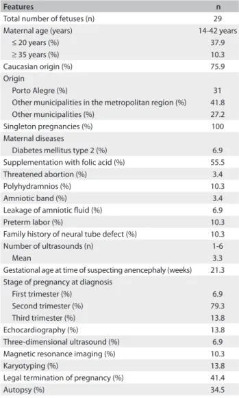
Table 1. Demographic data of the sample
Features n
Total number of fetuses (n) 29
Maternal age (years) 14-42 years
≤ 20 years (%) 37.9
≥ 35 years (%) 10.3
Caucasian origin (%) 75.9
Origin
Porto Alegre (%) 31
Other municipalities in the metropolitan region (%) 41.8
Other municipalities (%) 27.2
Singleton pregnancies (%) 100
Maternal diseases
Diabetes mellitus type 2 (%) 6.9
Supplementation with folic acid (%) 55.5
Threatened abortion (%) 3.4
Polyhydramnios (%) 10.3
Amniotic band (%) 3.4
Leakage of amniotic luid (%) 6.9
Preterm labor (%) 10.3
Family history of neural tube defect (%) 10.3
Number of ultrasounds (n) 1-6
Mean 3.3
Gestational age at time of suspecting anencephaly (weeks) 21.3 Stage of pregnancy at diagnosis
First trimester (%) 6.9
Second trimester (%) 79.3
Third trimester (%) 13.8
Echocardiography (%) 13.8
Three-dimensional ultrasound (%) 6.9
Magnetic resonance imaging (%) 10.3
Karyotyping (%) 13.8
Legal termination of pregnancy (%) 41.4
nine (31%) were referred from within the city and eight (27.2%) came from other municipalities in the state of Rio Grande do Sul. Regarding maternal occupation, the majority were housewives (44.8%). Two pregnant women (6.9%) had a history of working with chemical agents.
All cases in the sample consisted of singleton pregnancies (there were no twins). he number of pregnancies ranged from one to ive (mean: 2.3). hree pregnant women (10.3%) had a his-tory of one previous abortion. Maternal diseases were described in four cases (13.8%) and consisted of diabetes mellitus type 2 (n = 2), gestational diabetes mellitus (n = 1) and congenital cat-aract (n = 1). Both of the pregnant women with type 2 diabetes mellitus required insulin treatment. Data on the use of folic acid was reported in only nine cases (31%) and in ive, supplementa-tion was administered. However, this was done ater the second month of pregnancy in all cases. Regarding gestational exposure, ive pregnant women (17.2%) reported smoking, three (10.3%) alcohol intake and two (6.9%) use of illicit drugs (one case of
cocaine and one of marijuana) (Table 1).
he diagnosis of suspected anencephaly was made on average at a gestational age of 21.3 weeks (ranging from 12 to 34 weeks). he complementary examinations performed can be seen in
Table 1. here were no cases of chromosomal abnormalities. Seven fetuses had malformations that afected other organs and systems outside the central nervous system. hese included oral clets (n= 4), congenital heart defects (n = 2), microphthalmia (n = 2), hypertelorism (n = 1), hypotelorism (n = 1) and single umbilical artery (n = 1).
In 12 cases, there was a request for legal termination of preg-nancy. he time of discontinuation ranged from 18 to 30 weeks of gestation (mean of 24.3 weeks). he time between comple-tion of the applicacomple-tion and interrupcomple-tion ranged from 1 to 15 days (mean of 4.6 days). Only one fetus was evaluated ater the oicial legalization of pregnancy termination due to fetal anencephaly. Four fetuses already presented intrauterine fetal death at the time
of interruption (Figure 1). Among the pregnancies that were not
interrupted (n = 17), 4 (23.5%) evolved to intrauterine death. he gestational age of these cases ranged from 22 to 40 weeks. Among the pregnancies that were not interrupted and did not evolve to intrauterine death (n = 13), the majority (76.9%) were delivered vaginally, and the gestational age at birth ranged from 17 to 41 weeks (the average was 38.8 weeks, and there were four preterm cases). Birth weight ranged from 183 to 4,380 g (mean 2,392 g). All of these infants died during their irst week of life, and 11 of them (84.6%) died on the irst day.
Out of the whole sample, 16 fetuses (55.2%) were male.
In 10 cases (34.5%), an autopsy was performed (Table 1). None
of the fetuses underwent radiographic evaluation. Interestingly, one of the fetuses with anencephaly was born with the umbilical
cord attached to his cephalic pole. In 22 cases (75.9%), the diagnosis of anencephaly was conirmed. Other conditions that were diagnosed included amniotic band disruption com-plex (6.9%), microhydranencephaly/fetal brain disruption sequence (6.9%), merocrania (3.4%) and holoprosenceph-aly (3.4%). here was one case without a deinitive diagnosis (Table 2 and Figures 2 and 3).
In comparing the group of fetuses diagnosed with anenceph-aly (n = 22) with the rest of the sample (n = 7), no signiicant diferences were observed in relation to fetal sex (40.9% versus 57.1% females) (P = 0.6665); mother’s age at the time of preg-nancy (the averages were 24.2 years and 25.4 years, respectively); or frequency of primiparity (45.8% versus 71.4%, respectively; P = 0.3898). he timing of prenatal suspicion was also similar (in most cases and in both groups, this occurred during the sec-ond trimester of pregnancy: 77.3% versus 85.7%). However, there were two cases with anencephaly (9%) in which the diagnosis was suspected during the irst trimester of pregnancy. A family history of neural tube defects was observed only in cases of anen-cephaly (13.6%).
Most fetuses in both groups who were alive at birth died on the irst day of life: all nine of the infants with anencephaly (100%), and four out of the ive infants with another diagnosis. he only infant that lived beyond the irst day of life was diag-nosed with amniotic band disruption complex (presenting acra-nia; case 24) and survived for four days.
DISCUSSION
According to the Brazilian live birth information sys-tem (SINASC), the frequency of births of individuals with Figure 1. Kaplan-Meier curve showing the times of pregnancy termination in cases that were legally interrupted.
15
10
5
10 20 30 40
Time intervals - weeks
Fr
equenc
y
anencephaly in the state of Rio Grande do Sul in 2005 was 32 cases out of 147,199 births, i.e. there was a report of one case per 4,600 live births. his proportion is lower than what has been described in the literature in other countries, such as the United
States, as we noted before.1 hese frequencies may be inluenced
by termination of the pregnancy in cases of anencephaly, and by the supplementation with folic acid, which started at the begin-ning of the 2000s in Brazil. However, we believe that this result could be an underestimate.
During the period evaluated in the present study, at the Fetal Medicine Service of HMIPV, there were nine conirmed cases of anencephaly that were born alive (13 were interrupted or pre-sented intrauterine death). his service is a reference in the state of Rio Grande do Sul for pregnant women attended through the Brazilian National Health System (Sistema Único de Saúde, SUS). hus, many cases were born outside of the hospital, thus maybe indicating that many cases of anencephaly could have been born without this diagnosis having been made during pregnancy.
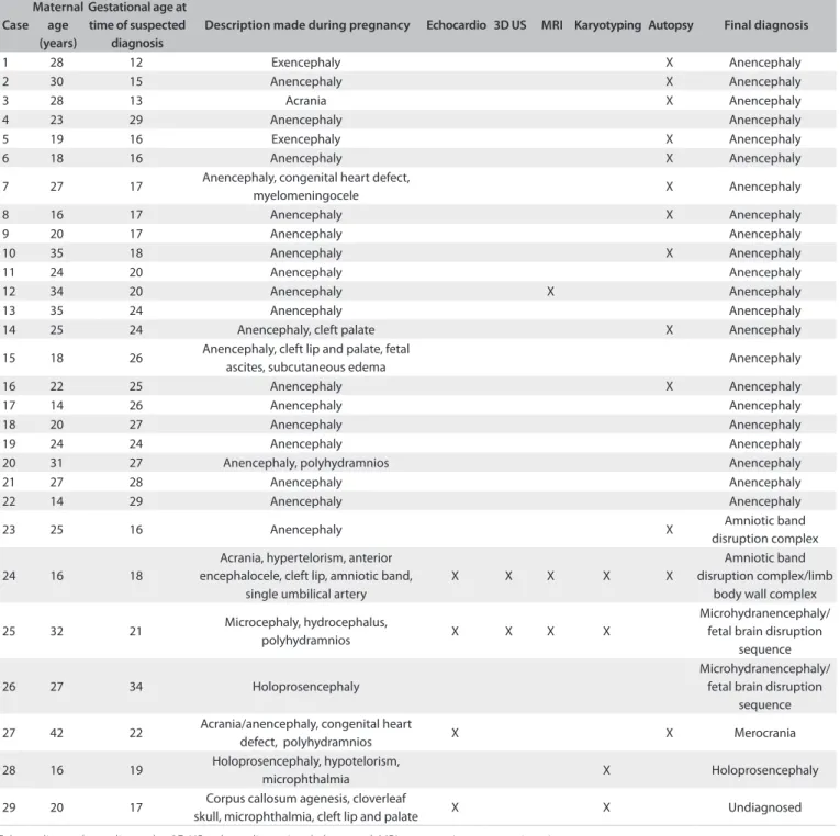
Case
Maternal age (years)
Gestational age at time of suspected
diagnosis
Description made during pregnancy Echocardio 3D US MRI Karyotyping Autopsy Final diagnosis
1 28 12 Exencephaly X Anencephaly
2 30 15 Anencephaly X Anencephaly
3 28 13 Acrania X Anencephaly
4 23 29 Anencephaly Anencephaly
5 19 16 Exencephaly X Anencephaly
6 18 16 Anencephaly X Anencephaly
7 27 17 Anencephaly, congenital heart defect,
myelomeningocele X Anencephaly
8 16 17 Anencephaly X Anencephaly
9 20 17 Anencephaly Anencephaly
10 35 18 Anencephaly X Anencephaly
11 24 20 Anencephaly Anencephaly
12 34 20 Anencephaly X Anencephaly
13 35 24 Anencephaly Anencephaly
14 25 24 Anencephaly, cleft palate X Anencephaly
15 18 26 Anencephaly, cleft lip and palate, fetal
ascites, subcutaneous edema Anencephaly
16 22 25 Anencephaly X Anencephaly
17 14 26 Anencephaly Anencephaly
18 20 27 Anencephaly Anencephaly
19 24 24 Anencephaly Anencephaly
20 31 27 Anencephaly, polyhydramnios Anencephaly
21 27 28 Anencephaly Anencephaly
22 14 29 Anencephaly Anencephaly
23 25 16 Anencephaly X Amniotic band
disruption complex
24 16 18
Acrania, hypertelorism, anterior encephalocele, cleft lip, amniotic band,
single umbilical artery
X X X X X
Amniotic band disruption complex/limb
body wall complex
25 32 21 Microcephaly, hydrocephalus,
polyhydramnios X X X X
Microhydranencephaly/ fetal brain disruption
sequence
26 27 34 Holoprosencephaly
Microhydranencephaly/ fetal brain disruption
sequence
27 42 22 Acrania/anencephaly, congenital heart
defect, polyhydramnios X X Merocrania
28 16 19 Holoprosencephaly, hypotelorism,
microphthalmia X Holoprosencephaly
29 20 17 Corpus callosum agenesis, cloverleaf
skull, microphthalmia, cleft lip and palate X X Undiagnosed
Echocardio = echocardiography; 3D US = three-dimensional ultrasound; MRI = magnetic resonance imaging.
Another point to be considered is the fact that because of the impossibility of treatment and lethality of anencephaly, it is a condition without investment. For this reason, perhaps, many cases prenatally diagnosed in hospitals other than HMIPV were not referred to our service, i.e. were handled locally.
Prenatal ultrasound is able to detect ossiied portions of the fetal skeleton from the end of the irst trimester of pregnancy onwards. In cases of anencephaly, the unprotected brain tissue undergoes gradual degeneration and destruction due to mechan-ical and chemmechan-ical trauma, thereby leading to complete or nearly
complete disappearance of the brain around the 14th week of
ges-tation. hus, at an early stage during the irst trimester of preg-nancy, there is still a variable amount of brain tissue. herefore, exencephaly is considered to be the best term to describe
anen-cephaly during this initial stage.8
Nowadays, prenatal detection of anencephaly by means of ultrasound is possible in almost 100% of cases. Moreover, during the last decade, it has become possible to diagnose this condition in the early stages of pregnancy: several case series and case reports have been published conirming the possibility of diagnosis from
the 10th week of gestation onwards.Anencephaly can be
diag-nosed by demonstrating the absence of a cranial vault. However, in the irst trimester, this can be particularly diicult because
of the variable quantity of brain tissue that may be present.4,9
In our sample, two cases of anencephaly were diagnosed during this gestational period. he diagnosis of suspected anenceph-aly was made on average at a gestational age of 21.3 weeks. We believe that this inding may be related to delay in the arrival of these fetuses for specialized evaluation. In addition, this may have important implications, especially with regard to termina-tion of pregnancy.
A
B
C
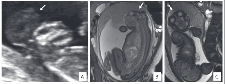
Figure 2. Two-dimensional ultrasound showing the proile of fetuses presenting exencephaly, i.e. the irst stage of anencephaly (case 1). Note also the presence of a signiicant amount of brain tissue (A). Appearance of one of the fetuses (case 12) with anencephaly (see arrows) evaluated through magnetic resonance imaging (B and C).
Figure 3. Ultrasound indings from a fetus (case 7) still in at the stage of exencephaly (A, see arrows) and with myelomeningocele (B).
A
B
BrainReduction in the crown-rump length or chin-vertex length and increased echogenicity of the amniotic luid are indings
that may assist in conirming the diagnosis of anencephaly.4,10,11
Surface images can also be obtained by means of
three-dimen-sional ultrasound.12 However, in our sample, this approach was
little used, as noted in Tables 1 and 2. Magnetic resonance
imag-ing (MRI) can also improve the accuracy of ultrasound when this
alone cannot provide all the answers.13 In our sample, the
appli-cability of MRI was quite evident, especially for the diferential
diagnosis (Tables 1 and 2).
Some authors have also been drawing attention to the impor-tance of the postmortem examination and simple radiographic studies for evaluating associated malformations and conirm-ing the prenatal indconirm-ings. hese are straightforward, inexpensive
and efective methods.6,14 In our sample, the autopsy was
impor-tant for deining the correct diagnosis in four cases (Table 2).
Unfortunately, none of our fetuses underwent radiographic eval-uation ater birth.
Some environmental factors are known to be associated with higher chances of occurrences of anencephaly, such as
pre-gestational diabetes and anticonvulsant drugs.1 In our sample,
we observed two cases of diabetes mellitus type 2. Anencephaly
has also been associated with twinning,15 but we did not ind any
cases of twins in our sample. Anencephaly has also been found as part of the clinical spectrum of other conditions, including some chromosomal abnormalities such as trisomy 18 (Edwards
syn-drome).16 In our sample, all four fetuses who underwent
karyo-typing showed a normal chromosomal constitution (Table 2).
hree fetuses in our sample had a family history of neural tube defects. Moreover, it was noteworthy that all the fetuses pre-sented a inal diagnosis of anencephaly. his information could be useful in evaluating suspected cases of anencephaly, since pos-itive indings of a family history of neural tube defects may favor a diagnosis of anencephaly.
he maternal complications reported in pregnancies of anencephalic fetuses include polyhydramnios, dysfunctional
labor and prolonged inducement.17 In our sample, we observed
one case of polyhydramnios among the 22 fetuses with a inal diagnosis of anencephaly. It is also noteworthy that the mothers in our sample were very oten of young age (less than 20 years)
(37.9%). his feature has been correlated with anencephaly7
(Tables 1 and 2).
The differential diagnosis of anencephaly includes a num-ber of conditions involving the skull vault and base. An amni-otic band is proposed as the cause of a variety of congenital anomalies, which include abnormalities of the skull and
face,18 and this was observed in two fetuses of our sample.
Interestingly, young maternal age, which is a feature related to
anencephaly, has also been associated with amniotic bands.19
Both mothers of the fetuses with amniotic bands were less
than 18 years of age (Table 2).
Acrania, as observed in one fetus of our sample (case 24), is a rare congenital anomaly in which the lat bones of the skull are partially or completely absent, with full but abnormal
develop-ment of the cerebral hemispheres.20,21 hree-dimensional
ultra-sound may contribute towards early detection of fetal acrania.20
Cases of acrania in which an amniotic band is present and the cephalic pole adheres to the placenta, as seen in our case 24, has
also been termed limb-body wall complex.21
Fetal brain disruption sequence is considered to be a rare cause of extreme microcephaly. Early death, as observed in our fetuses, is common. Some authors have suggested that difer-ent forms of vascular injury to the fetal brain can produce the
abnormalities observed in fetal brain disruption sequence.22
Some patients may present microhydranencephaly, with com-plete absence of cerebral hemispheres, as observed in our two fetuses. Microhydranencephaly has also been associated with an autosomal recessive pattern of inheritance and mutations
involving the gene NDE1.23
Merocrania, a defect observed in one fetus of our sample, refers to absence of the skull with the exception of the occipi-tal bone. he brain parenchyma is severely dysmorphic and cov-ered with a thin membrane. Merocrania results from a failure of migration of the mesenchyme under the ectoderm, and
associ-ated anomalies such as heart defects are common.24
In our sample, we found one case of holoprosencephaly, which is a malformation characterized by an abnormality of
separation of the cerebral hemispheres.Most patients present
chromosomal abnormalities or syndromic forms of
holopros-encephaly.25 In our case, we did not observe any presence of
additional malformations. However, this fetus did not undergo karyotype evaluation.
Genetic counseling on the risk of recurrence of fetal mal-formations is dependent on an accurate diagnosis. Ater a preg-nancy consisting of a case of anencephaly, the risk of recurrence in subsequent pregnancies is 2 to 5%. It is important to note that when anencephaly is part of the spectrum of a syndrome, the risk of recurrence will be applied to the syndrome itself. For example, in trisomy 18 cases, recurrence is considered extremely
rare, unlike what is seen with anencephaly.26 he risk of
recur-rence of acrania appears to be low.27 Regarding merocrania, its
pathogenesis is not clearly understood. Folic acid supplemen-tation is known to reduce recurrences of neural tube defects,
including anencephaly, by more than 50%.1,3 However, it was
it ater the second month of pregnancy, whereas closure of the neural tube takes place during the irst month.
Most children with anencephaly die in utero, and those who are born alive usually die within the irst week of life, as observed in our study. No fetal intervention is possible in
such cases, and treatment ater birth consists only of support.28
It was noteworthy in our study that the fetuses with diferential diagnoses of conditions other than anencephaly were also quite serious cases, and the observed length of survival was similar to that of the cases of anencephaly.
Termination of pregnancy in a case of an anencephalic fetus was recently authorized by the Federal Supreme Court of Brazil. In our sample, we identiied 11 cases of anencephaly that under-went legal termination of pregnancy and, in one of them, this occurred ater the implementation of the new law. he timing of
the termination was usually somewhat late, around the 24th week
of gestation (Figure 1), and this may relect a delay in the diagnosis.
It is important to note that in cases of fetuses with severe abnor-malities that are part of the diferential diagnoses of anencephaly, termination of pregnancy is not permitted by Brazilian law.
CONCLUSIONS
Diferent conditions involving the cranial vault may be confused with anencephaly, as seen in our sample. he diagnosis of anen-cephaly is usually made during the second trimester of
preg-nancy, around the 20th week. Factors associated with anencephaly
include young maternal age and pregestational diabetes mellitus. A family history of neural tube defects could be a useful ind-ing in evaluatind-ing suspected cases of anencephaly. Fetuses with a diagnosis of anencephaly, or even a suspected case of this, seem to have a poor prognosis. In our sample, all of them died during pregnancy or soon ater birth. Folic acid supplementation in the maternal diet seems to be insuicient, since only a few pregnant women reported using it, and they used it erroneously.
REFERENCES
1. Botto LD, Moore CA, Khoury MJ, Erickson JD. Neural-tube defects. N Engl J Med. 1999;341(20):1509-19.
2. Dolk H. EUROCAT: 25 years of European surveillance of congenital anomalies. Arch Dis Child Fetal Neonatal Ed. 2005;90(5):F355-8. 3. Gorgal R, Ramalho C, Brandão O, Matias A, Montenegro N. Revisiting
acrania: same phenotype, diferent aetiologies. Fetal Diagn Ther. 2011;29(2):166-70.
4. Johnson SP, Sebire NJ, Snijders RJ, Tunkel S, Nicolaides KH. Ultrasound screening for anencephaly at 10-14 weeks of gestation. Ultrasound Obstet Gynecol. 1997;9(1):14-6.
5. Harrington BJ, Horger EO, Edwards JG. A counseling dilemma involving anencephaly, acrania and amniotic bands. Genet Couns. 1992;3(4):183-6.
6. Keeling JW, Kjaer I. Diagnostic distinction between anencephaly and amnion rupture sequence based on skeletal analysis. J Med Genet. 1994;31(11):823-9.
7. Calzolari F, Gambi B, Garani G, Tamisari L. Anencephaly: MRI indings and pathogenetic theories. Pediatr Radiol. 2004;34(12):1012-6. 8. Timor-Tritsch IE, Monteagudo A. Transvaginal fetal neurosonography:
standardization of the planes and sections by anatomic landmarks. Ultrasound Obstet Gynecol. 1996;8(1):42-7.
9. Becker R, Mende B, Stiemer B, Entezami M. Sonographic markers of exencephaly at 9 + 3 weeks of gestation. Ultrasound Obstet Gynecol. 2000;16(6):582-4.
10. Sepulveda W, Sebire NJ, Fung TY, Pipi E, Nicolaides KH. Crown-chin length in normal and anencephalic fetuses at 10 to 14 weeks’ gestation. Am J Obstet Gynecol. 1997;176(4):852-5.
11. Caici D, Sepulveda W. First-trimester echogenic amniotic luid in the acrania-anencephaly sequence. J Ultrasound Med. 2003;22(10):1075-9; quiz 1080-1.
12. Downey DB, Fenster A, Williams JC. Clinical utility of three-dimensional US. Radiographics. 2000;20(2):559-71.
13. Ertl-Wagner B, Lienemann A, Strauss A, Reiser MF. Fetal magnetic resonance imaging: indications, technique, anatomical considerations and a review of fetal abnormalities. Eur Radiol. 2002;12(8):1931-40.
14. Ceylaner S, Ceylaner G, Günyeli I, et al. Postmortem evaluation of 220 prenatally diagnosed fetuses with neural tube defects: detection of associated anomalies in a Turkish population. Prenat Diagn. 2006;26(2):147-53.
15. Källén B, Cocchi G, Knudsen LB, et al. International study of sex ratio and twinning of neural tube defects. Teratology. 1994;50(5):322-31. 16. Rosa RF, Trevisan P, Rosa RC, et al. Trisomy 18 and neural tube defects.
Pediatr Neurol. 2013;49(3):203-4.
17. Jaquier M, Klein A, Boltshauser E. Spontaneous pregnancy outcome after prenatal diagnosis of anencephaly. BJOG. 2006;113(8):951-3. 18. Levy PA. Amniotic bands. Pediatr Rev. 1998;19(7):249.
19. Werler MM, Louik C, Mitchell AA. Epidemiologic analysis of maternal factors and amniotic band defects. Birth Defects Res A Clin Mol Teratol. 2003;67(1):68-72.
20. Liu Y, Zhou C, Wang H, Tang Z, Ding H. 3D ultrasound in assessment of growth and development of frontal lobes in children with perinatal brain injury. Conf Proc IEEE Eng Med Biol Soc. 2009:483-6.
21. Hunter AG, Seaver LH, Stevenson RE. Limb-body wall defect. Is there a defensible hypothesis and can it explain all the associated anomalies? Am J Med Genet A. 2011;155A(9):2045-59.
22. Corona-Rivera JR, Corona-Rivera E, Romero-Velarde E, et al. Report and review of the fetal brain disruption sequence. Eur J Pediatr. 2001;160(11):664-7.
24. Weissman A, Diukman R, Auslender R. Fetal acrania: ive new cases and review of the literature. J Clin Ultrasound. 1997;25(9):511-4. 25. Kauvar EF, Muenke M. Holoprosencephaly: recommendations for
diagnosis and management. Curr Opin Pediatr. 2010;22(6):687-95. 26. Rosa RF, Rosa RC, Zen PR, Graziadio C, Paskulin GA. Trisomy 18: review
of the clinical, etiologic, prognostic, and ethical aspects. Rev Paul Pediatr. 2013;31(1):111-20.
27. Bianca S, Ingegnosi C, Auditore S, et al. Prenatal and postnatal indings of acrania. Arch Gynecol Obstet. 2005;271(3):256-8. 28. Cook RJ, Erdman JN, Hevia M, Dickens BM. Prenatal management of
anencephaly. Int J Gynaecol Obstet. 2008;102(3):304-8.
Sources of funding: None
Conlict of interest: None
Date of irst submission: November 22, 2013
Last received: July 18, 2014
Accepted: August 26, 2014
Address for correspondence: Rafael Fabiano Machado Rosa Clinical Genetics UFCSPA-CHSCPA Rua Sarmento Leite, 245/403 Centro — Porto Alegre (RS) — Brasil CEP 90050-170