754 www.scielo.br/rsbmt
Revista da Sociedade Brasileira de Medicina Tropical 45(6):754-756, Nov-Dec, 2012
Short Communication
Address to: Dr. Reynaldo Dietze. NDI/UFES. Avenida Marechal Campos 1468, Maruípe, 29040-091 Vitória, ES, Brasil.
Phone: 55 27 3335-7208; Fax: 55 27 3335-7204
e-mail: rdietze@ndi.ufes.br
Received in 03/05/2011
Accepted in 30/09/2011
First descripion of autochthonous canine visceral leishmaniasis
in the metropolitan region of Vitória, State of Espírito Santo, Brazil
Marco André Loureiro Tonini
[1], Elenice Moreira Lemos
[1], Alexandre Barbosa Reis
[2],
Wendel Coura Vital
[2], Edelberto Santos Dias
[3]and Reynaldo Dietze
[1][1]. Núcleo de Doenças Infecciosas, Universidade Federal do Espírito Santo, Vitória, ES. [2]. Núcleo de Pesquisas em Ciências Biológicas, Universidade Federal de Ouro Preto, Ouro Preto, MG. [3]. Laboratório de Leishmanioses, Centro de Pesquisas René Rachou, Fundação Oswaldo Cruz, Belo Horizonte, MG.
ABSTRACT
Introducion: We invesigated autochthonous canine visceral leishmaniasis (CVL) in the metropolitan region of Vitória (MRV), an area in which a human case was previously reported. Methods: Serological, parasitological, and molecular tests were performed in 201 dogs. Results: Twenty-six (13%) and 12 (6%) dogs were ideniied as posiive using in-house enzyme-linked immunosorbent assay (ELISA) and rK39 tests, respecively. Two dogs had a posiive culture for
Leishmania chagasi, and 4 were polymerase chain reacion (PCR)-posiive for Leishmania spp. One posiive dog belonged to the aforemenioned paient. Conclusions: Although the responsible vector was not found, our results provide evidence of autochthonous CVL in the MRV, a non-endemic area for VL.
Keywords: Autochthonous canine visceral leishmaniasis. Metropolitan region of Vitória. Non-endemic area for visceral leishmaniasis
In the early 1950s, visceral leishmaniasis (VL) was typically a rural disease concentrated chiely in northeast Brazil1. However, since the
early 1980s the disease changed its epidemiological patern, and is now characterized by geographic expansion with a tendency towards urbanizaion2. Currently, 21 of the 27 states of Brazil in all 5 regions of
the country report autochthonous transmission of VL, encompassing 1,904 municipalities3. To date, autochthonous canine (CVL) and
human VL cases have been recorded in 11 capitals: São Luis, Teresina, Fortaleza, Natal, João Pessoa, Aracaju, Palmas, Campo Grande, Brasília, Belo Horizonte, and Rio de Janeiro, as well as in middle-sized towns with more than 100,000 inhabitants such as Montes Claros, Uberlândia, Sabará, Caxias, Araçatuba, Bauru, Piracicaba, and Varzea Grande3. Although the annual number of reported human VL cases
remain stable, an increase in the number of outbreaks is expected at some point in the future. This assumpion is based on an increase over the past 15 years of people at risk for infecion in new endemic areas, and also because there are an unknown number of areas in which CVL already exists without acive human transmission, an epidemiological hallmark that generally precedes human cases4.
In the State of Espírito Santo, 10 municipaliies located in the northwestern area are endemic for VL5. In 2006, a canine sera
screening study performed in stray dogs captured in the metropolitan region of Vitória (MRV), a VL-free area, detected 4.4% (7/158) of posiive animals by an immunochromatographic rK39 test (R Dietze: unpublished data). Although not parasitologically proven, these cases supported the hypothesis of the existence of autochthonous CVL transmission in the MRV. Eighteen months later, a patient from the municipality of Serra in the MRV died of VL. To invesigate the existence of CVL transmission in this city, we conducted an epidemiological study, which included serological, parasitological, and molecular assays in dogs from the urban area of Serra, and an entomological invesigaion in areas suspected of having canine cases.
During a 7-month period (November 2008 to May 2009), 201 dogs were screened for CVL infecion. These animals had been captured on the streets or brought in by their owners and were kept in the Zoonosis Control Center (ZCC) of Serra. Of these animals, 119 (59.2%) belonged to the Andre Carlone neighborhood where the human VL case was reported. Sera, plasma, and bufy coat samples were obtained from all 201 dogs and used to perform serological (Kalazar Detect Canine™ [InBios Internaional, Seatle, WA, USA], in house enzyme-linked immunosorbent assay [ELISA]), ELISA and indirect immunoluorescence tests (IIF; Biomanguinhos, Oswaldo Cruz Foundation, FIOCRUZ), and molecular assays (polymerase chain reacion [PCR]). Addiionally, bone marrow aspirates were also obtained from all Kalazar Detect™ posiive dogs, and aliquots were used for parasitological and molecular tests. Seroposiive stray dogs were necropsied and fragments of liver and spleen were used for parasitological conirmaion of VL. The Kalazar DetectTM test
was performed according to manufacturer’s instrucions. IIF and ELISA were conducted at the State Central Lab (LACEN) following manufacturer’s instrucions. The in-house ELISA was performed as described elsewhere6. Briely, ani-Leishmania immunoglobulin G
(IgG) was detected using soluble L. chagasi (MHOM/BR/1070/BH46) promasigotes as anigens.
Slides smears from bone marrow, liver, and spleen aspirates were stained with Dif Quick (Fisher Scieniic, Waltham, MA, USA) to detect Leishmania amasigotes. Pulverized spleen and liver fragments and bone marrow aspirates were inoculated into a biphasic culture medium (Novy-MacNeal-Nicolle and Liver Infusion Tryptose) to detect
Leishmania promasigotes.
A commercial kit (Wizard® Genomic DNA Puriicaion Kit, Promega, Madison, WI, USA) was used to extract DNA from Leishmania
promasigotes isolated in culture or from bone marrow and/or bufy coat. DNA was subjected to PCR using primers that amplify a 120-bp sequence of the conserved region of kDNA minicircles of Leishmania spp7.
Posiive controls of genomic DNA of Leishmania (Leishmania) chagasi
755
Tonini MAL et al - Canine visceral leishmaniasis in the metropolitan region of Vitória
The authors declare that there is no conlict of interest.
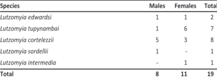
CONFLICT OF INTEREST ACKNOWLEDGMENTS TABLE 1 -Phlebotomine sand lies separated by gender and species, collected in
André Carlone and Carapina Grande neighborhoods, Serra, Espírito Santo, Brazil.
Species Males Females Total
Lutzomyia edwardsi 1 1 2
Lutzomyia tupynambai 1 6 7
Lutzomyia cortelezzii 5 3 8
Lutzomyia sordellii 1 - 1
Lutzomyia intermedia - 1 1
Total 8 11 19
polymorphism (RFLP) test was performed on the 120-bp fragments ampliied from DNA for Leishmania spp. ideniicaion8.
An entomological invesigaion using Centers for Disease Control (CDC) light traps was conducted in the André Carlone and Carapina neighborhoods, where human and canine cases of VL were recorded. The selecion of residences in which CDC traps were installed was based on the occurrence of CVL and the human VL cases as well as the presence of ecological and environmental condiions favoring the reproducion of phlebotomine sand lies, including the presence of organic material, plants, domesic animals, or poultry. The traps were installed between 5:00-6:00pm and removed at 8:00am the following day. Captures were performed at least once a week from September 2009 to February 2010. The insects that were collected were preserved in 70% ethanol and sent to the Leishmaniasis Laboratory at Rene Rachou Research Center for ideniicaion according to the protocol of Young and Duncan9.
The Animal Ethics Commitee of the Federal University of Espírito Santo approved the present study.
From November 2008 to May 2009, 201 dogs from the urban area of Serra municipality were screened for L. chagasi infecion. Out these, 38 were ideniied as posiive by at least 1 test: 13% (26/201) by in-house ELISA, 6% (12/201) by rK39 dipsick, and 3% (6/201) by PCR. Among these 38 dogs, 2 (1%) had a posiive culture, 6 (3%) were posiive by both PCR and rK39, and 3 (1.5%) were seroposiive by both in-house ELISA and rK39. Since the ELISA and IIF tests performed in the state lab (LACEN) provided negaive results in all animals, sera from all 38 posiive dogs were also retested at the Naional VL Reference Laboratory, Fundação Ezequiel Dias (FUNED), in Belo Horizonte. Only 6 animals were found to be posiive: 5 by IIF and 1 by ELISA.
The parasites isolated from bone marrow of the 2 dogs menioned above were ideniied as L. chagasi. Posiive PCR results in 4 dogs were ideniied as belonging to the Leishmania genus. Among the infected dogs, only 1 (rK39 and PCR posiive) showed clinical signs suggesive of CVL.
Although Lutzomyia longipalpis was not detected during the study, we were able to capture another 19 phlebotomine specimens (Table 1). All phlebotomines were found within a 700-m radius of the house of the VL paient.
The geographic spread and urbanization of VL in Brazil has been a challenge for the Brazilian VL Control Program. Due to this and in an atempt to slow down the spread of disease, expanded surveillance and control measures have also been implemented in areas without human or canine cases10. Our study was able to
idenify a new VL-endemic area, which should now be included in
the VL naional surveillance program. Numerous other silent areas may exist in Brazil, paricularly if we consider the low detecion rate of the tests (ELISA and IIF) used by the VL Naional program as a screening tool to idenify these areas. This new ideniied area is of great epidemiological importance, as it is part of the MRV that has 1.7 million inhabitants. As reported by others11, the urbanizaion of VL in
areas where human immunodeiciency virus (HIV) is also prevalent poses an addiional risk of disease reacivaion in paients co-infected with HIV. It has been shown that the rate of VL infecion/disease is about 20:112,13. In Brazil, approximately 3,500 VL cases have been
reported each year for the past 10 years10. Therefore, we can assume
that during this period, roughly 665,000 asymptomaic infecions occurred. This number needs to be considered in the epidemiologic equaion in areas in which HIV coexists in a reasonable prevalence. This scenario can be found in urban areas or when immigraion occurs in both direcions. The VL paient from our study had a posiive HIV test detected retrospecively during the invesigaion and could it well into any of these hypotheses. Thus, we cannot conirm autochthonous transmission since she was born in an endemic VL area (Barra de São Francisco) located in the northwest region of Espírito Santo where she oten used to visit. However, in favor of local transmission, we can argue that there was no reported VL cases in her hometown over the past 10 years14, and that both of her dogs, 1 with a posiive culture
and the other with a posiive rK39 test, were born and bred in Andre Carlone, Serra, and never let her house. The capture of a stray dog with a posiive culture for L. chagasi only 550m away from the VL paient residence and the inding of 4 PCR-posiive dogs in the surroundings of her dwelling reinforce the hypothesis of autochthonous transmission.
The absence of Lutzomyia longipalpis in our captures does not exclude its existence in the studied area. It is possible that this species could not be found due to the small number of capture atempts and the short capture ime (69 captures in 16 days during a 6-month period). Although Lutzomyia longipalpis was not encountered, others phlebotomine species were captured nearby the residence of the human VL case. Among the species captured, only Lutzomyia intermedia is of epidemiological importance, as it is considered a vector of American tegumentary leishmaniasis (ATL). Although there are no records of ATL in André Carlone, the presence of this species indicates a possibility of ATL transmission in this neighborhood.
In conclusion, despite the fact that the vector could not be found, the results of our study provide strong evidence of autochthonous transmission of CVL in Serra City. Due to the proximity of the municipalities of the MRV, there is an urgent need for a large-scale canine seroepidemiological investigation and a systematic phlebotomine capture study in addiional localiies in order to assess the extension of the infecion.
756
Rev Soc Bras Med Trop 45(6):754-756, Nov-Dec, 2012
REFERENCES
ABSTRACT IN pORTuGuESE
Primeira descrição de leishmaniose visceral canina
autóctone na Região Metropolitana de Vitória,
Estado do Espírito Santo, Brasil
Introdução: Descrevemos um foco de leishmaniose visceral canina (LVC) autóctone na Região Metropolitana de Vitória (RMV) onde um caso humano foi registrado anteriormente. Métodos: Testes sorológicos, parasitológicos e moleculares foram realizados em 201 cães. Resultados: Vinte e seis (13%) e 12 (6%) foram posiivos para um teste ELISA in house e rK39, respecivamente. Dois cães apresentaram cultura posiiva para Leishmania (Leishmania) chagasi e quatro PCR posiivo para Leishmania spp. Um dos cães posiivo pertencia ao paciente supracitado. Conclusões: Embora o vetor não tenha sido encontrado, nossos resultados fornecem evidências da LVC autóctone na RMV, área não-endêmica para leishmaniose visceral.
Palavras-chaves: Leishmaniose visceral canina autóctone. Região Metropolitana de Vitória. Área não-endêmica
para leishmaniose visceral.
1. Deane LM. Leishmaniose visceral no Brasil: Estudos sobre reservatórios e transmissores realizados no estado do Ceará. Rio de Janeiro: Serviço Nacional de Educação Sanitária; 1956.
2. Werneck GL. Geographic spread and urbanizaion of visceral leishmaniasis in Brazil. Introducion. Cad Saude Publica 2008; 24:2937-2940.
3. Maia-Elkyhoury ANS, Alves WA, Sousa-Gomes ML, Sena JM, Luna EA. Visceral leishmaniasis in Brazil: trends and challenges. Cad Saude Publica 2008; 24:2941-2947. 4. Galimberi MZ, Katz G, Camargo-Neves VL, Rodas LAC, Casanova C, Costa IP. Leishmaniose visceral americana no Estado de São Paulo. Rev Soc Bras Med Trop 1999; 32:217-218.
5. Secretaria de Saúde do Estado do Espírito Santo. Série histórica dos casos noiicados do período 1986-2004. Vitória: Programa de Controle das Leishmanioses. Secretaria do Estado do Espírito Santo; 2004.
6. Rosário EY, Genaro O, França-Silva JC, Costa RT, Mayrink W, Reis AB, et al. Evaluaion of enzyme-linked immunosorbent assay using crude Leishmania and recombinant anigens as a diagnosic marker for canine visceral leishmaniasis. Mem Inst Oswaldo Cruz 2005; 100:197-203.
7. Degrave W, Fernandes O, Campbell D, Bozza M, Loppes U. Use of molecular probes and PCR for detecion and typing of Leishmania – a mini review. Mem Inst Oswaldo Cruz 1994; 89:463-469.
8. Volpini AC, Passos VM, Oliveira GC, Romanha AJ. PCR-RFLP to idenify L. (V.) braziliensis and L. (L.) amazonensis causing American cutaneous Leishmaniasis. Act Tropica 2004; 90:31-37.
9. Young DG, Duncan MA. Guide to the ideniicaion and geographic distribuion of Lutzomyia sand lies in Mexico, the West Indies, Central and South America (Diptera, Psychodidae). Mem Am Entomol Inst 1994; 54:1-881.
10. Ministério da Saúde. Manual de Vigilância e Controle da Leishmaniose Visceral. Brasília: Secretária de Vigilância em Saúde; 2006.
11. Desjeux P, Alvar J. Leishmania/HIV co-infecions: epidemiology in Europe. Ann Trop Med Parasitol 2003; 97:3-15.
12. Badaro R, Jones TC, Carvalho EM, Sampaio D, Reed SG, Barral A, et al. New perspecives on a subclinical form of visceral leishmaniasis. J Infect Dis 1986; 154: 1003-1011.
13. Dietze R, Barros GB, Teixeira L, Harris J, Michelson K, Falqueto A, et al. Efect of Eliminaing Seroposiive Canines on the Transmission of Visceral Leishmaniasis in Brazil. Clin Infect Dis 1997; 25:1240-1242.