REVIEW ARTICLE
Non-colorectal intestinal tract carcinomas in inflammatory
bowel disease: Results of the 3rd ECCO Pathogenesis
Scientific Workshop (II)
Laurence Egan
a,⁎
, Renata D'Inca
b, Tine Jess
c, Gianluca Pellino
d,
Franck Carbonnel
e, Bernd Bokemeyer
f, Marcus Harbord
g, Paula Nunes
h,i,
Janneke Van der Woude
j, Francesco Selvaggi
k, John Triantafillidis
laDiscipline of Pharmacology & Therapeutics, NUI, Galway, Ireland
bDepartment of Surgical, Oncological and Gastroenterological Sciences, University of Padua, Italy
cDepartment of Epidemiology Research, Statens Serum Institut, National Health Surveillance & Research, Denmark
dGeneral Surgery Unit, Second University of Naples, Italy
eService de Gastroentérologie, CHU de Bicêtre, Assistance Publique-Hôpitaux de Paris, Université Paris Sud, France
fDepartment of General Internal Medicine I, Christian-Albrechts-University,
University Hospital Schleswig-Holstein, Kiel, Germany
gChelsea & Westminster Hospital, 369 Fulham Road, London, UK
hLisbon University & Higher School of Health Technology of Lisbon, Lisbon, Portugal iHospital Cuf Descobertas, Lisbon, Portugal
jErasmus Medical Centre, Rotterdam, the Netherlands kGeneral Surgery Unit, Second University of Naples, Italy
lDepartment of Gastroenterology and Center for IBD
“Saint Panteleimon”Hospital, Nicea, Greece
Received 2 April 2013; accepted 5 April 2013
Abstract
Patients with inflammatory bowel diseases (IBD) have an excess risk of certain gastrointestinal cancers. Much work has focused on colon cancer in IBD patients, but comparatively less is known about other more rare cancers. The European Crohn's and Colitis Organization established a pathogenesis workshop to review what is known about these cancers and formulate proposals for future studies to address the most important knowledge gaps. This article reviews the current state of knowledge about small bowel adenocarcinoma, ileo-anal pouch and rectal cuff cancer, and anal/perianal fistula cancers in IBD patients.
© 2013 European Crohn's and Colitis Organisation. Published by Elsevier B.V. All rights reserved.
Contents
1. Introduction. . . 20 2. Small bowel adenocarcinoma in Crohn's disease . . . 20 2.1. Epidemiology . . . 20
⁎ Corresponding author.
E-mail address:laurence.egan@nuigalway.ie(L. Egan).
1873-9946/$ - see front matter © 2013 European Crohn's and Colitis Organisation. Published by Elsevier B.V. All rights reserved. http://dx.doi.org/10.1016/j.crohns.2013.04.009
A v a i l a b l e o n l i n e a t w w w . s c i e n c e d i r e c t . c o m
2.2. Pathogenesis . . . 20
2.2.1. The implication of histological patterns . . . 21
2.2.2. Microsatellite instability . . . 21
2.2.3. c-K-ras mutations . . . 21
2.2.4. APC gene . . . 21
2.3. Risk factors . . . 21
2.4. Diagnosis. . . 21
2.4.1. Symptoms . . . 21
2.4.2. Imaging and endoscopy . . . 21
2.5. Treatment . . . 22
2.6. Prognosis . . . 22
3. Ileo-anal pouch and rectal cuff carcinoma . . . 23
3.1. Epidemiology . . . 23
3.1.1. Carcinoma . . . 23
3.1.2. Dysplasia . . . 23
3.2. Pathogenesis . . . 24
3.3. Risk factors . . . 24
3.3.1. Dysplasia or cancer in the resected colon . . . 24
3.3.2. Interval from the diagnosis of UC . . . 24
3.3.3. Pouchitis and mucosal changes . . . 24
3.3.4. Biomarkers and immunohistological markers . . . 25
3.3.5. Hand-sewn plus mucosectomy versus stapled anastomosis . . . 25
3.3.6. Extraintestinal manifestations . . . 25
3.4. Surveillance and diagnosis . . . 25
3.5. Survival . . . 25
4. Anal and fistula cancers . . . 26
4.1. Epidemiology . . . 26
4.2. Pathogenesis . . . 26
4.3. Risk factors . . . 26
4.4. Diagnosis. . . 26
4.5. Treatment . . . 26
Conflict of interest . . . 27
References. . . 27
1. Introduction
Inflammatory bowel diseases are strongly associated with carcinomas of the colorectum and, in patients with co-existing primary sclerosing cholangitis, with cholangiocarcinoma. The knowledge of these associations has provided impetus for researchers and clinicians to study the pathogenesis of those cancers, and to develop strategies for managing the elevated risk of those cancers in patients with IBD. In contrast, much less is known about associations between IBD and other more rare cancers of the lower intestinal tract.
We held two workshops to plan and develop a work program to review the current state of knowledge on small bowel adenocarcinoma, ileo-anal pouch and rectal cuff cancer, and anal/perianal fistula cancers in IBD patients. Authors worked together to review and summarize the literature and to identify the key knowledge gaps that could be filled through collaborative research through ECCO.
2. Small bowel adenocarcinoma in
Crohn's disease
2.1. Epidemiology
Small bowel malignancies represent 2% of all cancers.1,2 Adenocarcinoma accounts for 50% of small bowel malignancies,
followed by neuroendocrine tumors, lymphoma and sarcoma.3 Since Ginzburg et al.4first described the association of Crohn's disease (CD) and small bowel adenocarcinoma (SBA) in 1956, several case reports, case series and some case–control studies have been reported. Patients affected with CD are at higher risk of developing SBA.5–20Three meta-analyses have assessed the standardized incidence ratio (SIR) of SBA in patients with CD from population-based or referral center case series. They found that pooled SIRs were 27.1 (95% CI 14.9–49.2), 28.4 (95% confidence interval, 14.46–55.66), and 33.2 (15.9–60.9) respectively.20–22 One study, based upon a tertiary referral center, has found that the cumulative risk is 0.2% and 2.2% at 10 and 25 years since diagnosis, respectively.13,16
International case–control studies are advisable and the development of specific National and International Cancer Registries is to be encouraged.
2.2. Pathogenesis
2.2.1. The implication of histological patterns
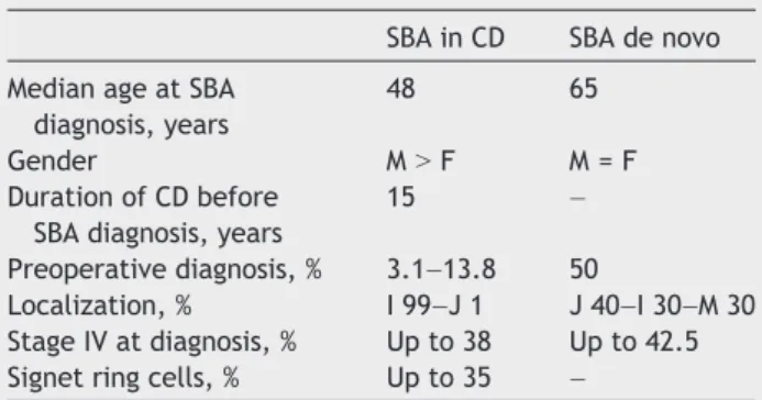
In the majority of the reported cases of SBA in CD, there is no description of histological subtype or molecular alter-ations, rending an insight on carcinogenic pathways difficult. SBA in CD shares some histological and molecular character-istics with colorectal carcinomas in UC. In a series of SBA published by Palascak-Juif et al., the rate of mucinous adenocarcinoma in CD was higher (30% vs 10%) and signet cell variety unique, when compared to small bowel adeno-carcinoma de novo (Table 1).16
2.2.2. Microsatellite instability
The data on microsatellite instability (MSI) in small bowel carcinomas are fewer than for colorectal cancer, with conflicting data published. One study reports the incidence of MSI as 20%.23 However, another study concludes that MSI is less common in CD than UC and this may reflect differences in cancer risk between these two forms of IBD.24 Further studies examining the expression and the presence or absence of mutations in these genes may provide insight into the mechanisms underlying the development of micro-satellite alterations in the setting of IBD.
2.2.3. c-K-ras mutations
Mutations of K-ras are accepted as common in colitis-associated colorectal neoplasia and can occur early in neoplastic progression. Rashid et al. found mutations in c-K-ras codons 12 and 13 in 43% of Crohn's associated with SBA. They also found that carcinomas with contiguous adenomas have the same c-K-ras mutation. Finally, they showed that small intestinal neoplasia is characterized by accumulation of multiple genetic alterations during progression through an adenoma–carcinoma sequence and an dysplasia– carcinoma sequence.25
2.2.4. APC gene
In contrast to sporadic colorectal carcinoma, the allelic losses of 5q (APC gene) and DCC regions (deleted in colon cancer gene region on q18) are uncommon events in CD and sporadic SBA, suggesting that genetic pathways involved in carcinomas of the small intestine are not identical to the colorectum.26,27
In conclusion, some of the available data indicate that the sequence inflammation–dysplasia–cancer might be
involved in the pathogenesis of SBA. The rarity of this neoplasm renders relevant study of pathogenesis difficult.
2.3. Risk factors
Risk factors associated with SBA include CD,2,5–7,9,12–14,16,18,20,28–31 celiac disease, hereditary gas-trointestinal cancer syndromes, immunodeficiency states and autoimmune disorders2,32; alcohol, cigarette smoking, and dietary habits (red meat, smoked, and salt-cured foods).29,33 The consumption of bread, pasta, rice, and sugar33,34have been also reported as correlated with SBA.
Risk factors for developing SBA in CD are less well defined, although several have been proposed:
- disease-related: distal jejunal/ileal localization, strictures and chronic penetrating disease, duration of disease
- patient-related: young age at diagnosis, male sex
- drug-related: corticosteroids, azathioprine, 6-mercaptopurine, anti-TNF-alpha agents
- surgery-related: small bowel bypass loops, strictureplasties, absence of resection
- environmental: occupational hazards/exposure to halogenated aromatic compounds with aliphatic amines, asbestos, and solvents.
All these factors have been variously proposed to have a carcinogenic potential.2,5–7,9,12–14,16,18–20,28,30,31,35Most are debated and results from single series are contrasting.
Concerning surgery, the role of strictureplasty in promot-ing SBA is unclear and not widely accepted.36The reason for SBA in strictureplasties could be due either to the develop-ment of carcinoma in an area of bowel previously treated by strictureplasty or to the potential for missing a malignancy at the time of strictureplasty.36–38
2.4. Diagnosis
Early diagnosis is the key to achieve successful treatment of SBA in CD. Dosset et al.39 were able to diagnose preopera-tively only 3.1% of SBA. A more recent study was more optimistic, but preoperative diagnosis did not even reach 14% in CD patients.40 Furthermore, nearly 30–35% of patients had metastatic disease (Stage IV) and 55% were found with positive nodes at diagnosis.32,33,40
2.4.1. Symptoms
Symptoms of SBA are nonspecific32 and are hard to distinguish from relapse of CD.16,19,33,39,40 Authors agree that two indicators may guide physicians and raise suspicion of SBA: intestinal obstruction not relieved by medical management and abrupt onset with severe symptoms after a period of quiescent disease.16,19,39–41
2.4.2. Imaging and endoscopy
Investigations also rarely succeed in differentiating active CD from SBA because strictures and masses are frequently seen in CD, due to its common complications.19,32 Radiological investigations such as CT, MR and barium radiography can detect strictures and masses but do not allow sampling of suspect areas.31 Similarly, small bowel capsule endoscopy does not allow tissue sampling and does not accurately localize and grade lesions.42
Table 1 Comparison between SBA in CD and SBA de novo.9,13,43,51,56
SBA in CD SBA de novo
Median age at SBA diagnosis, years
48 65
Gender MNF M = F
Duration of CD before SBA diagnosis, years
15 –
Preoperative diagnosis, % 3.1–13.8 50
Localization, % I 99–J 1 J 40–I 30–M 30 Stage IV at diagnosis, % Up to 38 Up to 42.5 Signet ring cells, % Up to 35 –
Most SBAs are located in the terminal or distal ileum and may be reached by a colonoscope, allowing mucosal biopsy for accurate diagnosis. For more proximal lesions, double balloon enteroscopy represents a promising tool for small bowel examination in patients with CD suspected of SBA. It is particularly useful in established CD as adhesions may limit other examinations (i.e. single balloon enteroscopy).42 How-ever, drawbacks must be taken into account: double balloon enteroscopy does not allow extra-intestinal assessment of the disease and requires extensive expertise and anesthesia42; also“in a patient with stricturing active CD, there appears to be a higher risk of complications”(EL4, RG C, Statement 3F, ECCO–OMED consensus on small bowel endoscopy in IBD).42
If the surgeon suspects that a patient is at higher risk of SBA (i.e. for presenting symptoms and/or duration of CD), it is useful to perform an intraoperative histologic examina-tion. Intraoperative enteroscopy may also be useful because it detects lesions inaccessible to conventional endoscopy, undetected in more than half of the patients prior to surgery.43–45 In the future, the usefulness of positron emission tomography CT in identifying, localizing and differentiating between CD and SBA should be tested.
In conclusion, as early diagnosis is of critical significance for the survival of the patient, clinicians should consider exploratory laparotomy as a possible diagnostic and thera-peutic approach in cases of CD patients with suspected SBA.
2.5. Treatment
Although there are very few data on the management of dysplasia, we encourage surgical resection whether low grade or high grade, when detected by endoscopic small bowel biopsies in areas of inflammation. Indeed, an association of dysplasia with synchronous SBA has been reported in surgical specimens46compatible with a dysplasia–carcinoma sequence. Similar to SBA de novo, surgery is the mainstay of treatment for SBA in CD, and is best performed as a radical resection. However, most SBAs in CD are diagnosed intraop-eratively39,40 and half of the carcinomas are not even suspected at operation and are diagnosed only upon microscopic examination in some series.47,48Early detection of SBA is therefore crucial to achieve radical treatment, and CD patients developing SBA commonly have a long history of repeated hospitalizations during which diagnosis of SBA was often overlooked and surgery was delayed.40
Both Solem et al.19 and the GETAID35 suggested that prolonged salicylate administration can decrease the risk of SBA in patients with longstanding ileal CD. The latter study also reported that surgery has a very important role in the potential prevention of SBA, thus advocating a role for prophylactic surgery.35In agreement with this observation, other authors reported that surgical resection can remove chronic inflamed segments which are highly associated with SBA development in these patients, and should be advocated especially in patients followed up for more than 10 years.16 SBA is rare and the number of patients to resect to prevent SBA is likely to be high, even in patients with longstanding CD lesions. By analogy with UC, it can be suggested to restrict prophylactic small bowel resection to patients with dysplasia on endoscopic biopsies. This suggested strategy still has to be validated.16,35,40
Operation for SBA in CD is the same as SBA de novo. Surgery should aim to obtain free resection margins, implying wide resection, and resection of a wedge of mesentery is necessary to capture the draining lymph node basin. Tumors with extensive local disease, metastases to multiple regional/ distant lymph nodes and/or the liver/peritoneal surface may be unresectable. For tumors of the jejunum and proximal ileum, wide local excision with lymphadenectomy is the procedure of choice.49–51 The number of lymph nodes retrieved has been used as a measure of quality in surgical care of bowel malignancies,52 but the optimal harvest of nodes for SBA has yet to be defined. Nicholl et al.53, by applying lymph node recovery as a surrogate for adequate surgical resection, suggested that SBA appears to be surgically undertreated. They proposed a threshold of 10 nodes to define an adequate resection, and found that majority of patients have few nodes resected, even in early-stage disease.
Adequate mesenteric resection is limited by the proxim-ity of the nodes or tumor to the superior mesenteric artery.31 Pancreaticoduodenectomy may be required for tumors in the second or third portion of the duodenum.1,31 Segmental resection of duodenal tumors generally corre-lates with a better overall survival compared with a Whipple procedure54although this observation is not confirmed in a series of 37 patients operated on for SBA at the Cleveland Clinic, where increased survival was found in patients who underwent Whipple resection.1
Distal ileal lesions may require ileocolectomy/right hemicolectomy31 to remove all nodes draining the tumor site. Patients who are considered incurable may undergo palliative resections, bypass procedures or double-balloon enteroscopy can be advocated to place self-extendable stents in stenosed bowel lumen with palliative intent.55
For metastatic disease and incomplete resection some authors advocated the use of radiotherapy or chemotherapy for SBA,31,50 but very few patients receive radiotherapy as part of their primary treatment.50,56,57Japanese investigators also explored the use of intraoperative radiotherapy, where a single dose of radiation can be given to a tumor bed with residual microscopic or macroscopic disease while shielding nearby organs.58 5-Fluorouracil is the most common chemo-therapy agent used, either as a single agent or in combination with other agents, including doxorubicin, cisplatin, levami-sole, and mitomycin.49,50,56,57,59,60 Average overall survival with metastatic disease is reported to increase from 9– 11 months to 17–20 months12 with less than 5% of patients surviving longer than 2 years in available series.12,49,50,56,59,60 There are several phase I and II trials on-going evaluating various radiotherapy and chemotherapy regimens for SBA. Patients affected with SBA in CD should be encouraged to enter future or on-going clinical trials.
2.6. Prognosis
SBA has an aggressive nature, and survival is poor.61–63The reported 5-year survival rates range between 20% and 30%.2,61 Lymph node involvement is reported to reduce 5-year survival from 60–70%51to 12–14%.51,64
as 9%.16,18,19,65,66 Nevertheless, Agrawal et al.67 reported survival up to 10 years for multiple patients initially diagnosed with stage II and III SBAs. The possibility of aggressive surgical care for early-stage SBA should not be dismissed.
3. Ileo-anal pouch and rectal cuff carcinoma
Restorative proctocolectomy with ileal pouch-anal anasto-mosis is the preferred surgical management of patients with UC who do not respond to intense medical treatment or who develop cancer or dysplasia. Because of the removal of the whole large bowel, this operation substantially reduces the risk of UC-associated dysplasia/neoplasia, although it does not completely abolish the risk of neoplasia of the pouch or rectal cuff.
In recent years, it has become apparent that the development of cancer in the pouch, although quite rare, is a reality. Various aspects concerning epidemiology, patho-genesis, diagnosis, and prevention strategies have been investigated. However, the available data are not enough to allow drawing firm conclusions and more work is necessary in order to accomplish this task.
3.1. Epidemiology
3.1.1. Carcinoma
Pouch-related carcinomas have been described with increasing frequency since the first report in 1984 in a number of case-reports and small case-series. During the last two years, three reviews have collected this literature aiming to analyze the clinicoepidemiological features, risk factors and prevention strategies for pouch carcinoma.68–70 In a recently published review including 12 retrospective series and 15 case-reports of IBD-associated colorectal cancer patients and those undergoing restorative proctocolectomy surgeries from 1984 to 2010, the authors found 43 cases of pouch-related cancers, of which 32 were located in the anal transitional zone (ATZ) and 11 in the pouch body. Most of the cases were adenocarcinomas with some of them being squamous cell carcinomas. Sixteen cases were described in retrospective series, 1 case was from a prospective study, and 26 from case-reports. Thirty patients underwent mucosectomy and 13 had stapled anastomoses.70
Alessandroni et al. described 50 reported cases in the international literature. Twenty-five of these cases appeared after mucosectomy and hand-sewn anastomosis and 25 after stapling technique. In 48% of them, dysplasia or cancer was already present at the time of the colectomy.68
Ault et al. described a series of five patients who developed adenocarcinoma in the middle of the pouch.69 Al Sukhni et al.71 estimated the risk of new cancer or recurrence in the pouch or rectal cuff in patients with UC who had undergone stapled ilea pouch-anal anastomosis versus mucosectomy with hand-sewn anastomosis. They noticed that performing a stapled ileal pelvic anal anastomosis does not appear to be inferior to mucosectomy and hand-sewn anastomosis in preventing cancer.
3.1.2. Dysplasia
Development of dysplasia could precede or appear con-currently with carcinoma. A number of descriptions have
dealt with the development of dysplasia in the pouch and cuff.
Das et al. noticed that dysplasia in the ileal reservoir is rare and was associated with histological type C changes with an atrophic mucosa, unremitting pouchitis, and the presence of sclerosing cholangitis. They reported 17 adeno-carcinomas: 9 in the residual anorectal mucosa, 7 in the reservoir, and 1 of their own. Since the time intervals from the onset of UC and from the restorative proctocolectomy to the development of cancer were 120 to 528 and 16 to 216 months respectively, they proposed that cancer appeared to be related to the duration of UC itself and not to the interval from operation.72
It has been suggested that patients with UC and a long-standing ileoanal pouch anastomosis could develop persistent severe mucosal atrophy which is a risk factor for neoplastic transformation of the pouch mucosa. Gullberg et al.73studied 7 patients with ileoanal pouch anastomosis and persistent mucosal atrophy (type C) and 14 controls with slight atrophy (type A). Dysplasia was found in 71% (5 out of 7 patients) in the type C group compared to none in the type A group (Pb0.001).
In another study referring to 138 patients who underwent restorative proctocolectomy for UC (n = 118), familial adenomatous polyposis (n = 10), Crohn's colitis (n = 2), and indeterminate colitis (n = 8), dysplasia was found in the colectomy specimens in 22 (18.3%) and invasive cancer in 8 (6.7%) of them. Median interval between proctocolectomy and pouch biopsy was 5.4 years. Indefinite dysplasia was found in only one out of 138 patients.74 Another study suggested that the development of dysplasia in ileal pouches is probably a rare event within 15–20 years of pouch surgery. In this 10-year study, the authors found only one case of multifocal, low-grade dysplasia (0.9%). Aneuploidy was found in this one and two more patients.75
In a group of 276 patients who had undergone restorative proctocolectomy between 1984 and 2009 Banasiewicz et al.76 found that pouchitis significantly increased the risk of low grade dysplasia (OR 13.48, 1.48–122.86 95% CI, P = 0.021); the time period preceding dysplasia was commonly shorter in patients with pouchitis, but three patients developed low grade dysplasia after 7, 15 and 16 years progressing to high grade dysplasia in the following 6–8 years. This observation points out the importance of long term follow-up, as dysplasia may occur many years after IPAA.
Three more studies also suggested that the risk of the development of dysplasia in the pouch is rather low. Börjesson et al. suggested that the risk of developing either dysplasia or cancer in the ileal pouch mucosa after restorative proctocolectomy for UC is low even after a long follow-up.77 In their study of 45 patients, who had been operated on with an ileo-anal pouch anastomosis with a time interval from the start of the disease until the end of follow-up of 24.8 years, no high-grade dysplasia or invasive carcinoma was found.
agreement between the two groups of pathologists regard-ing the absence of high-grade dysplasia and invasive carcinoma in the examined biopsies, although a significant disagreement in the frequency of low-grade and indefinite categories of dysplasia was seen.
ATZ dysplasia after ileal pouch-anal anastomosis is infrequent, most common in the first 2–3 years postopera-tively, and may apparently disappear on repeated biopsy. O'Riordain et al. followed 210 patients for a median time of 6.4 years and found 7 patients (3%) who developed dysplasia in the ATZ.80It is of interest that in 5 out of these 7 patients, dysplasia“disappeared”during further follow-up.
Patients with UC and primary sclerosing cholangitis with an ileal pouch are more prone to develop mucosal atrophy in the pouch (type C changes) and have a higher risk of neoplastic transformation in the pouch mucosa than patients with UC without sclerosing cholangitis. Stahlberg et al. studied 16 patients with UC and primary sclerosing cholangitis and 16 matched patients with UC without sclerosing cholangitis. Moderate or severe atrophy was more common in UC and primary sclerosing cholangitis compared to the control group (8 vs 2 pts). Low-grade dysplasia was found in 3 patients with sclerosing cholangitis and in 2 of the controls.81
A large study estimated the risk of the development of dysplasia in the ATZ and the outcome of a conservative management policy for ATZ dysplasia, with at least a 10-year follow-up after ileal pouch-anal anastomosis. One hundred and eight patients undergoing ATZ-sparing ileal pouch-anal anastomosis were submitted to anal transitional zone biopsies for at least ten years postoperatively. In 8 patients ATZ dysplasia posed the risk of dysplasia being significantly associated with preoperative diagnosis of cancer or dyspla-sia. Dysplasia was of high-grade in two patients, and low-grade in six patients. No association with age, gender, IBD duration and extent of IBD was found.82
In conclusion, the available data support the assumption that the development of carcinoma and dysplasia in the pouch and rectal cuff or ATZ of patients with UC undergoing ileo-anal pouch anastomosis is a reality. However, the risk of the appearance of dysplasia seems to be low even many years after the creation of the pouch.
3.2. Pathogenesis
The pathogenesis of pouch carcinoma is largely unknown. The most relevant proposed pathway is for the ileal pouch mucosa to go through the adaptational changes, then to proceed through the stages of inflammation (chronic pouchitis), dysplasia (of low and high degree), and subse-quently to adenocarcinoma. In many of the available studies, however, the dysplasia–cancer sequence is incon-clusive since the carcinoma might have developed from the ileal mucosa itself or from residual rectal mucosa.
In a study aiming to evaluate the prevalence and spectrum of inflammatory changes in the ileum in patients with UC, 200 consecutive patients were evaluated for a variety of pathologic features in the ileum and colon. Overall, 34 out of 200 (17%) UC patients had inflammatory changes in the ileum; 32 of these 34 patients (94%) had pancolitis, which was significantly higher than the rate of
pancolitis (39%) in patients without ileal disease (N = 166) (Pb0.001). The presence of inflammatory changes in the ileum had no effect on the prevalence of pouch dysplasia or cancer.83
Other pathogenetic mechanisms could be related to the residual diseased mucosa left behind. However, Kariv et al., in a study of 3203 patients who underwent restorative proctocolectomy with ileo-anal pouch anastomosis, found that mucosectomy did not protect against pouch neoplasia. In fact, 38 patients (1.2%) developed pouch neoplasia (11 adenocarcinoma, 1 lymphoma, and 2 squamous cell carci-noma) with cumulative incidence of 1.3% at 10, and 5.1% at 25 years respectively. Only the presence of preoperative neoplasia was associated with an increased risk of pouch neoplasia and thus, the performance of mucosectomy was not protective against the development of carcinoma.84
In conclusion, the available data are in favor of the assumption that although most of pouch neoplasia follows the chronic inflammation–dysplasia–cancer sequence, this is not the rule in all cases. Chronic inflammation, severe atrophy and previous diagnosis of cancer or dysplasia in the large bowel are probably involved in the pathogenesis of cancer appearing in the pouch.
3.3. Risk factors
Several risk factors for the development of pouch-related cancer have been considered.
3.3.1. Dysplasia or cancer in the resected colon
These features increase the likelihood of subsequent dysplasia. Most studies reported this finding.75,82,85Tsunoda et al.85reviewed 118 patients with colitis: 2 out of 3 patients with dysplasia were in a subgroup of 8 patients who had a carcinoma in the large bowel. Also Remzi et al.82found an association between dysplasia in the ATZ and cancer or dysplasia in the postoperative specimen, concluding that the carcinoma poses the anal mucosa at high risk. The presence of pre-existing dysplasia or cancer seems to be the most important predisposing risk factor for the development of neoplasia after ileo-anal pouch anastomosis, as 54–70% of patients developing pouch-related cancer had a dysplasia or cancer which was identified preoperatively or that was found in the operative specimen.72,75
3.3.2. Interval from the diagnosis of UC
The risk of pouch cancer, as well as that of colorectal cancer in UC, increases with time, probably in a similar fashion70,76,86and it is related to the anatomical extent of the disease in preoperative specimen.85Cancer is reported to occur at least 10 years after UC onset.70,72Disease onset is definitively more important than interval from IPAA and is to be considered when considering surveillance.
3.3.3. Pouchitis and mucosal changes
3.3.4. Biomarkers and immunohistological markers Coull et al.87 followed up for a median of 56 months 110 patients who underwent stapled ileo-anal pouch anastomosis for UC by cuff surveillance biopsies under general anesthesia; p53 was performed for each of most recent cuff biopsy. Neither carcinomas developed nor dysplasia or carcinoma was observed in any cuff biopsy during the follow-up period. The authors concluded that p53 is not useful in surveillance of cuff biopsies from patients who have undergone ileo-anal pouch anastomosis for UC. Several markers of inflammation are over expressed in ileal pouch, even without the endoscopic evidence of pouchitis.88 Vento et al.89 studied 42 patients with chronic pouchitis by endoscopy with biopsies performed after a median of 8.3 years from ileo-anal pouch anastomosis. No p53, abnormal DNA flow cytometry and dysplasia were observed. The authors concluded that patients undergoing ileo-anal pouch anastomosis for UC should not be intensively followed up with endoscopy during the first 10 years after surgery, on condition that hand-sewn anastomosis with mucosectomy was accurately performed.
3.3.5. Hand-sewn plus mucosectomy versus stapled anastomosis
Mucosectomy does not eliminate the potential for neoplastic transformation. In fact, studies reported cases of adenocarcinoma after ileo-anal pouch anastomosis occur-ring in patients who underwent mucosectomy.70,90Residual microscopic mucosal rectal islands have been reported in 20% of patients, so that a cancer can develop between the pouch and the muscle layers.
Debate exists as to the benefits of performing mucosectomy in pouch surgery. Tsunoda et al.85found dysplasia in 3 patients within the ATZ, but 2 out of 3 were in a subgroup of 8 patients who had a carcinoma in the large bowel. Remzi et al.82studied dysplasia of ATZ performing several biopsies in 289 pouch patients for a minimum follow-up of 10 years. ATZ dysplasia developed in 8 patients 4–123 months after surgery (median 9 months). The risk was associated with a cancer or dysplasia in the postoperative specimen. Four patients with low grade dysplasia treated expectantly showed no subsequent dysplasia. Coull et al.91 studied 136 patients for 56 months and showed no evidence of dysplasia in cuff biopsies. In 113 patients at 2.5 years of mean follow-up Thompson-Fawcett et al.92found no evidence of neoplastic change in 457 biopsy specimens. Although stapled IPAA seems to achieve similar oncological outcomes similar to that of mucosectomy,71,93 some authors72,89 recommended mucosectomy as a safer alternative. Functional concerns are to be considered too, but results are similar also in terms of function when the surgeon avoids excessive or prolonged dilation of the sphincters during mucosectomy.
3.3.6. Extraintestinal manifestations
Preoperative extraintestinal manifestations seem to be associated with higher risk of developing pouch-related malignancies. Interestingly, patients with extraintestinal manifestations are more likely to develop pouchitis and type C changes of the pouch.94–96 The likelihood of developing pouchitis or pouch dysplasia is higher in UC patients with primary sclerosing cholangitis regardless of the severity of liver disease.81,94
3.4. Surveillance and diagnosis
It early became apparent that restorative proctocolectomy does not completely abolish the risk of neoplasia of the pouch. Moreover, since not all pouch carcinomas follow the chronic inflammation–dysplasia–cancer sequence, the regular pouch endoscopy with biopsy, representing the current gold standard for surveillance, does not seem suitable for all cases. Moreover, because pouch endoscopy with biopsies still misses dysplasia, there is a debate on the optimal need and time interval of routine endoscopic surveillance. Subsequently, many authors suggest that surveillance endoscopy could be applied in patients at risk, such as those with a pre-colectomy diagnosis of UC-associated neoplasia, primary sclerosing cholangitis, type C ileal mucosal changes and unremitting pouchitis.70,78,97 If repeated biopsies confirm persistent dysplasia, mucosectomy with perineal pouch advancement and neo-ileal pouch-anal anastomosis are recommended.82
The optimum time interval for routine surveillance is unknown. Ault et al. suggest that patients with long-standing ileal pouches might benefit from routine surveillance of the pouch as often as every six months, which can be quickly and easily performed in the office using flexible endoscopy.69 Das et al. suggest that a surveillance program should begin 10 years from the onset of the disease by taking multiple biopsies of the ileal reservoir and the anorectal mucosa below the ileo-anal anastomosis.72
However, some authors suggest that routine pouch surveillance with biopsies may not be warranted.74 Um et al. proposed that there is little evidence to support routine biopsy of the ileal mucosa or the ATZ except in patients with predisposing factors.90 Little evidence to support routine biopsy of the ileal mucosa in UC patients was also proposed by both Herline et al.98and Borjesson et al.77
When performed, 4 quadrant biopsies should be taken from the upper and lower pouches, and a further 4 biopsies targeted at just below the anastomosis, in the ATZ. Anorectal mucosal biopsies should be taken under anesthe-sia, if necessary, as this area is sensitive. Biopsies should not include staple line, as their features of inflammation could lead to an incorrect diagnosis of pouchitis.
When available, the use of high magnification chromoscopic pouchoscopy is recommended. Hurlstone et al.99reported that it is a useful predictor of ATZ and cuff anatomy, allowing accurate biopsy targeting. This approach is to be encouraged, as the follow-up of patients with regard to the possible development of dysplasia is likely to be improved, as the technique has demonstrated morphological differences be-tween ATZ, anal columnar epithelium and the pouch mucosa.
In conclusion, there is no general agreement concerning the need for regular surveillance in these patients. However, the majority of reports suggest that regular endoscopic follow-up must be applied in patients at risk, such as those with a pre-colectomy diagnosis of UC-associated neoplasia, primary sclerosing cholangitis, patients with type C ileal mucosal changes, and patients with unremitting pouchitis.
3.5. Survival
pouch carcinoma, a fact that could be easily explained since the number of reported cases is quite small for drawing firm conclusions. However, from the case-reports that appeared in the literature it seems that the survival of these patients does not differ from patients with large bowel cancer being developed on the ground of IBD. Nevertheless, although the natural history and prognosis of pouch dysplasia are not clear, the mortality rate associated with pouch cancer, once diagnosed, appears to be high.97
Regarding the influence of the kind of anastomosis on the survival of the operated patients, it seems that stapled ileal-pelvic anal anastomosis is not inferior to mucosectomy and handsewn anastomosis. A retrospective analysis of patients with UC associated with colorectal dysplasia or cancer, who underwent ileal-pouch anal anastomosis either stapled (n = 59) or handsewn (n = 22), revealed that 52 had dysplasia and 29 had colorectal cancer at the time of surgery. During a median follow-up time of 6.3 years, nine patients died (11%). Both colorectal cancer-related deaths were in patients with hand-sewn anastomoses. However, the overall 5-year survival between the two groups did not significantly differ.71If repeated biopsies confirm persistent dysplasia, an ATZ excision with neo-ileal-pouch anal anasto-mosis is recommended.80
4. Anal and fistula cancers
4.1. Epidemiology
Anal cancer is rare, with an annual incidence of 2 cases per 100,000100 There does not appear to be an excess risk in patients with IBD. One of the largest case series examining the incidence in patients with IBD followed 9602 patients for a cumulative 99,229 years; 1.3 cases were expected, whereas two were discovered.101
However, severe complicated perineal CD may increase the risk for anal cancer. Five anal cancers were reported from a cohort of about 1250 patients seen at St Mark's Hospital, London between 1940 and 1992. All had long-standing severe anorectal disease, with strictures, abscesses, fistulae and proctitis.102 They were followed up for a mean duration of 18 years (22,500 patient years). Less than one patient would be expected to have developed anal cancer, therefore five cases suggest that there may be an association between complex perianal CD and anal malignancy, although this conclusion is confounded by referral bias as St Mark's Hospital is a tertiary referral center. Others have reported carcinoma associated with anorectal fistula103–106which is supported by a higher rate of oncogenic HPV serotypes in sporadic anal fistula and fissures. This could facilitate high risk HPV serotypes access to the epithelium.103 A Swedish study suggested anal cancer occurs at an earlier age and presents at a more advanced stage in CD patients.107
4.2. Pathogenesis
Squamous cell cancer accounts for the majority of cases of anal cancer, arising from the transitional or squamous cell mucosa. Adenocarcinoma, melanoma and rarely sarcoma can occur. Anal squamous cell carcinoma is strongly associated with human papilloma virus infection. As with cervical cancer, HPV
16 and HPV 18 are the most frequently associated oncogenic serotypes. Anal HPV infection occurred in 50% of a cohort of sexually active women over one year, with more than half spontaneously clearing the infection within one year.108About 1/3 of infections are with oncogenic HPV strains. The prevalence and clearance of anal HPV infection are similar in heterosexual men. However, men who have sex with men have more persistent infection, and a higher proportion of oncogenic serotypes, particularly with multiple partners and following receptive anal intercourse.109 Consequently the rate of anal cancer is much higher in men who have sex with men, particularly those with HIV infection where a rate of 131/ 100,000 person years has recently been reported.110
In conclusion, the available data indicate that HPV is responsible for the majority of anal cancers in IBD patients, which is a special interest from a preventive point of view.
4.3. Risk factors
The high rate in HIV + men who have sex with men may represent high risk behavior, however it does appear that chronic immune suppression (CD4b500 × 106/L) predisposes to progression to high grade anal-intraepithelial neoplasia (AIN) and anal cancer.111Lymphoma and squamous cell cancer occur more frequently with immune-suppression following solid organ transplantation. The rate of vulvar and anal cancers has been reported to increase 100-fold following renal transplantation.112The effect of immune suppression on anal cancer rates in IBD is largely unknown. The CESAME cohort113 did not demonstrate an association between thiopurine use and anal cancer in IBD patients although the crude risk in the subset of patients with perianal CD exposed to thiopurines was 0.42 per 1000 patient years, which is much higher than that expected. The TREAT and the ENCORE registries have not demonstrated an excess of anal cancers in patients treated with Infliximab.113,114
4.4. Diagnosis
Screening for AIN using high resolution anoscopy has been recommended in high risk individuals, such as HIV + men who have sex with men, women with high grade cervical dysplasia, those known to have AIN or with immune suppression following solid organ transplantation. Treating AIN can prevent its progression. Anal cancer should be suspected in IBD patients with exuberant perianal granulation tissue, persistent ulcer-ation, or anorectal pain or bleeding. The diagnosis is often delayed in IBD, due to similar nonspecific symptoms, with cancer assumed to be a benign stricture.
In conclusion, anoscopy represents the diagnostic tool of choice in daily clinical practice that should be performed if the patient develops suspicious symptoms.
4.5. Treatment
resection may be required due to the additional burden of longstanding complicated perianal CD.116,117
In summary, multimodal treatment of anal cancer in IBD is similar to that in sporadic cancer but at least proctectomy in CD, or total colectomy in UC, may be required depending on disease extent. Survival outcomes seem to be the same as in sporadic non-IBD cancer for the corresponding stage. Overall, the prognosis is often poor due to a delay in diagnosing anal cancer in chronic perianal disease.104
Text Box: Key steps that can be taken to address current knowledge gaps on non-colorectal intestinal tract carci-nomas in inflammatory bowel disease
• Establishment of international case–control
stud-ies and disease registrstud-ies to elucidate risk factors and natural history
• Testing of new diagnostic modalities for small bowel adenocarcinoma in Crohn's disease, such as positron emission tomography and double balloon enteroscopy
• Determination of optimum surveillance method and schedule in patients with ileo-anal pouch anastomosis
Conflict of interest
The authors declare no conflicts of interest.
References
1. Abrahams NA, Halverson A, Fazio VW, Rybicki LA, Goldblum JR. Adenocarcinoma of the small bowel: a study of 37 cases with emphasis on histologic prognostic factors.Dis Colon Rectum 2002;45:1496–502.
2. Delaunoit T, Neczyporenko F, Limburg PJ, Erlichman C. Pathogenesis and risk factors of small bowel adenocarcinoma: a colorectal cancer sibling.Am J Gastroenterol2005;100:703–10. 3. Chow JS, Chen CC, Ahsan H, Neugut AI. A population-based study of the incidence of malignant small bowel tumours: SEER, 1973–1990.Int J Epidemiol1996;25:722–8.
4. Ginzburg L, Schneider KM, Dreizin DH, Levinson C. Carcinoma of the jejunum occurring in a case of regional enteritis. Surgery1956;39:347–51.
5. Bernstein CN, Blanchard JF, Kliewer E, Wajda A. Cancer risk in patients with inflammatory bowel disease: a population-based study.Cancer2001;91:854–62.
6. Bernstein D, Rogers A. Malignancy in Crohn's disease. Am J Gastroenterol1996;91:434–40.
7. Ekbom A, Helmick C, Zack M, Adami HO. Extracolonic malignan-cies in inflammatory bowel disease.Cancer1991;67:2015–9. 8. Fielding JF, Prior P, Waterhouse JA, Cooke WT. Malignancy in
Crohn's disease.Scand J Gastroenterol1972;7:3–7.
9. Greenstein AJ, Sachar D, Pucillo A, Kreel I, Geller S, Janowitz HD, et al. Cancer in Crohn's disease after diversionary surgery. A report of seven carcinomas occurring in excluded bowel.Am J Surg1978;135:86–90.
10. Hawker PC, Gyde SN, Thompson H, Allan RN. Adenocarcinoma of the small intestine complicating Crohn's disease.Gut1982;23: 188–93.
11. Jess T, Winther KV, Munkholm P, Langholz E, Binder V. Intestinal and extra-intestinal cancer in Crohn's disease: follow-up of a population-based cohort in Copenhagen County, Denmark.Aliment Pharmacol Ther2004;19:287–93.
12. Kronberger IE, Graziadei IW, Vogel W. Small bowel adenocar-cinoma in Crohn's disease: a case report and review of literature.World J Gastroenterol2006;12:1317–20.
13. Lashner BA. Risk factors for small bowel cancer in Crohn's disease.Dig Dis Sci1992;37:1179–84.
14. Michelassi F, Testa G, Pomidor WJ, Lashner BA, Block GE. Adenocarcinoma complicating Crohn's disease. Dis Colon Rectum1993;36:654–61.
15. Munkholm P, Langholz E, Davidsen M, Binder V. Intestinal cancer risk and mortality in patients with Crohn's disease. Gastroenterology2005;105:1716–23.
16. Palascak-Juif V, Bouvier AM, Cosnes J, Flourie B, Bouche O, Cadiot G, et al. Small bowel adenocarcinoma in patients with Crohn's disease compared with small bowel adenocarcinoma de novo.Inflamm Bowel Dis2005;11:828–32.
17. Persson PG, Karlen P, Bernell O, Leijonmarck CE, Brostrom O, Ahlbom A, et al. Crohn's disease and cancer: a population-based cohort study.Gastroenterology1994;107:1675–9.
18. Ribeiro MB, Greenstein AJ, Heimann TM, Yamazaki Y, Aufses Jr AH. Adenocarcinoma of the small intestine in Crohn's disease. Surg Gynecol Obstet2004;173:343–9.
19. Solem CA, Harmsen WS, Zinsmeister AR, Loftus Jr EV. Small intestinal adenocarcinoma in Crohn's disease: a case–control study.Inflamm Bowel Dis2004;10:32–5.
20. von Roon AC, Reese G, Teare J, Constantinides V, Darzi AW, Tekkis PP. The risk of cancer in patients with Crohn's disease. Dis Colon Rectum2007;50:839–55.
21. Canavan C, Abrams KR, Mayberry J. Meta-analysis: colorectal and small bowel cancer risk in patients with Crohn's disease. Aliment Pharmacol Ther2006;23:1097–104.
22. Jess T, Gamborg M, Matzen P, Munkholm P, Sorensen TI. Increased risk of intestinal cancer in Crohn's disease: a meta-analysis of population-based cohort studies.Am J Gastroenterol2005;100: 2724–9.
23. Planck M, Ericson K, Piotrowska Z, Halvarsson B, Rambech E, Nilbert M. Microsatellite instability and expression of MLH1 and MSH2 in carcinomas of the small intestine.Cancer 1999;97: 1551–7.
24. Noffsinger A, Kretschmer S, Belli J, Fogt F, Fenoglio-Preiser C. Microsatellite instability is uncommon in intestinal mucosa of patients with Crohn's disease.Dig Dis Sci1992;45:378–84. 25. Rashid A, Hamilton SR. Genetic alterations in sporadic and
Crohn's-associated adenocarcinomas of the small intestine. Gastroenterology1997;113:127–35.
26. Arai M, Shimizu S, Imai Y, Nakatsuru Y, Oda H, Oohara T, et al. Mutations of the Ki-ras, p53 and APC genes in adenocarcinomas of the human small intestine.Int J Cancer1997;70:390–5. 27. Bell SM, Kelly SA, Hoyle JA, Lewis FA, Taylor GR, Thompson H,
et al. c-Ki-ras gene mutations in dysplasia and carcinomas complicating ulcerative colitis.Br J Cancer1991;64:174–8. 28. Valdes-Dapena A, Rudolph I, Hidayat A, Roth JL, Laucks RB.
Adenocarcinoma of the small bowel in association with regional enteritis. Four new cases.Cancer1976;37:2938–47. 29. Chow WH, Linet MS, McLaughlin JK, Hsing AW, Chien HT, Blot
WJ. Risk factors for small intestine cancer. Cancer Causes Control1993;4:163–9.
30. Ekbom A, Helmick C, Zack M, Adami HO. Increased risk of large-bowel cancer in Crohn's disease with colonic involve-ment.Lancet1990;336:357–9.
31. Feldstein RC, Sood S, Katz S. Small bowel adenocarcinoma in Crohn's disease.Inflamm Bowel Dis2008;14:1154–7. 32. Nesbit Jr RR, Elbadawi NA, Morton JH, Cooper Jr RA.
33. Dabaja BS, Suki D, Pro B, Bonnen M, Ajani J. Adenocarcinoma of the small bowel: presentation, prognostic factors, and outcome of 217 patients.Cancer2004;101:518–26.
34. Negri E, Bosetti C, La Vecchia C, Fioretti F, Conti E, Franceschi S. Risk factors for adenocarcinoma of the small intestine.Int J Cancer1999;82:171–4.
35. Piton G, Cosnes J, Monnet E, Beaugerie L, Seksik P, Savoye G, et al. Risk factors associated with small bowel adenocarcinoma in Crohn's disease: a case–control study.Am J Gastroenterol 2008;103:1730–6.
36. Partridge SK, Hodin RA. Small bowel adenocarcinoma at a strictureplasty site in a patient with Crohn's disease: report of a case.Dis Colon Rectum2004;47:778–81.
37. Jaskowiak NT, Michelassi F. Adenocarcinoma at a strictureplasty site in Crohn's disease: report of a case. Dis Colon Rectum 2001;44:284–7.
38. Menon AM, Mirza AH, Moolla S, Morton DG. Adenocarcinoma of the small bowel arising from a previous strictureplasty for Crohn's disease: report of a case.Dis Colon Rectum2007;50:257–9. 39. Dossett LA, White LM, Welch DC, Herline AJ, Muldoon RL,
Schwartz DA, et al. Small bowel adenocarcinoma complicating Crohn's disease: case series and review of the literature.Am Surg2007;73:1181–7.
40. Widmar M, Greenstein AJ, Sachar DB, Harpaz N, Bauer JJ. Small bowel adenocarcinoma in Crohn's disease.J Gastrointest Surg2011;15:797–802.
41. Louis E, Ancion G, Colard A, Spote V, Belaiche J, Hustinx R. Noninvasive assessment of Crohn's disease intestinal lesions with (18)F-FDG PET/CT.J Nucl Med2007;48:1053–9. 42. Bourreille A, Ignjatovic A, Aabakken L, Loftus Jr EV, Eliakim R,
Pennazio M, et al. Role of small-bowel endoscopy in the manage-ment of patients with inflammatory bowel disease: an interna-tional OMED–ECCO consensus.Endoscopy2009;41:618–37. 43. Hotokezaka M, Jimi SI, Hidaka H, Maehara N, Eto TA, Chijiiwa
K. Role of intraoperative enteroscopy for surgical decision making with Crohn's disease.Surg Endosc2007;21:1238–42. 44. Hotokezaka M, Jimi S, Hidaka H, Eto TA, Chijiiwa K.
Intraoperative enteroscopy in minimally invasive surgery. Surg Laparosc Endosc Percutan Tech2007;17:492–4. 45. Klein O, Colombel JF, Lescut D, Gambiez L, Desreumaux P,
Quandalle P, et al. Remaining small bowel endoscopic lesions at surgery have no influence on early anastomotic recurrences in Crohn's disease.Am J Gastroenterol1995;90:1949–52. 46. Sigel JE, Petras RE, Lashner BA, Fazio VW, Goldblum JR. Intestinal
adenocarcinoma in Crohn's disease: a report of 30 cases with a focus on coexisting dysplasia.Am J Surg Pathol1999;23:651–5. 47. Fleming KA, Pollock AC. A case of‘Crohn's carcinoma’. Gut
1975;16:533–7.
48. Wyatt AP. Regional enteritis leading to carcinoma of the small bowel.Gut1969;10:924–7.
49. Bauer RL, Palmer ML, Bauer AM, Nava HR, Douglass Jr HO. Adenocarcinoma of the small intestine: 21-year review of diag-nosis, treatment, and prognosis.Ann Surg Oncol1994;1:183–8. 50. Frost DB, Mercado PD, Tyrell JS. Small bowel cancer: a 30-year
review.Ann Surg Oncol1994;1:290–5.
51. Ouriel K, Adams JT. Adenocarcinoma of the small intestine. Am J Surg1984;147:66–71.
52. Wong SL, Ji H, Hollenbeck BK, Morris AM, Baser O, Birkmeyer JD. Hospital lymph node examination rates and survival after resection for colon cancer.JAMA2007;298:2149–54. 53. Nicholl MB, Ahuja V, Conway WC, Vu VD, Sim MS, Singh G.
Small bowel adenocarcinoma: understaged and undertreated. Ann Surg Oncol2010;17:2728–32.
54. Lowell JA, Rossi RL, Munson JL, Braasch JW. Primary adenocar-cinoma of third and fourth portions of duodenum. Favorable prognosis after resection.Arch Surg1992;127:557–60. 55. Lee H, Park JC, Shin SK, Lee SK, Lee YC. Preliminary study of
enteroscopy-guided, self-expandable metal stent placement
for malignant small bowel obstruction.J Gastroenterol Hepatol July 2012;27(7):1181–6.
56. Gillen CD, Wilson CA, Walmsley RS, Sanders DS, O'Dwyer ST, Allan RN. Occult small bowel adenocarcinoma complicating Crohn's disease: a report of three cases.Postgrad Med J1995;71:172–4. 57. Temple DF. Adenocarcinoma of small intestine (25 year review—
Roswell Park).J Fla Med Assoc1986;73:526–8.
58. Abe M, Takahashi M, Yabumoto E, Adachi H, Yoshii M, Mori K. Clinical experiences with intraoperative radiotherapy of locally advanced cancers.Cancer1980;45:40–8.
59. Overman MJ, Kopetz S, Lin E, Abbruzzese JL, Wolff RA. Is there a role for adjuvant therapy in resected adenocarcinoma of the small intestine.Acta Oncol2010;49:474–9.
60. Zaanan A, Costes L, Gauthier M, Malka D, Locher C, Mitry E, et al. Chemotherapy of advanced small-bowel adenocarcino-ma: a multicenter AGEO study.Ann Oncol2010;21:1786–93. 61. Delaunoit T, Neczyporenko F, Limburg PJ, Erlichman C. Small
bowel adenocarcinoma: a rare but aggressive disease. Clin Colorectal Cancer2004;4:241–8 [discussion 9-51].
62. Howe JR, Karnell LH, Menck HR, Scott-Conner C. The American College of Surgeons Commission on Cancer and the American Cancer Society. Adenocarcinoma of the small bowel: review of the National Cancer Data Base, 1985–1995.Cancer1999;86: 2693–706.
63. Jemal A, Siegel R, Ward E, Murray T, Xu J, Smigal C, et al. Cancer statistics.CA Cancer J Clin2006;56:106–30.
64. Adler SN, Lyon DT, Sullivan PD. Adenocarcinoma of the small bowel. Clinical features, similarity to regional enteritis, and analysis of 338 documented cases.Am J Gastroenterol1982;77: 326–30.
65. Collier PE, Turowski P, Diamond DL. Small intestinal adenocar-cinoma complicating regional enteritis.Cancer1985;55:516–21. 66. Zouhairi ME, Venner A, Charabaty A, Pishvaian MJ. Small bowel adenocarcinoma.Curr Treat Options Oncol2008;s9:388–99. 67. Agrawal S, McCarron EC, Gibbs JF, Nava HR, Wilding GE, Rajput
A. Surgical management and outcome in primary adenocarcino-ma of the sadenocarcino-mall bowel.Ann Surg Oncol2007;14:2263–9. 68. Alessandroni L, Kohn A, Capaldi M, Guadagni I, Scotti A,
Tersigni R. Adenocarcinoma below stapled ileoanal anastomo-sis after restorative proctocolectomy for ulcerative colitis. Updates Surg2012;64:149–52.
69. Ault GT, Nunoo-Mensah JW, Johnson L, Vukasin P, Kaiser A, Beart Jr RW. Adenocarcinoma arising in the middle of ileoanal pouches: report of five cases.Dis Colon Rectum2009;52:538–41. 70. M'Koma AE, Moses HL, Adunyah SE. Inflammatory bowel
disease-associated colorectal cancer: proctocolectomy and mucosectomy do not necessarily eliminate pouch-related cancer incidences.Int J Colorectal Dis2011;26:533–52. 71. Al-Sukhni W, McLeod RS, MacRae H, O'Connor B, Huang H,
Cohen Z. Oncologic outcome in patients with ulcerative colitis associated with dyplasia or cancer who underwent stapled or handsewn ileal pouch-anal anastomosis. Dis Colon Rectum 2010;53:1495–500.
72. Das P, Johnson MW, Tekkis PP, Nicholls RJ. Risk of dysplasia and adenocarcinoma following restorative proctocolectomy for ulcerative colitis.Colorectal Dis2007;9:15–27.
73. Gullberg K, Stahlberg D, Liljeqvist L, Tribukait B, Reinholt FP, Veress B, et al. Neoplastic transformation of the pelvic pouch mucosa in patients with ulcerative colitis.Gastroenterology 1997;112:1487–92.
74. Nilubol N, Scherl E, Bub DS, Gorfine SR, Marion J, Harris MT, et al. Mucosal dysplasia in ileal pelvic pouches after restor-ative proctocolectomy.Dis Colon Rectum2007;50:825–31. 75. Thompson-Fawcett MW, Marcus V, Redston M, Cohen Z, McLeod
RS. Risk of dysplasia in long-term ileal pouches and pouches with chronic pouchitis.Gastroenterology2001;121:275–81. 76. Banasiewicz T, Marciniak R, Paszkowski J, Krokowicz P,
risk of dysplasia after restorative proctocolectomy in patients with ulcerative colitis.Colorectal DisJanuary 2012;14(1):92–7. 77. Borjesson L, Willen R, Haboubi N, Duff SE, Hulten L. The risk of dysplasia and cancer in the ileal pouch mucosa after restorative proctocolectomy for ulcerative proctocolitis is low: a long-term term follow-up study.Colorectal Dis2004;6:494–8.
78. Veress B, Reinholt FP, Lindquist K, Lofberg R, Liljeqvist L. Long-term histomorphological surveillance of the pelvic ileal pouch: dysplasia develops in a subgroup of patients. Gastro-enterology1995;109:1090–7.
79. Hulten L, Willen R, Nilsson O, Safarani N, Haboubi N. Mucosal assessment for dysplasia and cancer in the ileal pouch mucosa in patients operated on for ulcerative colitis — a 30-year follow-up study.Dis Colon Rectum2002;45:448–52.
80. O'Riordain MG, Fazio VW, Lavery IC, Remzi F, Fabbri N, Meneu J, et al. Incidence and natural history of dysplasia of the anal transitional zone after ileal pouch-anal anastomosis: results of a five-year to ten-year follow-up. Dis Colon Rectum 2000;43: 1660–5.
81. Stahlberg D, Veress B, Tribukait B, Broome U. Atrophy and neoplastic transformation of the ileal pouch mucosa in patients with ulcerative colitis and primary sclerosing cholangitis: a case control study.Dis Colon Rectum2003;46:770–8.
82. Remzi FH, Fazio VW, Delaney CP, Preen M, Ormsby A, Bast J, et al. Dysplasia of the anal transitional zone after ileal pouch-anal anastomosis: results of prospective evaluation after a minimum of ten years.Dis Colon Rectum2003;46:6–13. 83. Haskell H, Andrews Jr CW, Reddy SI, Dendrinos K, Farraye FA,
Stucchi AF, et al. Pathologic features and clinical significance of“backwash”ileitis in ulcerative colitis.Am J Surg Pathol 2005;29:1472–81.
84. Kariv R, Remzi FH, Lian L, Bennett AE, Kiran RP, Kariv Y, et al. Preoperative colorectal neoplasia increases risk for pouch neoplasia in patients with restorative proctocolectomy. Gastroenterology2010;139:806–12 [12 e1-2].
85. Tsunoda A, Talbot IC, Nicholls RJ. Incidence of dysplasia in the anorectal mucosa in patients having restorative proctocolectomy. Br J Surg1990;77:506–8.
86. Haboubi N. Dysplasia in the ileal pouch revisited: what does it mean and what does it imply.Colorectal Dis2012;14:1–2. 87. Coull DB, Lee FD, Anderson JH, McKee RF, Finlay IG, Dunlop
MG. Long-term cancer risk of the anorectal cuff following restorative proctocolectomy assessed by p53 expression and cuff dysplasia.Colorectal Dis2007;9:321–7.
88. Romano M, Cuomo A, Tuccillo C, Salerno R, Rocco A, Staibano S, et al. Vascular endothelial growth factor and cyclooxygenase-2 are overexpressed in ileal pouch-anal anastomosis. Dis Colon Rectum2007;50:650–9.
89. Vento P, Lepisto A, Karkkainen P, Ristimaki A, Haglund C, Jarvinen HJ. Risk of cancer in patients with chronic pouchitis after restorative proctocolectomy for ulcerative colitis. Colorectal Dis2011;13:58–66.
90. Um JW, M'Koma AE. Pouch-related dysplasia and adenocarci-noma following restorative proctocolectomy for ulcerative colitis.Tech Coloproctol2011;15:7–16.
91. Coull DB, Lee FD, Henderson AP, Anderson JH, McKee RF, Finlay IG. Risk of dysplasia in the columnar cuff after stapled restorative proctocolectomy.Br J Surg2003;90:72–5. 92. Thompson-Fawcett MW, Rust NA, Warren BF, Mortensen NJ.
Aneuploidy and columnar cuff surveillance after stapled ileal pouch-anal anastomosis in ulcerative colitis.Dis Colon Rectum 2000;43:408–13.
93. Zmora O, Spector D, Dotan I, Klausner JM, Rabau M, Tulchinsky H. Is stapled ileal pouch anal anastomosis a safe option in ulcerative colitis patients with dysplasia or cancer. Int J Colorectal Dis2009;24:1181–6.
94. Lofberg R, Leijonmarck CE, Brostrom O, Hellers G, Tribukait B, Ost A. Mucosal dysplasia and DNA content in ulcerative colitis
patients with ileorectal anastomosis. Follow-up study in a defined patient group.Dis Colon Rectum1991;34:566–71. 95. Lofberg R, Liljeqvist L, Lindquist K, Veress B, Reinholt FP,
Tribukait B. Dysplasia and DNA aneuploidy in a pelvic pouch. Report of a case. Dis Colon Rectum 1991;34:280–3 [discussion 3-4].
96. Lohmuller JL, Pemberton JH, Dozois RR, Ilstrup D, van Heerden J. Pouchitis and extraintestinal manifestations of inflammato-ry bowel disease after ileal pouch-anal anastomosis.Ann Surg 2011:622–7 [discussion 7–9].
97. Liu ZX, Kiran RP, Bennett AE, Ni RZ, Shen B. Diagnosis and management of dysplasia and cancer of the ileal pouch in patients with underlying inflammatory bowel disease.Cancer2011;117: 3081–92.
98. Herline AJ, Meisinger LL, Rusin LC, Roberts PL, Murray JJ, Coller JA, et al. Is routine pouch surveillance for dysplasia indicated for ileoanal pouches.Dis Colon Rectum2003;46:156–9.
99. Hurlstone DP, Shorthouse AJ, Cross SS, Brown S, Sanders DS, Lobo AJ. High-magnification chromoscopic pouchoscopy: a novel in vivo technique for surveillance of the anal transition zone and columnar cuff following ileal pouch-anal anastomo-sis.Tech Coloproctol2004;8:173–8 [discussion 8].
100. Johnson LG, Madeleine MM, Newcomer LM, Schwartz SM, Daling JR. Anal cancer incidence and survival: the surveil-lance, epidemiology, and end results experience, 1973–2000. Cancer2004;101:281–8.
101. Frisch M, Johansen C. Anal carcinoma in inflammatory bowel disease.Br J Cancer2000;83:89–90.
102. Connell WR, Sheffield JP, Kamm MA, Ritchie JK, Hawley PR, Lennard-Jones JE. Lower gastrointestinal malignancy in Crohn's disease.Gut1994;35:347–52.
103. Ky A, Sohn N, Weinstein MA, Korelitz BI. Carcinoma arising in anorectal fistulas of Crohn's disease.Dis Colon Rectum1998;41: 992–6.
104. Slater G, Greenstein A, Aufses Jr AH. Anal carcinoma in patients with Crohn's disease.Ann Surg1984;199:348–50. 105. Smith R, Hicks D, Tomljanovich PI, Rajput A, Lele SB, Dunn KB.
Adenocarcinoma arising from chronic perianal Crohn's disease: case report and review of the literature.Am Surg2008;74:59–61. 106. Thomas M, Bienkowski R, Vandermeer TJ, Trostle D, Cagir B. Malignant transformation in perianal fistulas of Crohn's disease: a systematic review of literature.J Gastrointest Surg2010;14: 66–73.
107. Sjodahl RI, Myrelid P, Soderholm JD. Anal and rectal cancer in Crohn's disease.Colorectal Dis2003;5:490–5.
108. Frisch M, Glimelius B, van den Brule AJ, Wohlfahrt J, Meijer CJ, Walboomers JM, et al. Benign anal lesions, inflammatory bowel disease and risk for high-risk human papillomavirus-positive and -negative anal carcinoma.Br J Cancer1998;78:1534–8. 109. Shvetsov YB, Hernandez BY, McDuffie K, Wilkens LR, Zhu X, Ning
L, et al. Duration and clearance of anal human papillomavirus (HPV) infection among women: the Hawaii HPV cohort study. Clin Infect Dis2009;48:536–46.
110. Chin-Hong PV, Vittinghoff E, Cranston RD, Buchbinder S, Cohen D, Colfax G, et al. Age-specific prevalence of anal human papillomavirus infection in HIV-negative sexually active men who have sex with men: the EXPLORE study. J Infect Dis 2004;190:2070–6.
111. Silverberg MJ, Lau B, Justice AC, Engels E, Gill MJ, Goedert JJ, et al. Risk of anal cancer in HIV-infected and HIV-uninfected individuals in North America.Clin Infect Dis2012;54:1026–34. 112. Critchlow CW, Surawicz CM, Holmes KK, Kuypers J, Daling JR, Hawes SE, et al. Prospective study of high grade anal squamous intraepithelial neoplasia in a cohort of homosexual men: influence of HIV infection, immunosuppression and human papillomavirus infection.AIDS1995;9:1255–62.
114. Colombel J, Prantera C, Rutgeerts PJ. No new safety signals identified in Crohn's disease patients treated with infliximab in an interim review of the ENCORE registry.Gastroenterology 2008;134:A-472.
115. Lichtenstein GR, Feagan BG, Cohen RD, Salzberg BA, Diamond RH, Chen DM, et al. Serious infections and mortality in association with therapies for Crohn's disease: TREAT registry. Clin Gastroenterol Hepatol2006;4:621–30.
116. Green S, Stock RG, Greenstein AJ. Rectal cancer and inflamma-tory bowel disease: natural hisinflamma-tory and implications for radiation therapy.Int J Radiat Oncol Biol Phys1999;44:835–40. 117. Walgenbach S, Junginger T, Rothmund M, Nagel K. Crohn