einstein. 2012;10(2):253-4
LEARNING BY IMAGES
Transient global amnesia
Amnésia global transitória
Juliana Frota Guimarães1,Cristiane Wosny1,Alcino Alves Barbosa Junior1
1 Department of Diagnostic Imaging, Hospital Israelita Albert Einstein – HIAE, São Paulo (SP), Brazil.
Corresponding author: Juliana Frota Guimarães – Avenida Albert Einstein, 627/701 – Morumbi – Zip code: 05651-901 – São Paulo (SP), Brazil – Phone: (55 11) 2151-1233 – E-mail: [email protected] Received on: Mar 27, 2012 – Accepted on: Apr 23, 2012
A 65-year-old male patient with no past medical history, complains of sudden loss of memory that lasted about four hours. He was submitted to a magnetic resonance image (MRI) of the cranium, which showed no alterations. Follow-up MRI 48 hours later showed a diffusion restriction focus in the left hippocampus, consistent with the clinical hypothesis of transitory global amnesia (Figures 1 and 2).
Transient global amnesia (TGA) is a syndrome characterized by transient sudden loss of memory and
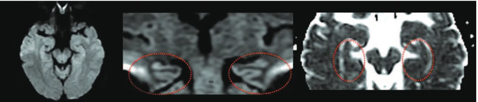
Figure 1. Diffusion sequences (axial and coronal, respectively) and ADC map (axial) on the first day of symptoms, highlighting the hippocampi (circles) that present with no foci of diffusion restriction
Figure 2. Axial and coronal slices of the diffusion sequence in which hypersignal foci (arrows) are seen, consistent with diffusion restriction in hippocampi. The last image corresponds to an axial slice of the ADC map, characterizing foci of diffusion restriction in the hippocampi
incapacity to acquire new information, lasting a few
hours. Complete remission occurs within 24 hours(1).
einstein. 2012;10(2):253-4
254 Guimarães JF, Wosny C, Barbosa Junior AA
These hyperintense hippocampal lesions are small in size (1 to 2 mm), with low apparent diffusion coefficients (ADC), and generally remit within two
weeks(2,5). However, these image changes in patients
with TGA, despite being seen in ischemia of arterial origin, are not specific and may occur due to prolonged ictal activity, multiple sclerosis, hypoglycemia, venous thrombosis, phenylketonuria, emotional stress, pain, sexual intercourse, and physical activity(1,3).
If the clinical presentation is typical, no additional evaluation is mandatory. In case of doubt, imaging studies may be necessary in order to exclude other differential
diagnoses(3). One must consider that amnesia may be a
part of the clinical picture of other diseases, hindering the distinction from TGA based merely on the clinical presentation(6). There are data that suggest hypoperfusion of the hippocampal region as cause of the disease, and the cerebral image study indicated should be MRI with the diffusion technique, emphasizing that during the first 24 hours, the lesion might not be detected(3,5).
AckNowLEdGMENt
To Dr. Ayrton Massaro, neurologist that assisteded this pacient.
REFERENcES
1. Quinette P, Guillery-Girard B, Dayan J, de la Sayette V, Marquis S, Viader F, et al. What does transient global amnesia really mean? Review of the literature and thorough study of 142 cases. Brain. 2006;129(Pt 7): 1640-58.
2. Godeiro-Junior C, de Miranda-Alves MA, Massaro AR. Diffusion magnetic resonance imaging in transient global amnesia. Arq Neuropsiquiatr. 2009; 67(1):130-1.
3. Berli R, Hutter A, Waespe W, Bachli EB. Transient global amnesia - not so rare after all. Swiss Med Wkly. 2009;139(19-20):288-92.
4. Sedlaezek O, Hirsch JG, Grips E, Peters CN, Gass A, Wöhrle J, et al. Detection of delayed focal MR changes in the lateral hippocampus in transient global amnesia. Neurology. 2004;62(12):2165-70.
5. Tong DC, Grossman M. What causes transient global amnesia? New insights from DWI. Neurology. 2004;62(12):2154-5. Comments on: Neurology. 2004; 62(12):2165-70.