Is there any association between National Institute of Health
category IV prostatitis and prostate-specific antigen levels
in patients with low-risk localized prostate cancer?
_______________________________________________
Omer Gokhan Doluoglu
1, Cavit Ceylan
2, Fatih Kilinc
1, Eymen Gazel
2, Berkan Resorlu
1, Oner Odabas
21 Department of Urology Clinic, Ankara Training and Research Hospital, , Ankara Turkey; 2 Department
of Urology Clinic of Yüksek Ihtisas Training and Research Hospital, Ankara, Turkey
ABSTRACT
ARTICLE
INFO
______________________________________________________________ ______________________ Purpose: We investigated the association between National Institute of Health category
IV prostatitis and prostate-specific antigen levels in patients with low-risk localized prostate cancer.
Materials and Methods: The data of 440 patients who had undergone prostate biopsies due to high PSA levels and suspicious digital rectal examination findings were re-viewed retrospectively. The patients were divided into two groups based on the presen-ce of accompanying NIH IV prostatitis. The exclusion criteria were as follows: Gleason score>6, PSA level>20ng/mL, >2 positive cores, >50% cancerous tissue per biopsy, urinary tract infection, urological interventions at least 1 week previously (cystoscopy, urethral catheterization, or similar procedure), history of prostate biopsy, and history of androgen or 5-alpha reductase use. All patient’s age, total PSA and free PSA levels, ratio of free to total PSA, PSA density and prostate volume were recorded.
Results: In total, 101 patients were included in the study. Histopathological examina-tion revealed only PCa in 78 (77.2%) patients and PCa+NIH IV prostatitis in 23 (22.7%) patients. The median total PSA level was 7.4 (3.5–20.0) ng/mL in the PCa+NIH IV prostatitis group and 6.5 (0.6–20.0) ng/mL in the PCa group (p=0.67). The PSA level was≤10ng/mL in 60 (76.9%) patients in the PCa group and in 16 (69.6%) patients in the PCa+NIH IV prostatitis group (p=0.32).
Conclusions: Our study showed no statistically significant difference in PSA levels between patients with and without NIH IV prostatitis accompanying PCa.
Key words:
Prostatitis; Prostatic Neoplasms; prostate-specific antigen (146-154) [Supplementary Concept]
Int Braz J Urol. 2016; 42: 346-50
_____________________
Submitted for publication: November 22, 2014
_____________________
Accepted after revision: June 09, 2015
INTRODUCTION
Prostate-specific antigen (PSA) is the most practical tumor marker used in the diagnosis of prostate cancer (PCa). Measurement of PSA levels has marked a new era in the diagnosis of PCa (1). PSA is a serine protease that is produced almost exclusively by the epithelial cells of the prostate. PSA is not a cancer-specific marker; rather, it is organ-specific (2). Therefore, its serum levels may
increase in nonmalignant conditions such as be-nign prostate hypertrophy (BPH) and prostatitis. The histological architecture of the prostate is dis-turbed in both PCa and prostatitis, causing grea-ter PSA leakage from the lumen of the prostatic glands into the circulation, increasing PSA levels.
unnecessary definitive treatment. Conservative treatment options such as active surveillance have been proposed to decrease the incidence of overtreatment in this subgroup of patients. The risk classification system for localized PCa des-cribed by D’Amico et al. (3), which defines low risk as a PSA level<10ng/mL, Gleason score≤6, and clinical stage of T1 to T2a, is one of the most frequently used risk classification systems to de-termine the need for active surveillance. The new National Comprehensive Cancer Network (NCCN) guidelines (2014 version 2) clinically divide lo-calized PCa into four groups based on patholo-gical results and clinical parameters: very low risk, low risk, intermediate risk, and high risk. In addition to the classiication by D’Amico et al. (3), the NCCN classiication adds a very-low-risk group deined as a PSA density (PSAD) <0.15ng/ mL/g, fewer than three positive prostate biopsy co-res, and≤50% cancerous tissue in any core. The cri -teria added to the low-risk group are a clinical stage of T1 to T2a, Gleason score≤6, and PSA<10ng/mL. Active surveillance is included in the treatment pro -tocol in both groups, and the treatment can be tai-lored according to the patient’s life expectancy (4).
Several studies have shown that active surveillance is associated with very low progres-sion and cancer-specific death rates in selected low-risk patients (5-7). This treatment option se-ems rational in low-risk patients with a life ex-pectancy of>10 years.
National Institute of Health (NIH) cate-gory IV (asymptomatic) prostatitis is described as the presence of inflammatory cells in the biopsy specimen secondary to a high PSA level in an asymptomatic patient (8-10). Considering that most patients with newly diagnosed PCa are in stage T1c and that asymptomatic prostatitis in-creases the PSA level, one wonders whether the addition of prostatitis to PCa further increases the PSA level. If this is the case, patients in the NCCN very-low-risk and low-risk groups may be reclassified in the intermediate-risk group only because of a PSA level>10ng/mL, and their tre-atment strategy may change completely. Thus, we investigated the effects of concurrent NIH IV prostatitis on PSA levels in patients with low--risk localized PCa.
MATERIALS AND METHODS
After approval from the local ethics com-mittee, the data of 440 patients who had undergone prostate biopsies due to high PSA levels and sus-picious digital rectal examination (DRE) findings in our hospital from 2012 to 2014 were reviewed retrospectively. The patients were divided into two groups according to the presence of accompanying NIH IV prostatitis. The exclusion criteria were as follows: Gleason score>6, PSA level>20ng/mL, >2 positive cores, >50% cancerous tissue per biopsy, urinary tract infection, urological interventions at least 1 week previously (cystoscopy, urethral ca-theterization, or similar procedures), a history of prostate biopsy, and a history of androgen or 5-al-pha reductase use.
The patient’s age, total PSA (tPSA) and free (fPSA) levels, f/tPSA, PSAD, and prostate volume (PV) were recorded. PSA was measured at least three times in all patients. The last pre-biopsy PSA measurement was used as the final value. Trans-rectal ultrasonography (TRUS)-assisted systema-tic 12-core prostate biopsy was performed in all patients. TRUS was performed using a LOG-13, 41123WS1 ultrasonography system (General Elec-tric) with a 6.5-MHz biplane transrectal probe. The patients were given prophylactic antibiotics 1 day before biopsy. PV was calculated using TRUS and the ellipsoid formula (height×width×length×0.52). The histopathological preparations were examined again for NIH IV prostatitis by an experienced uro-pathologist in the pathology clinic of our hospital. NIH IV prostatitis was determined by the presen-ce of inflammatory presen-cell infiltration in the prostate biopsy specimen in the absence of symptoms.
RESULTS
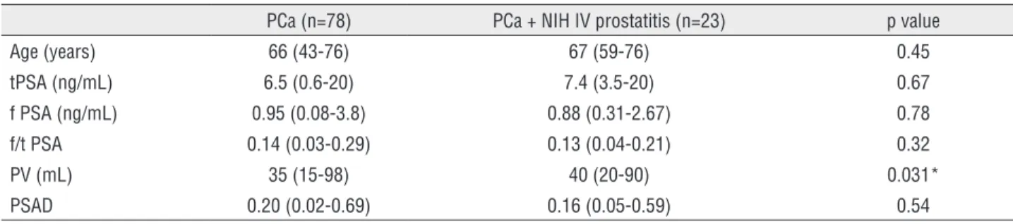
In total, 101 patients fulfilled the inclu-sion criteria and were included in the study. His-topathological examination revealed PCa only in 78 (77.2%) patients and PCa+NIH IV prostatitis in 23 (22.8%) patients. The median patient age was 66 (43–76) years in the PCa group and 67 (59–76) years in the PCa+NIH IV prostatitis group (p=0.45). The median tPSA level was 7.4 (3.5–20.0) ng/ mL in the PCa+NIH IV prostatitis group and 6.5 (0.6–20.0) ng/mL in the PCa group. Although the median PSA level was higher in the PCa+NIH IV prostatitis group, the difference between the groups was not statistically significant (p=0.67).The me-dian PV was 35 (15–98) mL in the PCa group and 40 (20–90) mL in the PCa+NIH IV prostatitis group (p=0.03). The difference between the groups was statistically significant. The data of the two groups are presented in Table-1. The PSA level was≤10ng/ mL in 60 (76.9%) patients in the PCa group and in 16 (69.6%) patients in the PCa+NIH IV prostatitis group (p=0.32). The difference between the groups was not statistically significant. In PCa group, 22 of 78 (28.2%) patients were very low risk patients, and 38 (48.7%) of them were low risk patients. There were 10 (43.5%) patients with very low risk, and 6 (26.1%) patients with low risk in PCa+NIH pros-tatitis group (p=0.15). Cancer was detected in one core in 49 of 78 (62.8%) patients in PCa group, in the same way there was tumor in one core in 18 of 23 (66.3%) patients in PCa+NIH IV prostatitits group (p=0.16). The mean percent of tumor in the cores was 20%±8.7 in PCa group while this value was found as 20%±9.3 in the other group (p=0.72).
DISCUSSION
Eighty-five percent of patients with PCa are diagnosed at>65 years of age (11). PCa is the most frequent visceral malignant neoplasm in adult males (12). After PSA measurement came into use, the incidence of locoregional disease in-creased and that of metastatic disease dein-creased (13). Most cases of PCa were diagnosed according to abnormal DRE findings, high PSA levels, or both in the 1980s and early 1990s. However, most cases diagnosed today are clinically nonpalpable
(stage T1c). The optimal treatment is chosen ac-cording to the stage of the disease. Therefore, for correct staging, it is important to know the factors that affect the parameters used for staging of PCa.
PSA is a serine protease belonging to the human kallikrein-3 group. It is an organ-specific marker that is produced primarily in the luminal epithelial cells of the prostate (14, 15). PSA pro-duction in PCa is not excessively higher than that of the normal prostate tissue (16). A high serum PSA level in patients with PCa is considered to be due to disruption of the cellular architecture of the gland (17). Loss of the barrier comprising the basal layer and basal membrane in the normal prostate gland results in leakage of PSA into the circula-tion. This may also be seen in other diseases of the prostate gland, such as prostatitis. Additionally, procedures such as prostate massage and prostate biopsy cause an increase in PSA level secondary to prostate manipulation (17, 18). Patients with BPH treated by finasteride or other 5-alpha reductase agents show an approximately 50% decrease in their PSA levels. Therefore, we excluded patients with a history of urological operations and those using agents that can affect the PSA level.
groups according to their preoperative PSA level, clinical stage, and Gleason score. Another frequen-tly used nomogram involves the Epstein criteria. The difference in this classification system is the absence of the PSA level among the criteria and the inclusion of the PSAD. This classification system indicates that the PSAD should be <0.15ng/mL in the low-risk group (19). We also investigated the effect of NIH IV prostatitis on PSAD in the present study and found similar PSADs in both groups (Ta-ble-1). This result indicates that NIH IV prostatitis does not affect PSAD.
NIH category IV prostatitis is an asympto-matic inflammatory prostatitis. Such patients may seek treatment for BPH, PCa, high serum PSA le-vels, or infertility. Inflammatory cells are seen in
prostate biopsy specimens, histopathological exa-mination of transurethral resection bites performed due to PCa or BPH, or microscopic examination of expressed prostatic secretion or semen.
Does the presence of NIH IV prostatitis incre-ase the PSA level in patients with localized PCa? In the present study, the median PSA level was higher in the PCa+NIH IV prostatitis group than in the PCa group; however, the difference was not statistically significant (7.4 (3.5–20.) versus 6.5 (0.6–20.0) ng/ mL, respectively; p 0.67). This finding was conside-red to indicate that NIH IV prostatitis accompanying PCa does not cause an additional increase in PSA compared with PCa alone. On the other hand, Agla-mis et al. (20) recently evaluated 198 patients with PCa and found that NIH IV prostatitis (Group 2) ac-companying PCa was associated with significantly
Table 1 . The comparison of age, tPSA and fPSA levels, f/tPSA ratio, and prostate volumes of the groups.
PCa (n=78) PCa + NIH IV prostatitis (n=23) p value
Age (years) 66 (43-76) 67 (59-76) 0.45
tPSA (ng/mL) 6.5 (0.6-20) 7.4 (3.5-20) 0.67
f PSA (ng/mL) 0.95 (0.08-3.8) 0.88 (0.31-2.67) 0.78
f/t PSA 0.14 (0.03-0.29) 0.13 (0.04-0.21) 0.32
PV (mL) 35 (15-98) 40 (20-90) 0.031*
PSAD 0.20 (0.02-0.69) 0.16 (0.05-0.59) 0.54
PSAD = PSA Density; * = statistically significant.
increased PSA levels. However, the PSA range was wider and the study population was more heteroge-neous in their study than in this work.
The PSA level increases as the PV incre-ases. Each 1-mL increase in the PV causes a 4% increase in the PSA level (21). Although the me-dian PV was higher in our PCa+NIH IV prostatitis group (40 versus 35mL, respectively; p=0.035), the PSA levels in this group were not different from those in the PCa group.
The absence of significantly higher PSA levels in the PCa+NIH IV prostatitis group in our study indicates that prostatitis accompanying PCa does not cause any additional increase in PSA le-vels. Therefore, it is not possible to evaluate low--risk patients as intermediatelow--risk patients.
Limitations of our study include the relati-vely small number of patients and its retrospective nature. Another limitation is selection bias. Some patients harboring occult low-grade PCa with or without inflammation do not undergo biopsies. On the other hand, the main strength of our study is inclusion of patients with stage cT1c to T2a PCa, a Gleason score≤6, ≤2 positive cores, and ≤50% can-cerous tissue per biopsy. Our study is also the first to evaluate the effect of NIH IV prostatitis on PSA levels in patients with low-risk localized PCa.
CONCLUSIONS
studies with larger patient cohorts are needed to clarify this issue.
ABBREVIATIONS
BPH = Benign prostate hypertrophy
DRE = Digital rectal examination
fPSA = Free prostate-specific antigen
NCCN = National Comprehensive Cancer Network
NIH = National Institute of Health
PCa = Prostate cancer
PSA = Prostate-specific antigen
PSAD = Prostate-specific antigen density
PV = Prostate Volume
tPSA = total Prostate-specific antigen
TRUS = Transrectal ultrasonography
CONFLICT OF INTEREST
None declared.
REFERENCES
1. Stamey TA, Yang N, Hay AR, McNeal JE, Freiha FS, Redwine E. Prostate-specific antigen as a serum marker for adenocarcinoma of the prostate. N Engl J Med. 1987;317:909-16.
2. Mottet N, Bastian PJ, Bellmunt J, van den Bergh RCN, Bolla M, van Casteren NJ et al. Guidelines on prostate cancer. In: European association of urology guidelines. Eur Assoc Urol. 2014; pp. 16.
3. D’Amico AV, Whittington R, Malkowicz SB, Schultz D, Blank K, Broderick GA, et al. Biochemical outcome after radical prostatectomy, external beam radiation therapy, or interstitial radiation therapy for clinically localized prostate cancer. JAMA. 1998;280:969-74.
4. Mohler JL, Kantoff PW, Armstrong AJ, Bahnson RR, Cohen M, D’Amico AV, et al. Prostate cancer, version 2.2014. J Natl Compr Canc Netw. 2014;12:686-718.
5. Klotz L, Zhang L, Lam A, Nam R, Mamedov A, Loblaw A.
Clinical results of long-term follow-up of a large, active surveillance cohort with localized prostate cancer. J Clin Oncol. 2010;28:126-31.
6. van den Bergh RC, Roemeling S, Roobol MJ, Aus G, Hugosson J, Rannikko AS, et al. Outcomes of men with screen-detected prostate cancer eligible for active surveillance who were managed expectantly. Eur Urol. 2009;55:1-8.
7. Soloway MS, Soloway CT, Williams S, Ayyathurai R, Kava B, Manoharan M. Active surveillance; a reasonable management alternative for patients with prostate cancer: the Miami experience. BJU Int. 2008;101:165-9.
8. Stancik I, Lüftenegger W, Klimpfinger M, Müller MM, Hoeltl W. Effect of NIH-IV prostatitis on free and free-to-total PSA. Eur Urol. 2004;46:760-4.
9. Tan JK, Png DJ, Liew LC, Li MK, Wong ML. Prevalence of prostatitis-like symptoms in Singapore: a population-based study. Singapore Med J. 2002;43:189-93.
10. Nickel JC. Prostatitis: lessons from the 20th century. BJU Int. 2000;85:179-85.
11. Ries LA, Eisner MP, Kosary CL, Hankey BF, Miller BA, Clegg L et al. eds: SEER Cancer Statistics Rewiev, 1975-2001. Bethesda, Md, National Cancer Institute, 2004. Available at: http://seer. cancer.gov/csr/1975_2001/
12. Jemal A, Tiwari RC, Murray T, Ghafoor A, Samuels A, Ward E, et al. Cancer statistics, 2004. CA Cancer J Clin. 2004;54:8-29. 13. Newcomer LM, Stanford JL, Blumenstein BA, Brawer
MK. Temporal trends in rates of prostate cancer: declining incidence of advanced stage disease, 1974 to 1994. J Urol. 1997;158:1427-30.
14. Diamandis EP, Yousef GM. Human tissue kallikrein gene family: a rich source of novel disease biomarkers. Expert Rev Mol Diagn. 2001;1:182-90.
15. Diamandis EP, Yousef GM, Luo LY, Magklara A, Obiezu CV. The new human kallikrein gene family: implications in carcinogenesis. Trends Endocrinol Metab. 2000;11:54-60. 16. Meng FJ, Shan A, Jin L, Young CY. The expression of a variant
prostate-specific antigen in human prostate. Cancer Epidemiol Biomarkers Prev. 2002;11:305-9.
17. Stamey TA, Yang N, Hay AR, McNeal JE, Freiha FS, Redwine E. Prostate-specific antigen as a serum marker for adenocarcinoma of the prostate. N Engl J Med. 1987;317:909-16.
18. Oesterling JE, Rice DC, Glenski WJ, Bergstralh EJ. Effect of cystoscopy, prostate biopsy, and transurethral resection of prostate on sérum prostate-specific antigen concentration. Urology. 1993;42:276-82.
19. Epstein JI, Walsh PC, Carmichael M, Brendler CB. Pathologic and clinical findings to predict tumor extent of nonpalpable (stage T1c) prostate cancer. JAMA. 1994;271:368-74.
20. Aglamis E, Tasdemir C, Ceylan C. The role of National Institute of Health category IV prostatitis in accurately staging the newly diagnosed prostate cancer. Ir J Med Sci. 2013;182:463-7. 21. Oesterling JE, Jacobsen SJ, Chute CG, Guess HA, Girman
CJ, Panser LA, et al. Serum prostate-specific antigen in a community-based population of healthy men. Establishment of age-specific reference ranges. JAMA. 1993;270:860-4.
_______________________ Correspondence address: