Rapid intraoperative tissue expansion with Foley catheter
in a challenging cripple Hypospadias
_______________________________________________
Murat Cakmak
1, Gulnur Gollu
1, Gonul Kucuk
1, Berktug Bahadir
11 Department of Pediatric Surgery, Ankara University Faculty of Medicine, Cebeci, Ankara
ABSTRACT
ARTICLE
INFO
______________________________________________________________ ______________________
Failed hypospadiass cases may result in hypovascular, scarred penis with residual peni-le chordee and peni-leave the patient with minimal residual skin for penipeni-le resurfacing and urethroplasty. Local tissue expansion has become a good alternative to provide skin for penis by using expanders however they require long periods of time for expansion. Besides, rapid tissue expansion was also described in different tissues.We used rapid intraoperative expansion technique by using a Foley catheter in a failed hypospadias case who had minimal residual skin secondary to infection and we concluded that ra-pid intraoperative tissue expansion with Foley catheter is an effective, feasible recons-tructive method for easy dissection and penile resurfacing in failed hypospadiass cases.
Key words:
Hypospadias; Urinary
Catheterization; Tissue Expansion
Int Braz J Urol. 2015; 41: 591-5
_____________________
Submitted for publication: July 01, 2014
_____________________
Accepted after revision: September 16, 2014
INTRODUCTION
The risk of complications such as fistu-la, infection, residual penile curvature in hypos-padiass surgery is relatively high even though perfect surgeries are performed; correction of the complications with secondary surgeries are still challenging especially in patients with scarred, inadequate penile tissue (1). It is reported that when we evaluate all kind of complications the rate is between 1-90%. Several failed hypospa-diass surgeries may result in hypovascular, shor-tened, scarred penis with penile chordee and leave the patient with minimal residual local skin for penile coverage and urethral reconstruction (2). In
PROCEDURE
A four-year old boy who was treated pri-marily a year ago in another center for penoscro-tal hypospadias encountered infection following the first surgery which resulted in scarred, inade-quate penile skin for reconstruction. The patient still showed penoscrotal hypospadias (Figure-1). By combining the advantages of rapid tissue ex-pansion and local tissue usage, we decided to use rapid intraoperative expansion technique using a Foley catheter in this failed hypospadias case who had inadequate, fragile, scarred penile skin. Informed consent was obtained from parents for publication.
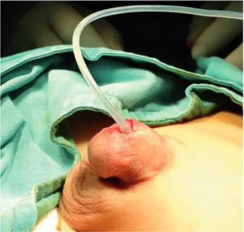
Under general anesthesia, following ap-propriate preoperative antibiotics, two incisions of 0.3 cm length were made to dorsum of penis at the corona, fashioned at 11 and 1 o’clock po-sition. Then the tips of two 8Fr Foley catheters were trimmed, keeping the balloons intact. After blunt dissection, the catheters were inserted under the dorsal penile skin (Figures 2-4). The balloons were filled with 3 ml saline until the flaps were blanched. Balloons were inflated synchronously for four times for three minutes with a two-minute rest period. Easy dissection of the skin from the dorsal and lateral sides of the penis was achie-ved without bleeding. Following tissue expansion, adequate penile tissue was achieved to cover the penile shaft through Snodgrass repair (Figure-5). In four months follow-up, the penis has an opti-mal esthetic appearance with no fistula and with good urethral calibration (Figures 6 and 7).
DISCUSSION
We used rapid intraoperative expansion technique by using Foley catheters in a failed hy-pospadias case who had minimal residual skin se-condary to infection. In the literature, Foley cathe-ter has been described as atool for rapid expansion in cleft palate repair, alopecia repair, tissue defect repair following mass excision such as basal cell carcinoma, melanocytic nevus from forehead, temple, eyelid, periorbital region and extremi-ties (4, 6-10). The aim of this paper is to describe the use of Foley catheter in cripple hypospadias
Figure 1 - Preoperative view.
Figure 2 - Intraoperative view. First Foley catheter was inserted under the dorsal penile skin at 11 o’clock position.
surgery for rapid intraoperative tissue expansion with easier dissection and less bleeding.
Tissue for resurfacement of penis can be obtained from local tissue or from extragenital areas such as buccal mucosa and skin. Extrage-nital skin grafts may result in loss of the graft, graft shrinkage, lack of sensation, hair bearing skin with different pigmentation (1, 4). Methods for local tissue resurfacement are dorsal relaxing incision, Z-plasty, rotation of a scrotal flap and burying the penis into the scrotum.
Tissue expansion is especially useful in those who lack enough penile skin and extrage-nital skin graft is not preferred. In the literature, authors reported that tissue expansion provides increased tissue area which is well-vascularized, sensate skin and excellent tissue match for pig-mentation, texture and thickness (2, 4, 8, 11). It was also used in staged hypospadiass repairs (13). It also avoids the need to transfer tissue over a distance. For these reasons, local tissue expansion has become a good alternative to provide skin for
Figure 4 - Intraoperative view. The Foley catheters were expanded under the dorsal penile skin.
Figure 5 - Postoperative view following penile reconstruction and urethroplasty.
penis (2). Another advantage of local tissue ex-pansion is the recruitment of additional skin with a similar distribution of androgen receptors for penile reconstruction as hypothesized by Kajba-fzadeh et al. (1). Recently, besides chronic tissue expansion, rapid tissue expansion technique has also been used in different parts of the body by some authors. According to Auletta et al. imme-diate tissue expansion has provided enough tis-sue to close defects up to 5cm in diameter with 16-6% tissue expansion and according to Baker and Johnson although actual amount of skin in-creased during expansion is uncertain, it has been estimated as 31% (3, 6, 13). Although the authors described expansion of 30%, our case had nearly 10-15% of expansion. The reason for this small amount may be the small dimension of the scar-red penis. Demirseren et al. described immediate tissue expansion with Foley catheter in a case of congenital melanocytic nevus and emphasized the advantage of omnidirectional expansion (7).
Expanders require long periods of time for expansion, may cause erosion, they are for single--use, expensive and difficult to use in children. Meanwhile, Foley catheters are readily available, inexpensive, disposable and practical for intrao-perative tissue expansion (1, 9, 14, 15). Chronic tissue expansion has a potential risk of infection in children as discussed by Friedman et al. howe-ver this wasn’t a significant problem in Mathews’ series (2, 11). Also, chronic tissue expansion can be slightly uncomfortable to the patient, may cre-ate an unnatural distortion of the dorsum of the penis, requires multiple outpatient clinic visits, re-peated inflations and may have psychological im-plications. According to Friedman et al. the major complications of chronic tissue expansion were implant exposure, deflations, wound dehiscen-ce and minor technical complications with port, hematoma and significant pain (11). The authors describe the method as three to four cycles of in-flation and dein-flation after placement of catheter for 3 to 5 minutes (3). Another advantage of rapid expansion is that it does not create a fibrous cap-sule in the expanded tissue because of brief
dura-tion avoiding retracdura-tion (4). Another advantage of this technique is to provide easier dissection with less bleeding in secondary cases and scarred tis-sues. By using this method, we were able to both dissect the dorsal and lateral penile skin easily wi-thout bleeding and also close the ventral defect.
The use of rapid intraoperative tissue ex-pansion in failed hypospadias cases is an effective and feasible reconstructive method which provi-des additional sensate, hair bearing local tissue with excellent match of texture, pigmentation and thickness to the native penis.
CONFLICT OF INTEREST
None declared.
REFERENCES
1. Kajbafzadeh AM, Sina A, Payabvash S. Management of multiple failed repairs of the phallus using tissue expanders: long-term postpubertal results. J Urol. 2007;177:1872-7. 2. Mathews R, Nelson CP, Gearhart JP, Vander Kolk CA. Tissue
expansion in management of failed phallic reconstruction: initial report of clinical series. Urology. 2005;66:180-4. 3. Johnson TM, Brown MD, Sullivan MJ, Swanson NA.
Immediate intraoperative tissue expansion. J Am Acad Dermatol. 1990;22:283-7.
4. Abramo AC, Viola JC, Angelo AJ. Intraoperative rapid expansion in cleft palate repair. Plast Reconstr Surg. 1993;91:441-5.
5. Vordermark JS. Tissue expansion for urethral and penile reconstruction. Dialogues Pediatr Urol. 1995;18:4.
6. Auletta MJ, Matarasso SL, Glogau RG, Tromovitch TA. Comparison of skin hooks and Foley catheters for immediate tissue expansion. J Dermatol Surg Oncol. 1993;19:1084-8. 7. Demirseren ME, Ceran C, Demirseren DD. Treatment of
a congenital melanocytic nevus on the forehead with immediate tissue expansion technique: a three-year follow-up. Pediatr Dermatol. 2012;29:621-4.
8. Foster JA, Scheiner AJ, Wulc AE, Wallace IB, Greenbaum SS. Intraoperative tissue expansion in eyelid reconstruction. Ophthalmology. 1998;105:170-5.
10. Cil Y, Emre A, Ozturk S. Dissection of the expander pocket for burn scar alopesia treatment with the aid of urologic instrument and a foley catheter. J Burn Care Res. 2008;29:681. 11. Friedman RM, Ingram AE Jr, Rohrich RJ, Byrd HS, Hodges
PL, Burns AJ, Hobar PC. Risk factors for complications in pediatric tissue expansion. Plast Reconstr Surg. 1996;98:1242-6.
12. Cakmak M, Vargel I, Soyer T, Cavusoglu T, Yazıcı I, Hancerliogulları O, et al. The use of tissue expander in the management of staged proximal hypospadias repair: report of case. Eur J Plast Surg. 2012;35:253-5.
13. Baker SR, Swanson NA. Rapid intraoperative tissue expansion in reconstruction of the head and neck. Arch Otolaryngol Head Neck Surg. 1990;116:1431-4.
14. Greenbaum SS. Intraoperative tissue expansion with the Foley catheter. J Dermatol Surg Oncol. 1993;19:1079-83. 15. Greenbaum SS, Greenbaum CH. Intraoperative tissue
expansion using a Foley catheter following excision of a basal cell carcinoma. J Dermatol Surg Oncol. 1990;16:45-8.
_______________________
Correspondence address: Murat Cakmak, MD, PhD Ankara Universitesi Tip Fakultesi Cocuk