ULTRASTRUCTURE OF OVARIAN FOLLICULAR
EPITHELIUM OF THE AMAZONIAN FISH
Pseudotylosurus
microps
(TELEOSTEI: BELONIDAE): MORPHOLOGICAL
AND HISTOCHEMICAL CHARACTERIZATION OF THE
INTERCELLULAR DEPOSITS
CRUZ-LANDIM, C.1 and CRUZ-HÖFLING, M. A.2
1Departamento de Biologia, Instituto de Biociências, Unesp, CEP 13506-900, Rio Claro, SP, Brazil 2Departamento de Histologia e Embriologia, Instituto de Biologia, Unicamp, CEP 13083-970, Campinas, SP, Brazil
Correspondence to: Carminda da Cruz-Landim, Departamento de Biologia, Instituto de Biociências, Unesp, C.P. 199, CEP l3506-900, Rio Claro, SP, Brazil, e-mail: [email protected]
Received October 6, 1999 – Accepted April 25, 2000 – Distributed February 28, 2001
(With 4 figures)
ABSTRACT
The present paper reports the presence of great quantities of electrondense intercellular material in the follicular epithelium of P. microps. The material apparently is uptaken from circulation and enter the follicle through the intercellular spaces accumulating in the epithelial median-apical intercellular spaces and in perioocytic space. The accumulation starts in the early growth of the primary oocyte and proceed until vitellogenesis. The possible chemical nature and function of the deposits are dis-cussed.
Key words: chorion, oocytes, oogenesis, electrondense deposits, follicle.
RESUMO
Ultra-estrutura do epitélio folicular do ovário do peixe amazônico Pseudotylosurus microps (Teleostei: Belonidae): caracterização morfológica e histoquímica dos
depósitos intercelulares
Neste trabalho é mostrada a grande quantidade de material eletrondenso intercelular no epitélio folicular de P. microps. Aparentemente, o material é captado da circulação e enviado para o folículo por meio dos espaços intercelulares, acumulando-se nos espaços intercelulares médio-apicais do epitélio e no espaço perioocítico. A acumulação é iniciada no oócito primário e prossegue até a vitelogênese. A natureza química desse material é discutida.
Palavras-chave: cório, oócitos, oogênese, depósitos eletrondensos, folículo.
INTRODUCTION
The ultrastructure of oogenesis in fishes have been studied by several authors concerning the developmental stages of oocytes growth (Anderson, 1974; Iwasaki, 1973; Bruslé, 1980; Cruz-Landim & Cruz-Höfling, 1979; Droller & Roth, 1966; Norrevang, 1968; Wourms, 1976; Shackley & King, 1977), the cellular envelopes of the
Sheldon, 1976; Tesoriero, 1978; Fleger, 1977). However although seems to have accordance among the authors about the phases of the oocyte growth and the description of the characteristic structures present in it the same do not happens concerning the egg cellular and acellular covers. In fact the structure of follicular epithelium seems to be very much variable from one group to another. In face of this it function, as well as the function of the thecal sheath is not well stablished. In the same way the acellular covers that invest the oocyte during it development and after ovulation do not have it origin and function well defineded.
These structures have, however, had a long-standing appeal to the biologists since questions as ovary steroid synthesis, and adaptation of oocy-tes to certains environment conditions have been attributed to the cellular and acellular coverings respectively.
This work intent to contribute to the heap of knowledge about the oocytes covers, when still in the ovary, in order to form a background to the understanding of the pendent questions. In this context, a morphological and histochemical charac-terization of “adorning” elements found in the interstices of the follicular epithelium was made.
MATERIAL AND METHODS
Fragments of ovaries of the telostean P. microps, collected in Solimões River (Amazon Basin) were fixed to optical and electron micro-scopic studies.
For electron microscopy the pieces were fixed in 2.5% glutaraldehyde buffered with 0.2 M sodium caccodilate, pH 7.2, containning 4% sucrose and post-fixed in 1% osmium tetroxide in the same buffer.
After fixation the pieces were dehydrated in increasing concentration series of ethanol and via propylene oxide, embedded in Apon-Araldite resin. Sections obtained with glass knives were double-contrasted with uranyl acetate and lead citrate. The examination and photography were made on a Zeiss EM9S2 electron microscope.
For light microscopy the ovary fragments were fixed in 10% neutral formaldehyde and emb-beded in paraffin. To the paraffin section were applied histochemical tests (see Table 1) according Pearse (1960) to show acidic and neutral
muco-polyssacharids (also the sulfated ones), reducing group as SH, NH groupments, cholesterol and elastic fibers.
RESULTS
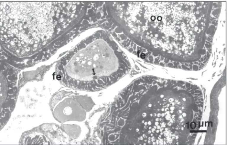
In the course of the study of P. microps ooge-nesis our attention was driven to peculiar features of its follicular epithelium. The follicular epithelium around the oocytes between the primary phase of oocyte development and early vitellogenesis accu-mulate in its intercellular spaces numerous poly-morphous bodies, in resin sections, stainable with toluidine blue and other acidic stains (Fig. 1). During the vitellogenic phase of the oocyte, the number and size of these structures is such that can even shade the epithelial cells to the light microscopy examen.
The origin and evolution of these material revealed to be dificult to follow, but as the ovary of P. microps is of asyncronous type several stages of accumulation can be seen at same time in differ-ent follicles.
Ultrastructure
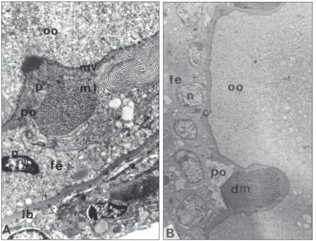
An attempt of follow the evolution of the material accumulated in the intercellular space of the follicular cells was made taken in account the growth phase of the oocyte enclosed in the follicle. By this approach it can be seen that when the oocy-te is in the perinucleolar phase and the follicular epithelium is still flat, the material appears only in the apical face, frequently occupying the space between the oocyte surface and the follicular cell top (perioocytic space) (Fig. 2A, B). In this stage the bodies appear made of reticular or concentric lamellae of electrondense material as in Fig. 2A or as compact electrondense masses (Fig. 2B).
In any case the amount of material accu-mulated is so, that the perioocytic space is enlarged and the follicular cells and oocytic surface de-formed (Figs. 1 and 2).
Fig. 1 — Micrograph of thick a section from material processed to electron microscopy stained with toluidine blue. Oo-cytes (oo) in different phases of oogenesis showing that in early oocyte development the dense material seem to enter the oocyte (arrows). fe = follicular epithelium.
to the oocyte cytoplasm or as in Fig. 2B, where the electrondense mass push the oocyte surface inward.
In this phase the follicular epithelium inter-cellular spaces are note widened but some electron-dense material may be seen fulfilling the basal portion of the cellular contacts (Fig. 2A, B).
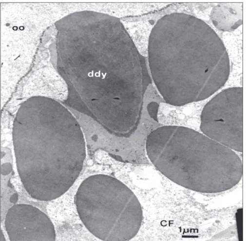
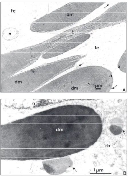
In vitellogenic oocytes the follicular epithe-lium have increased very much in height and the intercellular spaces between cells appear totally occu-pied by polimorphous dense bodies (Figs. 1, 3 and 4). The now electrondense bodies may appear homogeneously amorphous, and spherical, as in Fig. 3 or elongated and fibrillar, with differently structurated material aggregated to their periphery (Fig. 4A, B).
It seems that these bodies tend to coalesce and that the less dense material makes the link between them during the coalescence process. It is not ease to discerne the intercellular localization of this material because of its huge size that somewhat disorganized the epithelium. Where is possible to perceive the cellular contacts, as in basal side of the epithelium, they are closed. However even there can be seen electrondense material inside it and in connection with the dense bodies. In this phase the oocyte has already at least two layers of vitelline envelope around it and some-times can be seen the contacts between the dense bodies and the outer layer (still in deposition pro-cess) of this envelope (Fig. 3).
The dense bodies polymorphism may be due to different stages of their accumulation or mo-bilization. In fact, it can be seen, as already mentio-ned, the presence of less dense and organized material in the periphery of certain bodies, as well as it can be seen regions where the fibrillar arrange-ment is patent due both to the loose compactation of the fibrils and to the tangential section of the boundaries of them (Fig. 4A). Some granules present even paracrystalline organization being their fibrillar constituents disposed with highly regular periodicity (Fig. 4B).
The described material starts to accumulate during early perinucleolar phase of the oocyte and probably reachs their peak in early vitellogenic oocytes. However, even after the end of the ooge-nesis, when the mature egg have already completed the deposition of the acellular coverings, great amounts of the bodies are still present. When, after the end of the oocyte growth the follicular epithelium is disrupted the dense bodies seem remain entire.
Histochemistry
As seen in Table 1 the histochemical tests applied to the ovaries show that the material occuping the intercellular space of the follicular cells is negative to Alcian Blue, pH 0.5, 2.5 and Ninhidryn Schiff. It is positive to PAS and Schiff, and react strongly to ferric ferricyanide which indicates the presence of reducing groups.
Fig. 2 — Aspects of the material present in the perioocytic (po) space of follicles in the perinucleolar stage. A. Material lamelar (ml) and powder (p) apparently being incorporated by the oocyte (oo). B. Dense material (dm) compressing the oocyte (oo) surface. fe = follicular epithelium; po = perioocytic space; n = nuclei; c = oocyte envelope; mv = microvilli; lb = basal lamina.
TABLE 1 Histochemical results.
Reactions Reactive groups Reaction intensity
Schiff Reductor groups ±
Ninhidryn Schiff NH2 groups –
PAS Polysaccharids +
PAS + diastase Acid
Neutral
– –
PAS + sodium desoxicolate Lipids with vic-glicol groupments –
Toluidin blue Cholesterol –
Adams (ferric ferricianide) Reductor groups +
Adams + HgCl2 SH reductor groups + + +
Alcian blue pH 2.5 Proteoglicans –
Alcian blue pH 0.5 Sulfated proteoglicans –
Aldehide fucsin Elastic fibers –
Fig. 3 — Contact between the dense material in the perioocytic space with the outer layer of the chorion. Notice the coa-lescence of the deposits (ddy) by the adition a new material of different electron densities. CF = follicular cell.
presence of SH groupments, are responsible by the positivity. Also was demonstrated the absence of elastic fibers.
DISCUSSION AND CONCLUSIONS
The electrondense deposits that acccumulate in the intercellular spaces of follicular epithelium and in the perioocytic space tend to increase as the oocyte proceed in its development. This ma-terial seems to originate from substances intaken in the basal side of the follicular epithelium and travel to the apical one. The basal intercellular space between cells remain narrow during all the process although filled with electrondense material, showing that the material must enter this space slowly and accumulate apically. It is clear that the
material present in the perioocytic space reached this location coming through the intercellular space of follicular cells, since in no moment similar de-posits were seen inside the follicular cells.
It is known that some or all of the oocyte yolk has an exogenous origin, probably being synthe-sized in liver and transported to the ovary through circulation (Bruslé, 1980; Droller & Roth, 1966; Norrevang, 1968). The uptake of this vitellogenin material is made through the intercellular spaces of follicular cells and later by pynocitic activity on the oocyte surface. It is, therefore, possible that some of the electrondense material, mainly that one arising early in the oocyte development were of vitellogenic nature.
1967; Chaudry, 1956; Dumont & Brummett, 1980; Fleger, 1977; Tesoriero, 1978; Begovac & Wallace, 1989). The responsibility more frequently appoin-ted to this synthesis is to the follicular epithelium. However in this case there are some morphological evidence that the electrondense material can contribute to the formation of the outer envelo-pe of the oocyte, or chorion.
The chorion, of mature teleost eggs, is a pro-teinaceous covering. The paracrystalline pattern
assumed by some of the intercellular bodies seems to point to a proteinic nature, but the Ninhidryn Schiff reaction fail in show the presence of NH2 groupments in the material. This fail may be due to the lack of free NH2 groups. On the other hand, the presence of SH free radicals in this deposits are not well understood but they were already demonstrated in the chorion of marine teleosteans (Begovac & Wallace, 1989) and in the sea urchin they are responsible by the egg activation.
From the histochemical testes applied only polysaccharids, reductor groups and SH groups was demonstrated present in the intercellular de-posits. Therefore also the histochemical results indicate that the intercellular deposits could be formed by vitellogenic or by chorionic proteins. Whatever are the electrondense material func-tions, it is strange the great amounts of accumulation and their presence after the end of vitellogenesis is an accessory. However there is a possibility that the remaining material form some sort of appendages, that associated with the egg surface, may serve for eggs flutuation or fixation to a substrate, as verified in certain species (Anderson, 1966; Hart et al., 1984).
REFERENCES
ANDERSON, E., 1966, A study of the fibrillar appendages associated with the surface of eggs of the killifish, Fun-dulus heteroclitus. Anat. Rec., 154: 308-309. ANDERSON, E., 1967, The formation of the primary
envelo-pe during oocyte differentiation in teleosts. J. Cell Biol., 35: 193-212.
ANDERSON, E., 1974, Comparative aspects of the ultra-structure of the female gamete. Int. Rev. Cytol., 4(suppl.): 1-70.
BEGOVAC, P. C. & WALLACE, R. A., 1989, Major vitellins envelope proteins in piperfish oocytes originate within the follicle and are associated with the Z3 layer. J. Exp. Zool., 251: 56-73.
BRUSLÉ, S., 1980, Fine structure of early previtellogenic oocytes in Mugil (liza) auratis Risso, 1810 (Teleostei: Mugilidae). Cell Tissue Res., 207: 123-134.
BUSSON-MABILLOT, S., 1977, Un type particulier de sécretion exocrine: celui de l’appareil adhésif de l’veuf d’un poisson teléostéen. Biol. Cellulaire, 30: 233-244. CHAUDRY, H. S., 1956, The origin and structure of the zona pellucida in the ovarian eggs of teleosts. Z. Zellforsch., 43: 478-485.
CRUZ-HÖFLING, M. A. & CRUZ-LANDIM, C., 1990, The ultrastructure of the developmental stages of the oocytes of Astyanax bimaculatus (Teleostei: Characidae). Zool. Jb. Anat., 120: 1963-181.
CRUZ-HÖFLING, M. A. & CRUZ-LANDIM, C., 1992, Ul-trastructural studies on the follicular epithelium during oocyte growth in the Amazonian fish, Crenicichla johanna (Teleostei: Cichlidae). Zool. Jb. Ant., 122: 1-15. CRUZ-LANDIM, C. & CRUZ-HÖFLING, M. A., 1979, Comportamento de nucléolos e mitocôndrios durante a ovogênese de peixes teleósteos de água doce. Acta Ama-zônica, 9: 723-728.
CRUZ-LANDIM, C., 1990, Aspectos ultra-estruturais das cé-lulas produtoras de esteóides do ovário de Colossoma mitrei (Teleostei: Characidae). COLÓQUIO DA SOC. BRASIL. MICR. ELETR., 255-256.
CRUZ-LANDIM, C., SILVA DE MORAES, R. L. M. & CRUZ-HÖFLING, M. A., 1987, Aspectos ultra-estruturais das células foliculares de Crenicichla johanna
(Teleostei: Cichlidae). AN. XI COLÓQUIO DA SOC. BRAS. MICR. ELETR., Caxambú, MG, p. 107. DROLLER, M. J. & ROTH, T. F., 1966, An electron
micro-scope study of yolk formation during oogenesis in
Lebistes reticulatus uppyi. Cell Biol., 28: 209-232. DUMONT, J. N. & BRUMMETT, A. R., 1980, The vitelline
envelope, chorion and micropyle of Fundulus hetero-clitus eggs. Gamete Res., 3: 25-44.
FLEGER, C., 1977, Electron microscopic studies on the de-velopment of the chorion of the viviparous teleost Der-mogenys pusillus (Hemirhamphidae). Cell Trissue Res.,
179: 255-270.
HART, N. H., PIETRI, R. & DONOVAN, M., 1984, The structure of the chorion and associated surface filaments in Oryzias evidence for the presence of extracellular tu-bules. J. Exp. Zool., 230: 273-296.
HIROSE, K., 1972, The ultrastructure of the ovarian follicle of medaka (Oryzias latipes). Z. Zellforsch., 123: 316-329. IWASAKI, Y., 1973, Histochemical detection of 3 – hidroxysteroid dehydrogenase in the ovary of medaka,
Oryzias latipes, during annual reproductive cycle. Hokkaido Univ. Bull. Fac. Fish., 23: 177-184. KHOO, K. H., 1975, The corpus luteum of gold fish (
Caras-sius auratus L.) and its functions. Can. J. Zool., 53: 1306-1323.
LAMBERT, J. G. D., 1970, The ovary of the guppy Poecilia reticulta. The granulosa cells as sites of steroid biosyn-thesis. Gen. Comp. Endocrinol., 15: 464-476. LOPES, R. A., SANTOS, H. S. L., COSTA, J. R. V.,
PELI-ZARO, M. G. & CASTAGNOLI, N., 1982, Histochemical study of oocyte zona radiata of the lambari Astyanax bimaculatus lacustris Linnaeus, 1758 (Osteichthyes: Characidae). Zool. Anz., 208: 265-268.
NICHOLLS, T. J. & MAPLE, G., 1972, Ultrastructural ob-servations on possible sites of steroid biosynthesis in the ovarian follicular epithelium of two species of cichlid fish, Cichlasoma nigrofasciatum and Haplochomie multi-color. Z. Zellforsh., 128: 317-335.
NORREVANG, A., 1968, Electron microscopic morphology of oogenesis. Intern. Rev. Cytol., 23: 113-186. PEARSE, A. G. E., 1960, Histochemistry: Theoretical and
Applied. J. & A. Churchill Ltd., London, p. 998. SHACKLEY, S. E. & KING, P. E., 1977, Oogenesis in a
SHELTON, W. L., 1978, Fate of the follicular epithelium in
Dorosoma petenense (Pisces: Clupeidae). Copeia, 2: 237-244. TESORIERO, J. V., 1978, Formation of the chorion (Zona Pellucida) in the Teleost, Oryzias latipes. J. Ultr. Res.,
64: 315-326.
WOURMS, J. P., 1976, Annual fish oogenesis. I. Differen-tiation of the mature oocyte and formation of the primary envelope. Devel. Biol., 50: 338-354.
WOURMS, J. P. & SHELDON, H., 1976, Annual fish oogenesis. II. Formation of the secondary envelopes.