Craniofacial abnormalities among patients with Edwards Syndrome
Anormalidades craniofaciais em pacientes com Síndrome de Edwards
Anormalidades craniofaciales entre pacientes con síndrome de Edwards
Rafael Fabiano M. Rosa1, Rosana Cardoso M. Rosa2, Marina Boff Lorenzen3, Paulo Ricardo G. Zen4, Carla Graziadio5,
Giorgio Adriano Paskulin6
Instituição: Universidade Federal de Ciências da Saúde de Porto Alegre (UFCSPA) e Complexo Hospitalar Santa Casa de Porto Alegre (CHSCPA) 1Doutor pelo Programa de Pós-Graduação em Patologia da UFCSPA; Geneticista Clínico do Hospital Materno Infantil Presidente Vargas, Porto Alegre, RS, Brasil
2Aluna de Mestrado do Programa de Pós-Graduação em Patologia da UFCSPA, Porto Alegre, RS, Brasil
3Acadêmica do Curso de Medicina da UFCSPA, Porto Alegre, RS, Brasil 4Doutor pelo Programa de Pós-Graduação em Patologia da UFCSPA; Professor-Adjunto da Disciplina de Genética Clínica da UFCSPA, Porto Alegre, RS, Brasil
5Aluna de Doutorado do Programa de Pós-Graduação em Patologia da UFCSPA; Professora-Assistente da Disciplina de Genética Clínica da UFCSPA, Porto Alegre, RS, Brasil
6Doutor pelo Programa de Pós-Graduação em Genética e Biologia Molecular da Universidade Federal do Rio Grande do Sul (UFRGS); Professor-Associado da Disciplina de Genética Clínica da UFCSPA, Porto Alegre, RS, Brasil
ABSTRACT
Objective: To determine the frequency and types of crani-ofacial abnormalities observed in patients with trisomy 18 or Edwards syndrome (ES).
Methods: This descriptive and retrospective study of a case series included all patients diagnosed with ES in a Clini-cal Genetics Service of a reference hospital in Southern Brazil from 1975 to 2008. The results of the karyotypic analysis, along with clinical data, were collected from medical records.
Results: The sample consisted of 50 patients, of which 66% were female. The median age at first evaluation was 14 days. Regarding the karyotypes, full trisomy of chro-mosome 18 was the main alteration (90%). Mosaicism was observed in 10%. The main craniofacial abnormalities were: microretrognathia (76%), abnormalities of the ear helix/dysplastic ears (70%), prominent occiput (52%), posteriorly rotated (46%) and low set ears (44%), and short palpebral fissures/blepharophimosis (46%). Other uncom-mon — but relevant — abnormalities included: microtia (18%), orofacial clefts (12%), preauricular tags (10%), facial palsy (4%), encephalocele (4%), absence of external auditory canal (2%) and asymmetric face (2%). One pa-tient had an initial suspicion of oculo-auriculo-vertebral spectrum (OAVS) or Goldenhar syndrome.
Conclusions: Despite the literature description of a char-acteristic clinical presentation for ES, craniofacial alterations may be variable among these patients. The OAVS findings in this sample are noteworthy. The association of ES with OAVS has been reported once in the literature.
Key-words: chromosomes, human, pair 18; trisomy; chromosome aberrations; craniofacial abnormalities; Goldenhar syndrome.
RESUMO
Objetivo: Verificar a frequência e os tipos de alterações craniofaciais observadas em pacientes com trissomia do cromossomo 18 ou síndrome de Edwards (SE).
Métodos: Estudo descritivo e retrospectivo de uma série de casos que incluiu todos os pacientes diagnosticados com SE em um Serviço de Genética Clínica de um hospital de referência do sul do país, no período de 1975 a 2008. Os resultados da análise cariotípica, juntamente com dados clínicos, foram coletados a partir dos prontuários médicos.
Resultados: A amostra foi composta de 50 pacientes. Destes, 66% eram do sexo feminino. A mediana da idade no momento da primeira avaliação foi de 14 dias. Quanto aos cariótipos, a trissomia livre do cromossomo 18 foi a
al-Endereço para correspondência: Paulo Ricardo G. Zen
Rua Sarmento Leite, 245, sala 403 – Centro CEP 90050-170 – Porto Alegre/RS E-mail: [email protected]
Fonte financiadora: Coordenação de Aperfeiçoamento de Pessoal de Nível Superior (Capes)
Conflito de interesse: nada a declarar
teração principal (90%). Observou-se mosaicismo em 10%. As principais anormalidades craniofaciais observadas consis-tiram de microrretrognatia (76%), anormalidades de hélix das orelhas/orelhas displásicas (70%), occipital proeminente (52%), orelhas retrovertidas (46%) e baixo implantadas (44%) e fendas palpebrais pequenas/blefarofimose (46%). Outras anormalidades incomuns, mas relevantes, foram microtia (18%), fendas orofaciais (12%), apêndices pré-auriculares (10%), paralisia facial (4%), encefalocele (4%), ausência de conduto auditivo externo (2%) e assimetria de face (2%). Um dos pacientes apresentava suspeita inicial de espectro óculo-aurículo-vertebral (EOAV) ou síndrome de Goldenhar.
Conclusões: Apesar da descrição na literatura de quadro clínico usualmente característico para a SE, as alterações cranio-faciais podem ser variáveis nesses pacientes. Chamam atenção os achados pertencentes ao EOAV, sendo que a associação de SE com essa condição foi observada na literatura em um relato de caso.
Palavras-chave: cromossomos humanos par 18; trissomia; aberrações cromossômicas; anormalidades craniofaciais; síndrome de Goldenhar.
RESUMEN
Objetivo: Verificar la frecuencia y los tipos de alteracio-nes craniofaciales observadas en pacientes con trisomía del cromosoma 18 o síndrome de Edwards (SE).
Métodos: Estudio descriptivo y retrospectivo de una serie de casos que incluyó a todos los pacientes diagnosticados con SE en un Servicio de Genética Clínica de un hospital de referencia en el sur de Brasil, en el periodo de 1975 a 2008. Los resultados del análisis cariotípico, juntamente con datos clínicos, fueron recogidos a partir de los prontuarios médicos.
Resultados: La muestra fue compuesta por 50 pacientes. De estos, el 66% eran del sexo femenino. La mediana de edad en el momento de la primera evaluación fue de 14 días. Respecto a los cariotipos, la trisomía libre del cromosoma 18 fue la alteración principal (90%). Se observó mosaicismo en el 10%. Las principales anormalidades craniofaciales observadas consistieron en microrretrognatia (76%), anormalidades de hélix de las orejas/orejas displásicas (70%), occipital promi-nente (52%), orejas retrovertidas (46%) y bajo implantadas (44%) y hendiduras palpebrales/blefarofimosis (46%). Otras anormalidades poco comunes, pero relevantes, fueron microtia (18%), hendiduras orofaciales (12%), apéndices preauricula-res (10%), parálisis facial (4%), encefalocele (4%), ausencia de conducto auditivo externo (2%) y asimetría facial (2%).
Uno de los pacientes presentaba sospecha inicial de espectro óculo-auricular-vertebral (EOAV) o síndrome de Goldenhar.
Conclusiones: A pesar de la descripción en la literatura de cuadro clínico usualmente característico para la SE, las alteraciones craniofaciales pueden ser variables en esos pa-cientes. Llaman la atención los hallazgos pertenecientes al EOAV, siendo que la asociación de SE con esa condición se observó en la literatura en un relato de caso.
Palabras clave: cromosomas humanos par 18; trisomía; aberraciones cromosómicas; anormalidades craniofaciales; síndrome de Goldenhar.
Introduction
Edwards’ syndrome, also known as Trisomy 18, was irst described in 1960 by Edwards et al(1). It is considered the
second most frequently observed autosomal trisomy at birth (second only to Trisomy 21 – Down syndrome). Its prevalence is estimated at about 1 to 3,600–8,500 live births, and is predominant in female individuals(2-9). The syndrome is
char-acterized by a limited survival. Most fetuses with Edwards’ syndrome will die during fetal life, and among those who live, the median survival is usually varied from 2.5 to 14.5 days(2-8).
The syndrome has a quite broad spectrum of clinical mani-festations, and there is a description of more than 130 different anomalies in the literature to date, which may involve virtually all organs and systems. However, none of them is pathogno-monic, what may hinder the recognition of the syndrome, especially in patients without main classic indings(10,11).
Thus, the objective of this study was to determine the fre-quency of occurrence and the types of craniofacial alterations in a sample of patients with Edwards’ syndrome evaluated in a period of over 30 years.
Method
A retrospective and descriptive study of all patients diag-nosed with Edwards’ syndrome in the Clinical Genetics Service at Universidade Federal de Ciências da Saúde de Porto Alegre (UFCSPA)/Complexo Hospitalar Santa Casa de Porto Alegre (CHSCPA), a reference hospital in southern Brazil, in the pe-riod from 1975 to 2008. The patients described were also part of the work of Rosa et al(12) and Rosa et al(13), who described in
laboratory (Cytogenetics Laboratory of Universidade Federal de Ciências da Saúde de Porto Alegre - UFCSPA). The results, along with clinical data, were collected from the medical records. This study was approved by the Research Ethics Committee of the Hospital. The results were analyzed by two-tailed Fisher’s exact test, using the PEPI (Programs for Epidemiologists) (version 4.0). Signiicance was established at p<0.05.
Results
The sample was composed of 50 patients diagnosed with Edwards’ syndrome. Among these, 66% were female. The age at irst assessment ranged from 1 day to 16 years (median: 14 days). Regarding the results of karyotypes, the trisomy of chromosome 18 was the main abnormality, identiied in 90% of cases. The presence of chromosomal mosaicism (usually, lineage with full trisomy of chromosome 18 associated with a normal one) was veriied in 10% of patients. The mean of metaphase plaques analyzed was 25.
The main craniofacial abnormalities observed were micro-retrognatia (76%), abnormal ear helix/dysplastic ears (70%), prominent occipital (52%), Retroverted ears (46%) and low-set ears (44%), and blepharophimosis/short palpebral issures (46%) (Figure1). Another relevant abnormality, though less
frequent, included orofacial clefts (12%), ranging from cleft lip to bilateral cleft lip and palate. The unusual indings con-sisted of microtia (18%) (mostly bilateral – the unilateral cases were all on the right side), preauricular appendages (10%) (mostly unilateral and on the left side), facial paralysis(4%), encephalocele (4%) and absence of external auditory canal (2%). The encephalocele involved occipital region in both cases (Table 1). Suspected Edwards’ syndrome was observed in 34% of patients. One of them presented initial suspicion of oculo-auriculo-vertebral spectrum (OAVS), or Goldenhar syndrome, and had bilateral iris abnormality, micrognathia, preauricular left appendages, bilateral microtia and absence of the left external auditory canal. His chromosomal constitution was 47,XY,+18 (mosaicism not detected).
It was also evaluated whether the presence of mosaicism could be related to the clinical manifestations presented by patients. Thus, the frequencies of the major clinical indings observed in the sample (with levels above 40%) were compared between the group of patients with and without mosaicism. Among these, only the lack of association of short palpebral issures/ blepharophimosis with mosaicism was close to signii-cance (p=0.054 – there was no presence of mosaicism in any of the patients with this inding). Among the total, only two patients (4%) had simultaneous description of all of the most
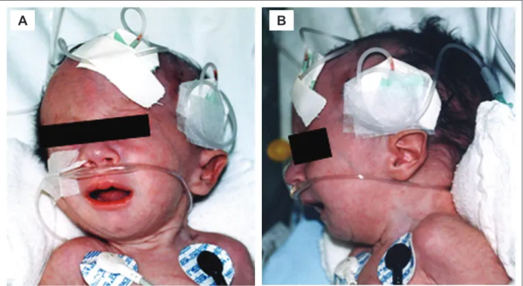
Figure 1 - Craniofacial indings of a patient with Edwards’ syndrome in the study sample. Note particularly micrognathia (B) and low-set ear faun (A and B)
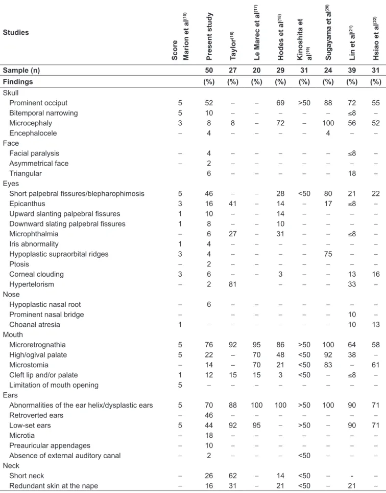
Table 1 - Frequency of craniofacial abnormalities observed in patients in the sample compared to those described in the literature Studies Sc o re Ma ri o n e t a l (1 5 ) Pr e s e n t s tu d y T a y lo r (1 6 ) L e Ma re c e t a l (1 7 ) H o d e s e t a l (1 8 ) K in o s h ita e t a l (1 9 ) Su g a y a m a e t a l (2 0 ) L in e t a l (2 1 ) H s ia o e t a l (2 2 )
Sample (n) 50 27 20 29 31 24 39 31
Findings (%) (%) (%) (%) (%) (%) (%) (%)
Skull
Prominent occiput 5 52 – – 69 >50 88 72 55
Bitemporal narrowing 5 10 – – – – – ≤8 –
Microcephaly 3 8 8 – 72 – 100 56 52
Encephalocele – 4 – – – – 4 – –
Face
Facial paralysis – 4 – – – – – ≤8 –
Asymmetrical face – 2 – – – – – – –
Triangular 6 – – – – – 18 –
Eyes
Short palpebral issures/blepharophimosis 5 46 – – 28 <50 80 21 22
Epicanthus 3 16 41 – 14 – 17 ≤8 –
Upward slanting palpebral issures 1 10 – – 14 – – – –
Downward slating palpebral issures 1 8 – – 10 – – – –
Microphthalmia – 6 27 – 31 – – ≤8 –
Iris abnormality 1 4 – – – – – – –
Hypoplastic supraorbital ridges 3 4 – – – – 75 – –
Ptosis – 2 – – – – – – –
Corneal clouding 3 6 – – 3 – – 13 16
Hypertelorism – 2 81 – – – 33 –
Nose
Hypoplastic nasal root – 6 – – – – – – –
Prominent nasal bridge – – – – – – 10 –
Choanal atresia 1 – – – – – – 10 13
Mouth
Microretrognathia 5 76 92 95 86 >50 100 64 58
High/ogival palate 5 22 – 70 48 <50 92 38 –
Microstomia – 14 – 70 21 <50 83 – 61
Cleft lip and/or palate 1 12 15 15 3 <50 – ≤8 –
Limitation of mouth opening 5 – – – – – – – –
Ears
Abnormalities of the ear helix/dysplastic ears 5 70 88 100 100 >50 100 90 71
Retroverted ears – 46 – – – – – – –
Low-set ears 5 44 92 95 – >50 – 90 71
Microtia – 18 – – – – – – –
Preauricular appendages – 10 – – – – – – –
Absence of external auditory canal – 2 – – – <50 – – –
Neck
Short neck – 26 62 – 14 <50 – - –
frequent indings. No patient with abnormal iris (especially coloboma), cleft lip and/or palate, and microtia presented mosaicism. On the other hand, the latter was observed in the only case with facial asymmetry of the study. The patient presented a chromosomal constitution with a predominance of normal lineage (47,XY,+18[5]/46,XY[35]).
Discussion
In this sample, the main chromosomal constitution observed was the full trisomy of chromosome 18, which was consistent with the literature, which describes a frequency around 95%. Mosaicism is considered a rare inding and was observed in 10% of patients in this study. This also applies to translocations. However, even with no cases of this chromosomal abnormality in the sample, its importance should be highlighted, especially with regard to genetic counseling, once the karyotype assessment of parents is indicated in cases of translocation, in order to verify whether one of them has the same chromosomal abnormality(14).
Despite reports in the literature of a clinical condition usually characteristic of Edwards’ syndrome, craniofacial al-terations may show variable in these patients. In this sample, only 4% of subjects had simultaneously all the main indings. Analyzing the studies described in the literature, a wide varia-tion in the frequency of some of these abnormalities can also be observed (Table 1)(15-22). The main changes observed both
in this work and in the literature were microretrognathia and dysplastic ears/ear helix abnormalities. Those are described in usually 80% of patients. In several studies, this inding of ear abnormality is reported in all subjects(17,18,20). These
altera-tions present maximum punctuation in the scoring system of Marion et al(15), a tool developed especially for neonatologists
in order to try to optimize the recognition of individuals with Edwards’ syndrome (Table 1)(15-22). Other indings
com-monly observed in this study and in the literature consisted of prominent occiput, small palpebral issures/blepharophimosis, low-set ears, short neck (often with excess skin or webbed) and high/ogival palate. The inding of posteriorly rotated ears was common in this evaluation, being veriied in 46% of patients. However, there was no description of the frequency of this alteration in other studies (Table 1)(15-22).
The frequency of hypoplastic supraorbital ridges appears to have been underestimated in the sample, probably due to a mea-surement bias. In the literature review, only one study describes its frequency. Sugayama et al(20) reported it in 75% of cases. This
may also apply to indings of hypertelorism and limitation of mouth opening, an abnormality that is more functional rather
than structural. The frequency of microcephaly veriied in the present study was also noteworthy. While many studies reported values above 50%(18,20-22), the frequency observed here, such as
in the study by Taylor(16), was of 8%, which may be related to
the deinition of microcephaly used. It was considered micro-cephaly not only when the measure of the head circumference was lower than the second percentile for the age of the patient, but also below that percentile for size/length (i.e., both absolute as relative microcephaly). There is no description of the way this variable was assessed in the other studies. Cleft lip and/or palate is described usually in less than 15% of patients, which was in line with the ratio found here. Encephalocele, in turn, is considered an uncommon inding. There was a description of only one patient with this inding in the review by Moore et al(23) and other case in the series of Sugayama et al(20). In both
reports, such as in this sample, encephalocele presented involve-ment of the occipital region.
In clinical practice, there is the idea that individuals with mosaicism tend to have a milder clinical presentation. In the literature there are no comparisons between the anomalies identiied in individuals with or without mosaicism. Anyhow, in the analysis performed in this study, there were no differ-ences between these two subgroups of patients. Only for the presence of short palpebral issures/blepharophimosis, there were values close to signiicance, suggesting that this inding may be less frequent in individuals with mosaicism. However, one of the limitations of this analysis was the low number of patients with this chromosomal constitution (n=5). Perhaps studies with a larger number of individuals may prove the existence of differences between the subgroups.
The presence of indings related to OAVS, such as facial asymmetry, microtia, and pre-auricular appendages, was noteworthy(24-26), and their association with Edwards’
syn-drome was already reported in the literature(27). Therefore, the
OAVS could be considered a potential differential diagnosis for Edwards’ syndrome. It is an etiologically heterogeneous condition and highly variable phenotypically, characterized by the involvement of the structures originating in the irst branchial arches, in particular. The indings related to OAVS include anomalies, normally asymmetric, of ears, face, eyes, and spine; and there are different types of criteria for the diagnosis described in the literature. Strömland et al, for instance, considered necessary for the diagnosis of OAVS the presence of normal chromosomal constitution and phenotypic abnormalities involving at least two of the following areas: oro-craniofacial, ocular, auricular, and vertebral(28). There are
References
1. Edwards JH, Harnden DG, Cameron AH, Crosse VM, Wolff OH. A new trisomic syndrome. Lancet 1960;1:787-90.
2. Rosa RF, Rosa RC, Lorenzen MB, de Moraes FN, Graziadio C, Zen PR et al.
Trisomy 18: experience of a reference hospital from the south of Brazil. Am J Med Genet A 2011;155A:1529-35.
3. Carter PE, Pearn JH, Bell J, Martin N, Anderson NG. Survival in trisomy 18. Life tables for use in genetic counselling and clinical paediatrics. Clin Genet 1985;27:59-61.
4. Young ID, Cook JP, Mehta L. Changing demography of trisomy 18. Arch Dis Child 1986;61:1035-6.
5. Goldstein H, Nielsen KG. Rates and survival of individuals with trisomy 13 and 18. Data from a 10-year period in Denmark. Clin Genet 1988;34:366-72. 6. Root S, Carey JC. Survival in trisomy 18. Am J Med Genet 1994;49:170-4. 7. Embleton ND, Wyllie JP, Wright MJ, Burn J, Hunter S. Natural history of trisomy
18. Arch Dis Child Fetal Neonatal Ed 1996;75:F38-41.
8. Rasmussen SA, Wong LY, Yang Q, May KM, Friedman JM. Population-based analyses of mortality in trisomy 13 and trisomy 18. Pediatrics 2003;111:777-84. 9. Weber WW. Survival and the sex ratio in trisomy 17-18. Am J Hum Genet
1967;19:369-77.
10. Jones KL. Smith’s recognizable patterns of human malformation. 6th ed.
Philadelphia: Elsevier Saunders; 2006.
11. Zen PR, Rosa RF, Rosa RC, Dale Mulle L, Graziadio C, Paskulin GA. Unusual clinical presentations of patients with Patau and Edwards syndromes: a diagnostic challenge? Rev Paul Pediatr 2008;26:295-9.
12. Rosa RF, Rosa RC, Lorenzen MB, Zen PR, de Oliveira CA, Graziadio C et al. Limb abnormalities on trisomy 18: evidence for early diagnosis. J Pediatr
(Rio J) 2012;88:401-5.
13. Rosa RF, Rosa RC, Lorenzen MB, de Oliveira CA, Graziadio C, Zen PR et al.
Trisomy 18: frequency, types, and prognosis of congenital heart defects in a Brazilian cohort. Am J Med Genet A 2012;158A:2358-61.
14. Chen H. Atlas of genetic diagnosis and counseling. Humana Press: New Jersey; 2006.
15. Marion RW, Chitayat D, Hutcheon RG, Neidich JA, Zackai EH, Singer LP et al. Trisomy 18 score: a rapid, reliable diagnostic test for trisomy 18. J Pediatr
1988;113:45-8.
16. Taylor AI. Autosomal trisomy syndromes: a detailed study of 27 cases of Edwards’ syndrome and 27 cases of Patau’s syndrome. J Med Genet 1968;5:227-52. 17. Le Marec BM, Lair JC, Kérisit J, Le Mée F, Sénécal J. 20 cases of trisomy 18.
Sex-ratio in relation to age of the mother. Ann Pediatr (Paris) 1977;24:125-36.
18. Hodes ME, Cole J, Palmer CG, Reed T. Clinical experience with trisomies 18 and 13. J Med Genet 1978;15:48-60.
19. Kinoshita M, Nakamura Y, Nakano R, Morimatsu M, Fukuda S, Nishimi Y et al. Thirty-one autopsy cases of trisomy 18: clinical features and pathological
indings. Pediatr Pathol 1989;9:445-57.
20. Sugayama SM, Kim CA, Utagawa CY, Albano LM, Bertola DR, Koiffmann CP
et al. Estudo genético-clínico de 24 pacientes com trissomia 18 (síndrome de
Edwards). Pediatria (São Paulo) 1999;21:133-43.
21. Lin HY, Lin SP, Chen YJ, Hung HY, Kao HA, Hsu CH et al. Clinical characteristics
and survival of trisomy 18 in a medical center in Taipei, 1988-2004. Am J Med Genet A 2006;140A:945-51.
22. Hsiao CC, Tsao LY, Chen HN, Chiu HY, Chang WC. Changing clinical presentations and survival pattern in trisomy 18. Pediatr Neonatol 2009;50:147-51.
23. Moore CA, Harmon JP, Padilla LM, Castro VB, Weaver DD. Neural tube defects and omphalocele in trisomy 18. Clin Genet 1988;34:98-103.
24. Rosa RF, Dall’agnol L, Zen PR, Pereira VL, Graziadio C, Paskulin GA. Oculo-auriculo-vertebral spectrum and cardiac malformations. Rev Assoc Med Bras 2010;56:62-6.
25. Rosa RF, Zen PR, Flores JA, Golendziner E, Pilla CB, Roman T et al.
Oculo-auriculo-vertebral spectrum in patients with congenital heart defects. Arq Bras Cardiol 2010;95:436-9.
26. Rosa RF, Silva AP, Goetze TB, Bier Bde A, Almeida ST, Paskulin GA et al. Ear
abnormalities in patients with oculo-auriculo-vertebral spectrum (Goldenhar syndrome). Braz J Otorhinolaryngol 2011;77:455-60.
27. Greenberg F, Herman G, Stal S, Gruber H, Ledbetter DH. Chromosome abnormalities associated with facio-auriculo-vertebral spectrum. Am J Med Genet1988;31:170.
28. Strömland K, Miller M, Sjögreen L, Johansson M, Joelsson BM, Billstedt E
et al. Oculo-auriculo-vertebral spectrum: associated anomalies, functional
deficits and possible developmental risk factors. Am J Med Genet A 2007;143A:1317-25.
29. Verloes A, Seret N, Bernier V, Gonzales M, Herens C, Koulischer L. Branchial arch anomalies in trisomy 18. Ann Genet 1991;34:22-4.
30. Wright CG, Brown OE, Meyerhoff WL, Rutledge JC. Inner ear anomalies in two cases of trisomy 18. Am J Otolaryngol 1985;6:392-404.
31. Zen PR, Rosa RF, Rosa RC, Graziadio C, Paskulin GA. New report of two patients with mosaic trisomy 9 presenting unusual features and longer survival. Sao Paulo Med J 2011;129:428-32.
affecting individuals with Edwards’ syndrome; however, these are rare. Verloes et al described three patients, two with ex-treme microtia and absence of the external auditory canal, and one with hemifacial microsomia(29). Inner ear abnormalities,
such as absence of cochlear nerve ibers and utriculoendolym-phatic valve, were described by Wright et al(30). Interestingly,
although there were no reports of patients with Edwards’ syndrome with auricular appendages, the sample pre-sented 5 patients (10%) with this inding. Facial asymmetry, a common alteration in the OAVS, has been associated to the presence of mosaicism in the literature(31), as observed in the
only individual of the sample with this inding.
Therefore, craniofacial abnormalities are common in indi-viduals with Edwards’ syndrome. However, classic indings, such as dysplastic ears, micrognathia, and prominent occiput may not be present, hampering the syndrome’s recognition.
Therefore, health professionals who might come across such syndrome, especially pediatricians and neonatologists, should be aware of this variability, besides the possibility of unusual clinical indings such as microtia, preauricular appendages, encephalocele, and absence of external auditory canal. Hence the importance of evaluation by karyotype testing, in order to identify these patients and, consequently, conduct the ap-propriate monitoring of these individuals and their families, including genetic counseling.