Efects of passive mobilization on acute hemodynamic
responses in mechanically ventilated patients
Efeitos da mobilização passiva nas respostas hemodinâmicas
agudas em pacientes sob ventilação mecânica
INTRODUCTION
Critically ill patients who require sedation and mechanical ventilation are restricted to bed for long periods. his coninement is a risk factor for dysfunction in various organ systems, often becoming more severe than the underlying condition itself.(1) In the intensive care unit (ICU), patients’ limbs
are routinely mobilized by the unit’s physiotherapists, aiming to preserve the range of motion, improve or preserve soft tissue length, maintain muscle
condition and reduce the risk of thromboembolism.(2) Mechanical stress
from limb mobilization may alter hemodynamic responses such as heart rate
(HR), blood pressure (BP) and myocardial oxygen consumption (mVO2).(3)
Cardiac output depends on the interaction between two main functions: 1) heart function, which is determined by HR, contractility and pre- and afterload, and 2) return function, which is determined by the volume of
Eliane Regina Ferreira Sernache de Freitas1, Renata Serrou da Silva
Bersi2, Mariana Yuri Kuromoto2,
Silviane de Camargo Slembarski2,
Ana Paula Ayumi Sato2, Marcela
Quadros Carvalho3
1. Intensive Care Unit, Hospital Santa Casa de Londrina - Londrina (PR), Brazil; Course of Physiotherapy, Universidade Norte do Paraná - UNOPAR - Londrina (PR), Brazil. 2. Course of Physiotherapy, Universidade Norte do Paraná - UNOPAR - Londrina (PR), Brazil. 3. Intensive Care Unit, Hospital Santa Casa de Londrina - Londrina (PR), Brazil.
ABSTRACT
Objective: To assess the efects of passive mobilization on acute hemodynamic responses in mechanically ventilated patients.
Methods: his cross-sectional, quantitative, observational study enrolled patients who were admitted to the intensive care unit, sedated and mechanically ventilated. he infusion of sedative and analgesic drugs aimed to maintain a Ramsay scale sedation level of 4 to 6. Passive mobilization consisted of hip and knee lexion-extension movements for ive minutes. After 10 minutes of rest, an additional ive minutes of lexion-extension passive movements was performed for the shoulders. Hemodynamic assessments (heart rate and systolic, diastolic and mean blood pressure) were performed one minute before the mobilization protocol and one minute
after each phase. he double product and myocardial oxygen consumption were calculated using appropriate formulas.
Results: A total of 13 patients (69.2% male, with a mean age of 69.1 ± 15.8 years) were admitted from June to December, 2011. Passive mobilization led to statistically signiicant increases in heart rate, double product and myocardial oxygen consumption. However, mean blood pressure was not signiicantly altered.
Conclusions: Our results suggest that passive mobilization of mechanically ventilated and sedated patients is safe and provides beneicial efects on acute hemodynamic parameters, particularly heart rate, although mean blood pressure is not signiicantly altered.
Keywords: Hemodynamics; Intensive care unit; Artiicial respiration
Study conducted in the Intensive Care Unit of Hospital Santa Casa de Londrina - Londrina (PR), Brazil.
Conlicts of interest: None.
Submitted on January 11, 2012 Accepted on March 13, 2012
Corresponding author:
Eliane Regina Ferreira Sernache de Freitas
Rua Belo Horizonte, 540 – Apto 11 Zip Code: 86020-060 - Londrina (PR), Brazil.
venous return, venous drainage resistance and right
atrial pressure.(4,5) It has also been demonstrated that
the distension and shortening of muscle ibers may activate mechanoreceptors, leading to cardiovascular adjustments via parasympathetic inhibition and sympathetic activation.(4-8)
he potential beneits of exercise for inactive ICU patients have been reported previously.(9,10) Passive lower
limb mobilization in critically ill patients prevents muscle ibers atrophy,(11) increases oxygen consumption (VO
2) and
reduces venous blood oxygen saturation (SVO2), most likely
due to an increased oxygen extraction rate (O2ER) and
cardiac index.(12) However, the physiological mechanisms
behind hemodynamic responses to passive mobilization in mechanically ventilated patients are not fully known.
his study aimed to assess the efects of passive mobilization on acute hemodynamic responses in mechanically ventilated patients.
METHODS
he study protocol was approved by the ethics committee of Irmandade Santa Casa de Londrina (ISCAL) (project #380/11), and written informed consent forms were signed by designated family members for each patient. he study design was a cross-sectional, quantitative, observational format following the Strengthening the Reporting of Observational
Studies in Epidemiology (STROBE) criteria.(13)
he patients included were aged over 18 years who were maintained on pressure-controlled-mode mechanical ventilation (MV) (Newport Wave E200, Newmed, CA, USA) for more than 48 hours with the following parameters: a positive end-expiratory
pressure (PEEP) between 5 and 8 cmH2O, a tidal
volume between 6 and 8 mL/kg and an inspired oxygen
fraction (FiO2) of 21-50%. he infusion of sedative
and analgesic drugs aimed at a sedation level between 4 (brisk response to stimulus) and 6 (no response to
painful stimulus) on the Ramsay scale.(14) All patients
were administered vasoactive drugs, and mean blood pressure (MBP) was maintained above 60 mmHg. he following subjects were excluded from the study: hemodynamically unstable patients (MBP < 60 mmHg), patients showing agitation during the maneuvers, and those with resistance to movement, dropping oxygen saturation (< 90%), an intra-aortic balloon, complex arrhythmias, neurologic and/or motor deicits or musculoskeletal limitations preventing the protocol-determined movements.
Passive mobilization (PM) protocol
he patients were maintained in a supine position, with the bed head raised at 30%. PM consisted of hip and knee lexion-extension movements for ive minutes (90º lexion). After 10 minutes of rest, an additional ive minutes of lexion-extension passive movements was performed for the shoulders (90º). PM was simultaneously performed by two physiotherapists alternating lexion and extension of the right and left limbs at a frequency of 30 movements/minute. To maintain a steady frequency, a metronome was used (KORG MA-30, Japan). Hemodynamic assessments (measurements of HR and systolic (SBP), diastolic (DBP) and mean (MBP) blood pressures) were performed one minute before the mobilization protocol and one minute after the end of each phase.
Clinical signs were continuously monitored using a multi-parameter monitor (Dixtal DX 2010 - Dixtal, Manaus, Brazil) to provide electrocardiogram (ECG), HR, SBP, DBP and MBP measurements. he variable double product (DP) was calculated as the product of SBP and HR (DP = SBP × HR), and myocardial
oxygen consumption (mVO2) was calculated using the
formula mVO2 = (DP × 0.0014) - 6.3.(15) Demographics
and Acute Physiology Chronic Health Evaluation II
(APACHE II) scores(16) were recorded for all patients.
Drug infusions and mechanical ventilator settings were constant during the protocol.
Statistical analysis
he normality of the data was conirmed using the Kolmogorov-Smirnov test. Categorical variables are presented as absolute values and proportions, and continuous variables are reported as the means with standard deviations (±SD). he assessments before and after PM were compared using the pairwise samples parametric Student’s t-test. A one-way ANOVA was used to compare pre- and post-PM assessments for the upper and lower limb mobilizations. Diferences between categorical variables were compared using the Chi-squared test. he Statistical Package for Social Sciences (SPSS 17.0) software was used for the analyses, and a value of p < 0.05 was considered to be signiicant.
RESULTS
Brazil. Table 1 lists the patients’ baseline characteristics. he Ramsay scale was adopted to assess the level of consciousness. One patient was categorized as Ramsay 4, three as Ramsay 5 and nine as Ramsay 6.
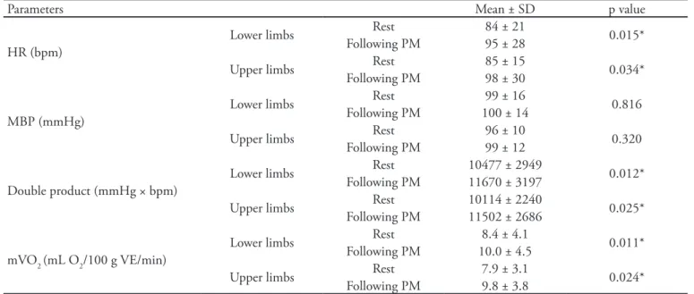
Table 2 presents the patients’ hemodynamic parameters according to the protocol applied to the upper and lower limbs. Statistically signiicant diferences were found for HR (lower limbs, p = 0.015; upper limbs, p = 0.034), DP (lower limbs, p = 0.012;
upper limbs, p = 0.025) and mVO2 (lower limbs, p =
0.011; upper limbs, p = 0.024). Immediately after PM, MBP showed a mild but not statistically signiicant increase.
When the responses to the lower and upper limb mobilizations were compared, no diference was found for the pre- and post-PM assessed variables (Table 3).
No adverse events, such as desaturation or agitation, were recorded during the PM maneuvers.
Table 2 - Hemodynamic parameters following passive lower and upper limb mobilizations
Parameters Mean ± SD p value
HR (bpm)
Lower limbs Rest 84 ± 21 0.015*
Following PM 95 ± 28
Upper limbs Rest 85 ± 15 0.034*
Following PM 98 ± 30
MBP (mmHg)
Lower limbs Rest 99 ± 16 0.816
Following PM 100 ± 14
Upper limbs Rest 96 ± 10 0.320
Following PM 99 ± 12
Double product (mmHg × bpm)
Lower limbs Rest 10477 ± 2949 0.012* Following PM 11670 ± 3197
Upper limbs Rest 10114 ± 2240 0.025* Following PM 11502 ± 2686
mVO2 (mL O2/100 g VE/min)
Lower limbs Rest 8.4 ± 4.1 0.011*
Following PM 10.0 ± 4.5
Upper limbs Rest 7.9 ± 3.1 0.024*
Following PM 9.8 ± 3.8
HR - heart rate; MBP - mean blood pressure; mVO2 - myocardial oxygen consumption; PM - passive mobilization; SD - standard deviation.
Table 1 - Baseline characteristics of the study patients
Characteristics Result p value
Age (years) 69.1 ± 15.8 (41 - 88) 0.4979
Gender
Male 9 (69.2)
0.9992
Female 4 (30.8)
APACHE II score 25 ± 4.1 (18 - 33) 1.000
Risk of death 52.2 ± 13.4 (29.1 - 78.6) 0.9098
Glasgow score 5.5 – 1.0 (4 - 8) 0.8971
Ramsay score 5.0 – 0.58 (4 - 6) 1.0000
Mechanical ventilation time 10.5 ± 8.6 (2 - 30) 0.9216 Diagnosis
Septic shock 03 (23.1)
0.0861
Pneumonia 06 (46.1)
Chronic obstructive pulmonary disease 03 (23.1) Postoperative (abdominal) 01 (7.7)
DISCUSSION
In this study, passive lower and upper limb mobilizations promoted signiicant increases in HR,
DP and mVO2, but not in MBP. he physiological
mechanisms of hemodynamic responses during passive mobilization are not fully known. Although HR increases with cardiac output (CO) during exercise, CO increase should not be interpreted as resulting only from increased HR but rather from the interaction of two main factors: 1) heart function, which is determined by HR, contractility and pre- and afterload, and 2) return function, which is determined by the volume of venous blood return, resistance to venous
return and right atrial pressure.(4,5) Although passive
mobilization produces no muscle contraction, Doppler tests conducted for the assessment of active and passive kinesiotherapy have shown an increased venous blood low from the sural pump during passive kinesiotherapy that was well above the baseline.(17)
Muscle tension caused by passive movements can also lead to increased HR due to the activation of tendinous
mechanoreceptors.(8,18) Prior studies have shown that
simultaneous muscle stretching and shortening, as in typical passive mobilization, causes the activation of mechanoreceptors and type III ibers, which can induce vagal activation and stimulate baroreceptors, thereby contributing to the overall cardiovascular response.(19,20)
he mobilization of large muscle groups (such as the hip, knee or shoulder) is another factor likely contributing to the increased HR observed in our study. A similar observation was recently reported by Farinatti et al.,(21) who assessed cardiovascular responses to static
lexibility with passive movements in healthy subjects. heir results showed that HR was consistently higher
with hip lexion (involving the ischiotibial muscles, a large group) than with ankle dorsal lexion (involving the gastrocnemius, a small group). Additionally
supporting our indings, these authors(21) also reported
that DP increased with the passive mobilization of both small and large muscle groups, concluding that passive stretching of muscle groups may afect cardiovascular
responses. Gladwell and Coote(22) also assessed HR and
SBP during one-minute passive and sustained sural triceps stretching, reporting signiicantly increased HR but not SBP, which are indings similar to our own.
Recently Magder (2012)(23) reviewed the
physiological mechanisms regulating HR and the relevance of mobilizing critically ill patients, concluding that HR may be interpreted within the overall hemodynamic condition of the patient and that HR and CO regulation during exertion is relected in a range of physiological responses, but that HR is the major cardiovascular system component responsible for
adjusting CO.(24) Additionally, when the HR response
is limited by underlying disease or pharmacological interventions, changes in the systolic volume may act to compensate for such limitations. However, this ability may in turn be limited by the passive illing of the left ventricle.
In our study, MBP was not observed to signiicantly increase in response to passive mobilization. his observation may be explained by noting that increased peripheral vascular resistance is also inluenced by sustained muscle contraction, consequently increasing BP.(25) However, our protocol used a low cycle frequency,
maintaining 30 movement cycles/minute for ive minutes without active contraction.
Based on our indings, the increased DP may have been caused by increased HR, as no signiicant BP
Table 3 - Comparison of hemodynamic parameters following passive lower and upper limb mobilization
Parameters
Lower limbs Mean ± SD
Upper limbs
Mean ± SD p value
HR (bpm) Rest 84 ± 21 85 ± 15 0.3445
Following PM 95 ± 28 98 ± 30
MBP (mmHg) Rest 99 ± 16 96 ± 10 0.9025
Following PM 100 ± 14 99 ± 12
Double product (mmHg × bpm) Rest 10477 ± 2949 10114 ± 2240 0.5840 Following PM 11670 ± 3197 11502 ± 2686
mVO2 (mL O2/100 g VE/min) Rest 8.4 ± 4.1 7.9 ± 3.1 0.5895 Following PM 10.0 ± 4.5 9.8 ± 3.8
increase was observed,(26) shown by the MBP results.
he DP is usually used to estimate cardiac workload
during aerobic and strength exercises.(24) However,
several authors have reported that during static lexibility exercises, DP may reach levels that are similar to the levels found during high-intensity, low-repetition resistance exercise.(27,28)
Comparative studies between upper and lower limb exertion tests have shown that at the same level of workload, the CO may be similar that for lower
loads.(29-31) his diference may explain our results when
comparing the responses for the mobilizations of the upper and lower limbs, where no diferences was found for any of the variables, as the loads were the same for both the upper and lower limbs (30 movements/ minute).
Cardiac metabolism is inluenced both by chrono- and inotropisms, and both have an inluence on
myocardial overload and oxygen demand.(32) Myocardial
oxygen demand may be measured as myocardial oxygen
consumption (mVO2), which is determined by the
interactions among myocardial tension, heart muscle contractility and HR. All of these factors change during physical exertion, increasing the myocardial requirements for nutrients and oxygen and causing
increased coronary low.(33) A linear correlation between
mVO2 and coronary blood low has been demonstrated,
thus providing information on cardiac overload, i.e., the work performed by the heart to meet the body’s demand. he product of HR and SBP, the DP, is highly correlated with mVO2 (r2=0.88).(32) Hellerstein
& Wenger(15) described a mathematical function to
convert DP into mVO2 (mVO2 = (DP × 0.00014) -
6.3), allowing the estimation of cardiac efort. his calculation was applied in our study; the results suggest that due to increased HR, which in turn inluenced the DP, heart muscle overload occurred, with a consequently
signiicant increase in mVO2. he hemodynamic
and metabolic efects of cyclic passive lower limb mobilization in mechanically ventilated patients was previously assessed by Savi et al.,(12) who reported that
all ive assessed patients displayed increased oxygen
consumption (VO2) concomitant with a drop in venous
blood oxygen saturation (SVO2), most likely due to an
increased oxygen extraction rate (O2ER) and cardiac
index (CI). he authors additionally concluded that cyclic passive lower limb mobilization may inluence the hemodynamic and metabolic conditions of sedated patients who are dependent on mechanical ventilation.(12)
Additionally, aiming to assess exertion induced
hemodynamic changes, Bittencourt et al. (2008)
evaluated HR, DP, BP and mVO2 in 11 male subjects. In
agreement with our indings, their results demonstrated that physical activity signiicantly changes HR, DP and mVO2.(34)
Our study had several limitations: 1) the small sample size may have inluenced the lack of statistical signiicance for MBP and 2) the study included patients with Ramsay levels of 4 to 6. Ramsay 4 patients may have had a certain degree of active/assisted muscle activity; however, no comparative intergroup analysis was conducted due to the small sample size.
CONCLUSION
Our results suggest that passive mobilization of the lower and upper limbs produces acute hemodynamic efects in mechanically ventilated and sedated patients, particularly increasing HR; however, MBP was not signiicantly afected. It should be noted that considering the assessed variables, no changes were observed that may be considered dangerous according to the available literature. Our procedure resulted in beneicial heart muscle overload in critically ill mechanically ventilated patients.
his work was an initial study, and further studies will be necessary to explore this subject more deeply.
RESUMO
Objetivo: Avaliar as respostas hemodinâmicas agudas da mobilização passiva de pacientes sob ventilação mecânica.
Métodos: Estudo de investigação clínica do tipo transversal, quantitativa e observacional. Incluindo pacientes internados na unidade de terapia intensiva, sedados e sob ventilação mecânica. A infusão de drogas sedativas e analgésicas visava o grau de seda-ção de 4 a 6 de acordo com a escala de Ramsay. A mobilizaçao passiva consistiu em movimentos de lexo-extensão de quadril e joelho durante cinco minutos. Após 10 minutos de repouso, fo-ram realizados mais cinco minutos de mobilização passiva com lexo-extensão de ombro. As mensurações hemodinâmicas (fre-qüência cardíaca, pressão arterial sistólica e diastólica e pressão arterial média) foram realizadas 1 minuto antes da realização do protocolo e no primeiro minuto após o término. O duplo produto e a medida do consumo ou captação de oxigênio pelo miocárdio foram obtidas por meio de fórmulas.
do duplo produto e do consumo ou captação de oxigênio pelo miocárdio com diferença estatisticamente signiicante. Entre-tanto a pressão arterial média não apresentou diferença signi-icativa.
Conclusões: Os resultados sugerem que a mobilização passi-va de membros inferiores e superiores em pacientes sedados sob
ventilação mecânica inluencia de forma segura nos efeitos he-modinâmicos agudos, particularmente na frequência cardíaca, porém sem alterar signiicativamente a pressão arterial média.
Descritores: Hemodinâmica; Unidades de terapia intensiva; Respiração artiicial
REFERENCES
1. Krasnof J, Painter P. he physiological consequences of bed rest and inactivity. Adv Ren Replace her. 1999;6(2):124-32. Review.
2. Koch SM, Fogarty S, Signorino C, Parmley L, Mehlhorn U. Efect of passive range of motion on intracranial pressure in neurosurgical patients. J Crit Care. 1996;11(4):176-9. 3. Rassier DE, MacIntosh BR, Herzog W. Length dependence
of active force production in skeletal muscle. J Appl Physiol. 1999;86(5):1445-57.
4. Gladwell VF, Fletcher J, Patel N, Elvidge LJ, Lloyd D, Chowdhary S, Coote JH. he inluence of small ibre muscle mechanoreceptors on the cardiac vagus in humans. J Physiol. 2005;567(Pt 2):713-21.
5. Fisher WJ, White MJ. Training-induced adaptations in the central command and peripheral relex components of the pressor response to isometric exercise of the human triceps surae. J Physiol. 1999;520 Pt 2:621-8.
6. Drew RC, Bell MP, White MJ. Modulation of spontaneous barorelex control of heart rate and indexes of vagal tone by passive calf muscle stretch during graded metaborelex activation in humans. J Appl Physiol. 2008;104(3):716-23. 7. Fisher JP, Bell MP, White MJ. Cardiovascular responses to
human calf muscle stretch during varying levels of muscle metaborelex activation. Exp Physiol. 2005;90(5):773-81. 8. Hayes SG, Kindig AE, Kaufman MP. Comparison between
the efect of static contraction and tendon stretch on the discharge of group III and IV muscle aferents. J Appl Physiol. 2005;99(5):1891-6.
9. Martin UJ, Hincapie L, Nimchuk M, Gaughan J, Criner GJ. Impact of whole-body rehabilitation in patients receiving chronic mechanical ventilation. Crit Care Med. 2005;33(10):2259-65.
10. Norrenberg M, De Backer D, Moraine JJ. Oxygen consumption can increase during passive leg mobilization. Intensive Care Med 1995;21(Suppl):S177.
11. Griiths RD, Palmer TE, Helliwell T, MacLennan P, MacMillan RR. Efect of passive stretching on the wasting of muscle in the critically ill. Nutrition. 1995;11(5):428-32. 12. Savi A, Maia CP, Dias AS, Teixeira C. Efeitos
hemodinâmicos e metabólicos da movimentação passiva dos membros inferiores em pacientes sob ventilação mecânica. Rev Bras Ter Intensiva. 2010;22(4):315-20. 13. Vandenbroucke JP, von Elm E, Altman DG, Gotzsche
PC, Mulrow CD, Pocock SJ, Poole C, Schlesselman JJ, Egger M; STROBE Initiative. Strengthening the Reporting of Observational Studies in Epidemiology (STROBE): explanation and elaboration. Epidemiology. 2007;18(6):805-35.
14. Ramsay MA, Savege TM, Simpson BR, Goodwin R. Controlled sedation with alphaxalone-alphadolone. Br Med J. 1974;2(5920):656-9.
15. Hellerstein HK, Wenger NK. Rehabilitation of coronary patient. New York: John Willey; 1974.
16. Knaus WA, Draper EA, Wagner DP, Zimmerman JE. APACHE II: a severity of disease classiication system. Crit Care Med. 1985;13(10):818-29.
17. Campos CC, Albuqerque PC, Braga IJ. Evaluation of venous low volume of the calf muscle pump by Doppler ultrasound during active and passive kinesiotherapy: a pilot study. J Vasc Bras. 2008;7(4):325-32.
18. Drew RC, McIntyre DB, Ring C, White MJ. Local metabolite accumulation augments passive muscle stretch-induced modulation of cardiac but not carotid-vasomotor barorelex sensitivity in man. Exp Physiol. 2008;93(9):1044-57.
19. Kaufman MP, Hayes SG. he exercise pressor relex. Clin Auton Res. 2002;12(6):429-39. Review.
20. Baum K, Selle K, Leyk D, Essfeld D. Comparison of blood pressure and heart rate responses to isometric exercise and passive muscle stretch in humans. Eur J Appl Physiol Occup Physiol. 1995;70(3):240-5.
21. Farinatti PT, Soares PP, Monteiro WD, Duarte AF, Castro LA. Cardiovascular responses to passive static lexibility exercises are inluenced by the stretched muscle mass and the Valsalva maneuver. Clinics (Sao Paulo). 2011;66(3):459-64.
22. Gladwell VF, Coote JH. Heart rate at the onset of muscle contraction and during passive muscle stretch in humans: a role for mechanoreceptors. J Physiol. 2002;540(Pt 3):1095-102.
23. Magder SA. he ups and downs of heart rate. Crit Care Med. 2012;40(1):239-45.
24. Simão R, Fleck SJ, Polito M, Monteiro W, Farinatti P. Efects of resistance training intensity, volume, and session format on the postexercise hypotensive response. J Strength Cond Res. 2005;19(4):853-8.
Hum Hypertens. 2000;14(5):317-20.
26. Barbosa P, Santos FV, Neufeld PM, Bernardelli GF, Castro SS, Fonseca JHP, et al. Efeitos da mobilização precoce na resposta cardiovascular e autonômica no pós-operatório de revascularização do miocárdio. ConScientiae Saúde. 2010;9(1):111-7.
27. DeBusk RF, Valdez R, Houston N, Haskell W. Cardiovascular responses to dynamic and static efort soon after myocardial infarction. Application to occupational work assessment. Circulation. 1978;58(2):368-75. 28. Longhurst JC, Stebbins CL. he power athlete. Cardiol
Clin. 1997;15(3):413-29. Review.
29. Bevegard S, Freyschuss U, Strandell T. Circulatory adaptation to arm and leg exercise in supine and sitting position. J Appl Physiol. 1966;21(1):37-46.
30. Vokac Z, Bell H, Bantz-Holter E, Rodahl K. Oxygen uptake/heart rate relationship in leg and arm exercice,
sitting and standing. J Appl Physiol. 1975;39(1):54-9. 31. Stenberg J, Astrand PO, Ekblom B, Royce J, Saltin B.
Hemodynamic response to work with diferent muscle groups, sitting and supine. J Appl Physiol. 1967;22(1):61-70. 32. Miranda H, Rangel F, Guimarães D, Dantas EH,
Novaes J, Simão R. Veriicação da frequência cardíaca, pressão arterial e duplo-produto em diferentes posições corporais no treinamento de força. Rev Treinam Desport. 2006;7(1):68-72.
33. Fletcher GF, Balady GJ, Amsterdam EA, Chaitman B, Eckel R, Fleg J, et al. Exercise standards for testing and training: a statement for healthcare professionals from the American Heart Association. Circulation. 2001;104(14):1694-740. 34. Bittencourt PF, Sad S, Pereira R, Machado M. Efects of