ISSN/$–see front matter © 2013 Sociedade Brasileira de Ortopedia e Traumatologia. Published by Elsevier Editora Ltda. All rights reserved. doi: 10.1016/j.rboe.2011.05.001
www.rbo.org.br/
*Corresponding author at: Rua Prof. Otávio Coelho de Magalhães, 111, bl. C, 2º piso, Mangabeiras, CEP: 30210-300, Belo Horizonte, MG, Brazil.
E-mail: [email protected] article info
Article history:
Received January 24 2011 Approved May 28 2011
Keywords: Shoulder Arthroscopy Osteoarthritis
Original Article
Videoarthroscopic treatment of glenohumeral osteoarthritis
Glaydson Gomes Godinho
1*, Flavio Marcio Lago Santos
2, Flavio Oliveira França
3,
José Márcio Alves Freitas
4, Fabrício Augusto Silva Mesquita
5, Thiago Serpa de Azevedo Silva
5 1Head of the Shoulder Surgery and Rehabilitation Service, Orthopedic Hospital (HO), Hospital Belo Horizonte (HBH) and Lifecenter Hospital(HLC), Belo Horizonte, MG, Brazil.
2Surgeon in the Shoulder Surgery and Rehabilitation Service, HLC, Belo Horizonte, MG, Brazil. 3Surgeon in the Shoulder Surgery and Rehabilitation Service, HO and HLC, Belo Horizonte, MG, Brazil. 4Surgeon in the Shoulder Surgery and Rehabilitation Service, HO and HBH, Belo Horizonte, MG, Brazil.
5Resident Physician (R4) in the Shoulder Surgery and Rehabilitation Service, HO, HBH and HLC, Belo Horizonte, MG, Brazil.
Work performed at the Shoulder Surgery and Rehabilitation Service, Orthopedic Hospital and Hospital Belo Horizonte, MG, Brazil.
a b s t r a c t
Objetive: To evaluate possible benefits obtained through the use of surgical videoarthrosco-py in the management of glenohumeral osteoarthritis.
Methods: We evaluated 37 patients (38 shoulders) who underwent through surgical videoar-throscopy in the period between November 1999 and May 2009 (minimum follow-up of two years). Twenty five patients attend for revaluation and thirteen were interviewed by telephonic contact. Functional assessments were performed (UCLA, Constant, and measu-rement of range of motion –ROM-), as well as pre and post surgical radiographics. We eva-luated the influence of the following factors in the final results: the presence of chondral lesions, joint space narrowing, osteophyte presence, associated injuries (rotator cuff torn or instability), and follow-up. Among those patients interviewed by phone we evaluated the satisfaction level and if they would submit themselves again to the surgical procedure. Results: It was observed significant gain towards to the function (UCLA) and the internal rotation, as well as the association between dissatisfaction and pre surgical joint space reduced. Among the operated patients, 84% were satisfied with the results and 86.6% would repeat the procedure. Conclusion: Surgical videoarthroscopy presents a relevant role in management of the glenohu-meral osteoarthritis, providing improvement of functional results and high levels of satisfaction. © 2013 Sociedade Brasileira de Ortopedia e Traumatologia. Published by Elsevier
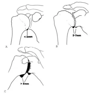
Fig. 1 - Samilson and Prieto classification:12 A) Mild arthrosis – lower osteophyte of the humeral head and/or glenoid smaller than 3 mm; B) Moderate arthrosis – lower osteophyte of the humeral head and/or glenoid measuring 3 to 7 mm, with gentle irregularity of the joint surface; C) Advanced arthrosis – lower osteophyte of the humeral head and/or glenoid larger than 7 mm, reduction of the joint space and bone sclerosis.
Introduction
Osteoarthritis of the glenohumeral joint is not uncommon and may affect more than 20% of the elderly population. Its therapeutic management begins with conservative methods, with the aims of alleviating painful symptoms and improving range of motion. Lifestyle changes, analgesic and anti-inflammatory medication, physiotherapy, joint infiltrations with corticoids and viscosupplementation have been mentioned in the literature.1-5
When conservative methods fail, total arthroplasty or hemiarthroplasty provide significant relief of painful symptoms and functional improvement, particularly in more elderly populations (over the age of 60 years). However, in younger populations (under the age of 50 years) that are active, these procedures do not present the same results, due mainly to the high functional demands made by this age group, their functional expectations and the length of survival of the implants, especially the glenoid component.1,4,6,7
Among patients with this profile, arthroscopic management may provide relief for painful symptoms and functional improvements. However, it is incapable of restoring joint cartilage that presents lesions.8,9 The arthroscopic procedures
of lavage and debridement provide satisfactory short-term results for 70 to 88% of these patients.8-10
The aim of the present study was to evaluate the results from videoarthroscopic treatment among patients with glenohumeral osteoarthritis.
Methodology
A retrospective survey was conducted among the patients with conditions of glenohumeral osteoarthritis (primary or secondary) who underwent operations arthroscopically, performed by the orthopedic shoulder group of Belo Horizonte between November 1999 and May 2009, with a minimum follow-up of two years. We identified 65 patients and 70 shoulders operated. Five patients (seven shoulders) were excluded from the study due to death; three patients were excluded because their cases evolved to arthroplasty; 18 patients (20 shoulders) were excluded because they could not be contacted; and two patients (two shoulders) were excluded because they refused to supply data for the investigation. Thirteen patients (14 shoulders) were unable to come for a physical examination and were interviewed by means of the telephone.
Out of the 37 patients (38 shoulders), 23 were male and 14 were female, with a mean age of 58.3 years (range: 33 to 80 years). The mean length of follow up was 5.13 years (range: 2 to 11 years). There were 28 operations on right shoulders and 10 on left shoulders; 26 cases involved the dominant arm and 12 were on the non-dominant arm. The initial mean range of motion was: 143.5° of active anterior elevation (EAA), 155° of passive anterior elevation (EAP), 50.13° of external rotation with the arm beside the body (RL I), 72.3° of external rotation
with the arm abducted at 90° (RL II) and internal rotation (RM) with a mean limitation of five vertebral levels. Out of the total, 33 shoulders presented associated diseases: 22 rotator cuff injuries, 10 Bankart lesions, three Slap lesions and one case dysplasia of the proximal humerus.
The patients selected were evaluated before and after the operation. The preoperative evaluations were done by reviewing the medical files and the initial radiographs, and the following data were gathered: age, gender, dominance, side affected, ranges of motion (EAA, EAP, ER I, ER II and IR), radiographic evaluation of the joint space (in the true anteroposterior and simple axillary lateral views),11
radiographic classification according to Samilson and Prieto12
(Fig. 1), function evaluation using the University of California
at Los Angeles (UCLA) score and presence of associated lesions.
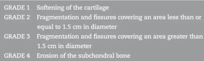
We reviewed the operative records of each patient, thus obtaining the classification of the chondral lesion as described by Outerbridge13 (Fig. 2), and the surgical procedures
years of follow-up with those with less than five years of follow-up. We also evaluated the influence of the length of follow-up on the functional results, in comparing the groups of chondral lesions (mild to moderate versus advanced), size of osteophyte (Samilson 1 and 2 versus Samilson 3) and joint space (preserved versus reduced), since a difference in length of follow-up between the groups compared might directly influence the results.
In relation to the associated lesions, we identified two groups of patients: one with rotator cuff injuries and the other with instability (Bankart or Slap). Two patients (two shoulders) who presented both instability and rotator cuff injury in association were taken to belong to the group of rotator cuff injuries.
Among the patients who said that they were dissatisfied in the postoperative subjective UCLA evaluation, we analy- zed which preoperative factors (degree of chondral degeneration, Samilson classification stage and joint space) contributed towards that level of satisfaction, along with the influence of the length of follow-up.
The statistical analysis was done using the resources of the PASW statistical software, version 18. The results were described in terms of descriptive measurements for quantitative variables and frequency tables for the qualitative variables analyzed.
The significance of the chondral degeneration, Samilson and Prieto stage, joint space, length of follow-up and resection of the osteophyte among the patients with Samilson and Prieto stage 3, in relation to the functional result, along with the influence of the length of follow-up on the patients level of satisfaction, was evaluated using nonparametric Mann-Whitney tests.
The differences between the preoperative and postoperative ranges of motion and UCLA scores were assessed using the nonparametric Wilcoxon test.
Contingency tables were used to correlate the patients’ degree of satisfaction with the preoperative factors of chondral degeneration, Samilson and Prieto stage and joint space. Fisher’s chi-square test was used to investigate the statistical significance of associations between these variables.
To compare the means of the Constant and UCLA variables of the group of patients with associated pathological conditions present (rotator cuff injury or instability) with those of the patients in the full sample, the t test for one sample was used. In this study, the specific value to be tested was the calculation of the general mean of 24 patients for the Constant and UCLA variables.
In all the statistical tests used, the significance level was taken to be 5%. Thus, associations were considered to be statistically significant if the p value was less than 0.05.
Results
Among the 24 patients (24 shoulders) on whom the functional results were analyzed, the preoperative mean UCLA score was 16 and the postoperative score was 28, with a significant difference between them (p = 0,000) (Fig. 3). The mean postoperative Constant score was 71.8.
GRADE 1 Softening of the cartilage
GRADE 2 Fragmentation and fissures covering an area less than or equal to 1.5 cm in diameter
GRADE 3 Fragmentation and fissures covering an area greater than 1.5 cm in diameter
GRADE 4 Erosion of the subchondral bone
Fig. 2 - Outerbridge classification13 developed for patellar
chondromalacia.
were not assessed separately, since these were performed on limited numbers of patients (four and three, respectively).
The postoperative evaluations were made by two independent examiners who had not participated in the surgical procedures. Twenty-four patients came for the examination (24 shoulders), at which they underwent assessments of range of motion (EAA, EAP, ER I and ER II by means of a goniometer and IR by means of the difference in vertebral level achieved, between the operated and contralateral sides) and the length of postoperative follow-up, with radiographic evaluation (preservation or non-preservation of the joint space and the Samilson and Prieto classification) and functional evaluation by means of the Constant and UCLA scores. The patients were also asked whether they would go through the same surgical procedure again if necessary.
The 13 patients (14 shoulders) who were unable to come for the physical examination were interviewed over the telephone and were evaluated regarding their current degree of satisfaction (satisfied or dissatisfied, in accordance with the UCLA score) and whether they would go through the same surgical procedure again. One patient in this group had undergone treatment in both shoulders: we considered evaluating each shoulder separately, since we judged that the subjective evaluation on one would not influence that of the other.
The preoperative and postoperative ranges of motion and functional evaluations were compared.
We investigated the influence of the degree of chondral degeneration, the size of the humeral or glenoid osteophyte (in accordance with the Samilson and Prieto classification), degree of preservation of the preoperative joint space, the length of postoperative follow-up and the presence of rotator cuff injuries or instability associated with glenohumeral osteoarthritis in postoperative functional assessments (Constant and UCLA).
We divided the occurrences of chondral degeneration into two groups: one with Outerbridge grades 1, 2 and 3 (mild to moderate chondral degenerations) and the other with Outerbridge grade 4 (advanced chondral degenerations). In relation to the size of the osteophyte, one group included patients with osteophytes smaller than 8 mm (mild and moderate arthrosis; Samilson and Prieto stages 1 and 2) and the other group included patients with osteophytes larger than or equal to 8 mm (advanced arthrosis; Samilson and Prieto stage 3). Among the patients in the second group (osteophytes larger than 8 mm), the influence of resection of the osteophyte on the postoperative functional result was analyzed.
the patients with advanced degeneration, 4.53 years; this difference was not significant (p = 0.402) (Fig. 4).
Thirteen of the 24 patients presented reduced joint space (less than 2 mm) and 11, preserved joint space in the preoperative radiographic evaluation. In the preoperative functional evaluation, the UCLA of the first group was 15 and of the second group, 21, without any significant difference between these values (p = 0.081). After the operation, the patients with reduced joint space presented UCLA of 26
These 24 patients presented the following mean preoperative ranges of motion: active anterior elevation = 160°, passive anterior elevation = 160°, external rotation with arm beside the body = 50°, external rotation with the arm abducted at 90° = 70° and medial rotation limited to six vertebral levels. The postoperative values were as follows: active anterior elevation =155°, passive anterior elevation = 160°, external rotation with arm beside body = 45°, external rotation with arm abducted at 90° = 78° and internal rotation limited to three vertebral levels. With the exception of the internal rotation (p = 0.043), there were no difference between the preoperative and postoperative range of motion measurements (Table 1).
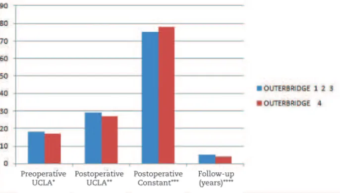
Among the 24 patients, 12 presented mild to moderate chondral degeneration (Outerbridge 1, 2 and 3) and the other 12 presented advanced chondral degeneration (Outerbridge 4). In the preoperative functional evaluation, the UCLA score of the first group was 18.5 and of the second group, 17, and there was no significant difference between these values (p = 0.706). After the operation, the patients with mild to moderate abnormalities presented UCLA of 29.5 and Constant of 75. The patients with advanced abnormalities presented UCLA of 27 and Constant of 78. The differences between the two groups were not significant (p = 0.367 and p = 0.862). The mean length of follow-up for the patients with mild to moderate degeneration was five years and for Fig. 3 - Difference in UCLA score between pre and
postoperative evaluations. *p < 0.05.
EAA EAP ER I ER II IR (NV)
Preoperative 160° (38.098) 160° (31.021) 50° (21.110) 70° (27.544) 6 (2.64)
Postoperative 155° (36.235) 160° (23.175) 45° (29.167) 78° (28.746) 3.5 (3.09)
p = 0.455 p = 0.836 p = 0.178 p = 0.454 p = 0.043
The values in parentheses are the standard deviations of the means for each range of motion. The p values in bold indicate significant differences. EAA: active anterior elevation; EAP: passive anterior elevation; ER I: external rotation with arm beside body; ER II: external rotation with arm at 90° of abduction; IR: internal rotation; NV: difference in vertebral levels.
Table 1 - Pre and postoperative ranges of motion.
Fig. 4 - Influence of the degree of chondral degeneration on the functional results. *p = 0.706; **p = 0.367; ***p = 0.862; ****p = 0.402.
and Constant of 79, while the patients with preserved joint space presented UCLA of 31 and Constant of 74. The differences between the two groups were not significant (p = 0.155 and p = 0.663). The mean length of follow-up among the patients with reduced joint space was 3.8 years and it was five years among the patients with preserved joint space. This difference was not significant (p = 0.522) (Fig. 5).
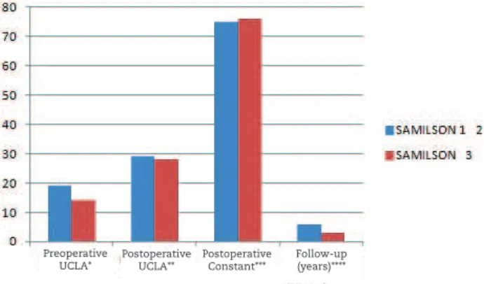
Among the 24 patients, 12 presented lower osteophytes smaller than 8 mm (Samilson and Prieto stages 1 and 2), while the other 12 had osteophytes larger than or equal to 8 mm (Samilson and Prieto stage 3). In the preoperative functional evaluation, the UCLA of the first
Preoperative Postoperative
Preoperative UCLA*
Postoperative Constant***
Follow-up (years)**** Postoperative
Fig. 5 - Influence of the joint space on the functional results from the preoperative radiographic evaluation. *p = 0.081;
**p = 0.153; ***p = 0.663;****p = 0.523. Fig. 6 - Influence of the stage of the Samilson classification on the functional results. *p = 0.582; **p = 0.727;
***p = 0.772;****p = 0.236.
Out of the 38 shoulders included in this study (24 shoulders among the patients examined and 14 shoulders among the 13 patients interviewed over the telephone), 32 (84%) presented satisfactory results in the subjective assessment. Also in this evaluation, there were no significant differences regarding the degree of chondral degeneration (p = 0.645), Samilson classification stage (p = 1.000) or length of follow-up (p = 0.542) between the shoulders with satisfactory and unsatisfactory results (Tables 4 and 5). The mean length of follow-up among the shoulders with satisfactory results in the subjective assessment was 4.75 years and among the unsatisfactory ones, 5.63 years, without any significant difference (p = 0.542). There was an association between unsatisfactory results in the subjective assessment and the presence of reduced preoperative joint space (p = 0.024) (Table 6).
Among the 37 patients included in the study, 33 (89%) would go through the surgical procedure again.
Fig. 7 - Influence of the resection of osteophytes on the functional results of patients with Samilson stage 3. *p = 0.798; **p = 0.730; ***p = 0.864.
group was 19.5 and of the second group, 14.5, without a significant difference between these values (p = 0.582). After the operation, the patients with osteophytes smaller than 8 mm presented UCLA of 29.5 and Constant of 75.5, while the patients with osteophytes larger than or equal to 8 mm had UCLA of 28 and Constant of 76.5. The differences between the two groups were not significant (p = 0.727 and p = 0.772). The mean length of follow-up among the patients with osteophytes smaller than 8 mm was 5.7 years and, among the patients with osteophytes larger than 8 mm, 3.3 years. This difference was not significant (p = 0.236) (Fig. 6).
Among the 12 patients with osteophytes larger than or equal to 8 mm (Samilson and Prieto stage 3), eight underwent resection of the osteophyte and four did not undergo this procedure. The preoperative UCLA scores (15.5 and 15.5), postoperative UCLA scores (25 and 26.5) and postoperative Constant scores (69 and 70.5) did not present any significant differences (p = 0.798, 0.730 and 0.864) (Fig. 7).
Among the 24 patients, 13 presented lengths of follow-up less than or equal to five years and 11 presented follow-ups longer than five years. The preoperative UCLA scores (15 and 15), postoperative UCLA scores (28 and 28) and postoperative Constant scores (77 and 74) did not present any significant differences (p = 0.931, 0.907 and 0.642).
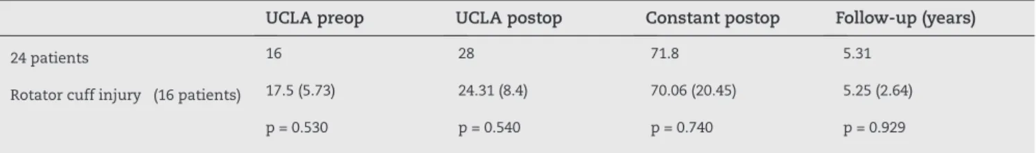
For these 24 patients, the mean preoperative UCLA and postoperative Constant were respectively, 16.6, 25.6 and 71.9; the mean length of follow-up among this population was 5.3 years. Of these patients, 16 presented rotator cuff injuries associated with osteoarthritis and had preoperative UCLA = 17.5, postoperative UCLA = 24.3 and postoperative Constant = 70.6; the mean length of follow-up among these patients was 5.25 years. There was no significant difference between these values and those of the compete group (p = 0.503, 0.540, 0.740 and 0.929) (Table 2).
The five patients in whom the associated pathological condition was instability presented preoperative UCLA = 14.8, postoperative UCLA = 25 and postoperative Constant = 74.4. The mean length of follow-up among these patients was 4.84 years. There was no significant difference in these values in comparison with the complete group (p = 0.403, 0.860, 0.647 and 0.413) (Table 3).
Preserved joint space
Reduced joint space
Osteophyte not resected
Osteophyte resected
Preoperative UCLA*
Preoperative UCLA*
Preoperative UCLA* Postoperative
UCLA**
Postoperative UCLA**
Postoperative UCLA** Postoperative
Constant***
Postoperative Constant***
Postoperative Constant*** Follow-up
(years)****
Table 5 - Association between degree of satisfaction and the preoperative Samilson stage (Fisher chi-square test).
Table 6 – Association between degree of satisfaction and the preoperative joint space.
Satisfaction
Samilson preop
Total p-value
1,2 3
Dissatisfied
Cases 3 3 6 1,000
% 50.0% 50.0% 100.0%
Satisfied Cases 9 11 20
% 45.0% 55.0% 100.0%
Total Cases 12 14 26
% 46.2% 53.8% 100.0%
Satisfaction
Preop joint space
Total p-value
Preserved Reduced
Dissatisfied
Cases 0 6 6 0.024
% 0% 100.0% 100.0%
Satisfied Cases 11 9 20
% 55.0% 45.0% 100.0%
Total Cases 11 15 26
% 42.3% 57.7% 100.0%
UCLA preop UCLA postop Constant postop Follow-up (years)
24 patients 16 28 71.8 5.31
Rotator cuff injury (16 patients) 17.5 (5.73) 24.31 (8.4) 70.06 (20.45) 5.25 (2.64)
p = 0.530 p = 0.540 p = 0.740 p = 0.929
The values in parentheses are the standard deviations of the means for each value.
Table 2 - Functional results among the patients with rotator cuff injury and in the complete group (t test).
UCLA preop UCLA postop Constant postop Follow-up (years)
24 patients 16 28 71.8 5.31
Instability
(5 patients) 14.8 (7.05) 25 (7.52) 74.4 (11.8) 4.84 (1.15)
p = 0.603 p = 0.860 p = 0.647 p = 0.413
The values in parentheses are the standard deviations of the means for each value.
Table 3 - Functional results among the patients with instability and in the complete group (t test).
Table 4 - Association between degree of satisfaction and degree of chondral degeneration (Fisher chi-square test).
Satisfaction
Outerbridge
Total p-value
1,2,3 4
Dissatisfied
Cases 2 4 6 0.645
% 33.3% 66.7% 100.00%
Satisfied Cases 11 9 20
% 55.0% 45.0% 100.0%
Total Cases 13 13 26
Discussion
Glenohumeral osteoarthritis may result in significant functional incapacity. From the patient’s perspective, the impact from this pathological condition is comparable to that of chronic comorbidities such as congestive heart failure, diabetes and coronary diseases.1
Clinically, such patients present with pain, which may interfere with their nighttime rest; and also with overall loss of range of motion with occasional blockade, which may be due to free intra-articular bodies.2 Pain at the extremities
of movements may result from impact syndrome, whereas pain in the middle of the range, particularly below shoulder level is associated with mechanical symptoms.14
In physical examinations, the symptoms of chondral lesions may resemble those of other intra-articular or extra-articular diseases, such as subacromial impact, tenosynovitis of the biceps and labral lesions.3,14,15 On
inspection, muscle hypotrophy and bone prominences are searched for, and the scapular-thoracic rhythm is assessed. The range of motion, both passive and active, generally presents limitations.3,14,15 Ellman described a
compression-rotation test that helps to differentiate chondral lesions from impact syndrome: a internal and external rotation maneuver with the arm beside the body, at the same time as performing compression of the humeral head in the direction of the glenoid, which is done before and after bursal infiltration using lidocaine. The symptoms that are alleviated on the second occasion are related to impact syndrome.14
In radiographic evaluations, glenohumeral osteoarthritis is classically characterized by asymmetrical reduction of the joint space, subchondral sclerosis, cyst formation and osteophyte formation (in the humeral head or glenoid). Free bodies can be seen inside the joint.2 Samilson and
Prieto developed a classification system that was originally described for degenerative joint alterations resulting from arthropathy due to instability, which today is applied to arthrosis of other etiologies. The classification takes into consideration the size of the osteophyte, whether it is located inferiorly on the humeral head or on the glenoid, and any presence of irregularities on the joint surface, observed in anteroposterior radiographic view of the glenohumeral joint. The arthrosis is mild if the osteophyte is smaller than 3 mm, moderate if between 3 and 7 mm, in association with mild irregularity of the joint surface, and severe if greater than 7 mm, in association with diminished joint space and bone sclerosis.12 The size of the osteophyte
is correlated negatively with range of motion.16
In arthroscopic evaluations, chondral lesions are classified in accordance with the system proposed by Outerbridge.13 Grade 1 represents softening of the cartilage.
Grade 2 presents fragmentation and fissures covering an area less than or equal to 1.5 cm in diameter. Grade 3 presents fragmentation and fissures covering an area greater than 1.5 cm in diameter. Grade 4 presents erosion of the subchondral bone. These chondral lesions can be found in 5% to 17% of routine arthroscopic evaluations.3,4
Total arthroplasty or hemiarthroplasty provides significant pain relief and functional improvement, especially in more elderly populations (over the age of 60 years).1,4,6,7 On the other hand, Sperling et al.17
observed that among patients under the age of 50 years who underwent hemiarthroplasty or total arthroplasty of the shoulder, the rate of unsatisfactory results was approximately 56%, thus suggesting that for this group of patients, another therapeutic approach should be used.
Several studies in the literature have demonstrated good results from arthroscopic approaches for treating glenohumeral osteoarthritis. Ogilvie-Harris and Wiley15
conducted a retrospective analysis on 439 patients who underwent arthroscopic shoulder surgery and found 54 cases of glenohumeral osteoarthritis. Of these, 29 presented associated diseases. These patients underwent removal of the arthroscopic debris and chondral fragments, and synovectomy. Satisfactory results were achieved in two-thirds of the patients with slight degenerative alterations (superficial lesions in the joint cartilage) and on one-third of the patients with severe degenerative alterations (with exposure of the subchondral bone).
Ellman et al.14 reported on a group of 18 patients with
degenerative joint diseases the clinically resembled impact syndrome. Among these 18, ten came to a diagnosis of glenohumeral osteoarthritis even before the operation. During the operation, no cases of complete rotator cuff injury were found, but partial joint lesions were found in five shoulders (three A1 and two A3). The arthroscopic procedures consisted of debridement of the unstable cartilaginous fragments, removal of free bodies and partial synovectomy, Subacromial decompression was performed in 15 patients. Among the 18 patients, ten presented a minimum duration of symptom relief greater than six months. Richards and Burkart18 presented preliminary
results from arthroscopic debridement associated with release of the rotator interval and capsulotomy, for treating glenohumeral osteoarthritis. In addition to pain reduction, there were increases in anterior elevation, external rotation and internal rotation. The alleviation of the painful symptoms was due to elimination of the joint debris and diminution of the joint contact pressure.
Weinstein et al.10 followed up 25 patients for 12
months, who had undergone arthroscopic debridement of glenohumeral osteoarthritis. Nine of these patients presented an associated disease. The procedures for treating the osteoarthritis consisted of arthroscopic lavage, debridement of labral and cartilaginous lesions, removal of free bodies, partial synovectomy and resection of the osteophyte, in addition to treatment for the associated diseases. At the end of the follow-up, it was observed that 8% of the results were excellent, 72% good and 20% unsatisfactory. There was no statistical correlation between good results and the degree of radiographic alterations and degenerative joint alterations. Pain was the most important factor in evaluating the patients.
Cameron et al.9 retrospectively analyzed 61 patients
patients were divided according to the location of the lesion (humeral, glenoid or bipolar) and the size of the osteochondral defect (greater than or less than 2 cm2). The
indication for capsulotomy was a restriction of more than 15° in any plane of the range of motion. Improvements in painful symptoms were observed in 88% of the patients, based on a visual analogue pain scale and on the increase in the score of the American Shoulder and Elbow Surgeons
(ASES). Among the patients, 87% stated that they would undergo this surgical procedure again, if necessary. The location and size of the lesions did not have any influence on the improvements in pain and functional scores.
Kerr and McCarty19 analyzed 19 patients (20 shoulders)
who underwent arthroscopic debridement to treat glenohumeral osteoarthritis. No difference in functional results was found between the patients with mild-to-moderate degenerative alterations (Outerbridge 2 and 3) and those with advances degenerative alterations (Outerbridge 4). However, patients with unipolar impairment presented better results than did those with bipolar impairment.
Van Thi el et al .2 0 f ol lowed u p 71 patient s w ith
glenohumeral osteoarthritis who underwent arthroscopic debridement. Of these, 22% evolved to arthroplastic procedures after a mean of 10 months of follow-up, w h i l e 7 8 % c o n t i nu e d w i t h o u t a r t h ro p l a s t y ove r a follow-up of 27 months. The group of patients who did not evolve to arthroplasty presented larger joint spaces and lower stages in the Samilson classification from preoperative radiographs and, at the end of the follow-up, better functional results and fewer painful symptoms. In this group of patients, 87% said that they would undergo this procedure again.
In our series of patients, we obtained a significant difference in UCLA scores from before to after the operation (p = 0.000), and this was concordant with previous studies in relation to functional improvement. The mean postoperative Constant score was 71.8, which was considered satisfactory. We did not find any relationship between the functional results and the degree of chondral degeneration (p = 0.367 and 0.862 for the postoperative UCLA and Constant scores), and this was concordant with what had previously been reported by Weinstein et al.,10
Kerr and McCarty19 and Cameron et al.9 The reduction in
joint space also did not influence the functional results (p = 0.153 and 0.663 for the postoperative UCLA and Constant scores), thus resembling the findings of Van Thiel et al.20 There was a tendency (p = 0.081) for the preoperative
UCLA to be greater in the patients with preserved joint space. We did not find any correlation between the Samilson classification stages (osteophyte size) and the functional results (p = 0.727 for the postoperative UCLA and Constant scores), which was concordant with the reports of Weinstein et al.10 Although the radiographic classification
used in that study had been drawn up at their own clinic, it resembled the Samilson classification with regard to progression of the osteophyte. On the other hand, Van Thiel et al.20 presented better functional results among patients
with lower Samilson stages in the preoperative radiographic evaluation. Among our patients with osteophytes larger
than 8 mm, there was no influence on the functional results caused by resecting the osteophyte (p = 0.730 and 0.864 for the postoperative UCLA and Constant scores). Neither Van Thiel et al.20 nor Weinstein et al.10 mentioned any influence
from resecting the osteophytes on their results.
Our sample presented a mean follow-up of 5.13 years, with a range from 2 to 11 years. There was no difference in the functional results between the group of patients with less than five years of follow-up and those with more than five years of follow-up (p = 0.907 and 0.642 for the postoperative UCLA and Constant scores), thus suggesting that the improvement in functional results could be long-lasting. The length of follow-up also did not interfere with the functional evaluation when we took into account the degree of chondral degeneration, Samilson classification stage or joint space. In relation to the length of follow-up, our study differs from the remainder of the literature, in which the length of follow-up was a maximum of two years.20
We found high incidence of rotator cuff injuries and instability associated with glenohumeral arthrosis. An association between glenohumeral arthrosis and both intra and extra-articular disease had already been mentioned in the studies by Ogilvie-Harris and Wiley15 and Ellman et al.14
Although our sample was of limited size, we did not find any influence from these diseases on the functional result (postoperative UCLA and Constant, with p = 0.540 and 0,740 in patients with rotator cuff injuries, and p = 0.860 and 0.647 in patients with associated instability). In relation to the influence of rotator cuff lesions on treatments for glenohumeral osteoarthritis, Wirth et al.21 observed that small lesions,
independent of whether they had been repaired concomitantly with the arthroplastic procedure, did not interfere with the final result from hemiarthroplasty. Iannotti and Norris22
analyzed the influence of preoperative factors on the results from shoulder arthroplasty for treating glenohumeral arthrosis, and found that small repairable rotator cuff injuries that were limited to the supraspinatus did not affect the postoperative score of the American Shoulder and Elbow Surgeons (ASES). In our series of patients, all the associated rotator cuff lesions were successfully repaired and, although the treatment type was different, the results were concordant with what had been proposed by Iannotti and Norris22 and Wirth et al.,21 regarding
the presence of repairable lesions of the rotator cuff associated with glenohumeral osteoarthritis.
Millett and Gaskill23 presented their preliminary results.
presented painful limitation of the range of motion, along with pain on the posterior face of the shoulder, thus suggesting axillary nerve compression. After arthroscopic debridement and complete resection of the osteophyte (Fig. 9), this patient evolved with improvement of the range of motion and painful symptoms. Differently to what was proposed by Millet, we did not do any intraoperative controls using fluoroscopy. At the end of the surgical procedure, radiography was performed in true anteroposterior view, in order to verify the resection of the lower osteophyte. We also did not perform additional decompression of the axillary nerve, and resection of the osteophyte was sufficient for improving the compressive symptoms. After five years of follow-up, the patient is satisfied with the procedure that was performed, with few painful symptoms and the following range of motion: EAA = 170°, ER I = 30°, ER II = 70° and IR = 5th lumbar vertebra (Fig. 10).
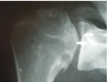
Fig. 8 - A) Radiograph in true AP view on the right shoulder, showing large lower osteophyte in the humeral head, preservation of the joint space and presence of metal anchors from previous surgery on the glenoid; B) NMR T2 image with fat suppression – presence of large lower osteophyte and subchondral cysts in the humeral head; tendon of supraspinatus preserved in its insertion.
Fig. 9 - Radiograph in true AP view on the right shoulder. Postoperative control demonstration complete resection of the lower osteophyte of the humeral head and preservation of the joint space.
Fig. 10 - After five years of follow-up, the patient still presented good range of motion; A) External rotation I, B) External rotation II and C) Internal rotation.
R E F E R E N C E S
1. Denard PJ, Wirth MA, Orfaly RM. Management of
glenohumeral arthritis in the young adult. J Bone Joint Surg Am. 2011;93(9):885-92.
2. Chong PY, Srikumaran U, Kuye IO, Warner JJP. Glenohumeral arthritis in the young patient. J Shoulder Elbow Sur. 2011;20(2 Suppl):S30-40.
3. Boselli KJ, Ahmad CS, Levine WN. Treatment of glenohumeral arthrosis. Am J Sports Med. 2010;38(12):2558-72.
4. McCarty LP 3rd., Cole BJ. Nonarthroplasty treatment of glenohumeral cartilage lesions. Arthroscopy. 2005;21(9):1131-48. 5. Strauss EJ, Hart JA, Miller MD, Altman RD, Rosen JE.
Hyaluronic acid viscosupplementation and osteoarthritis: current uses and future directions. Am J Sports Med. 2009;37(8):1636-44.
6. Savoie FH 3rd., Brislin KJ, Argo D. Arthroscopic glenoid resurfacing as a surgical treatment for glenohumeral arthritis in the young patient: midterm results. Arthroscopy. 2009;25(8):864-71.
7. Millet PJ, Huffard BH, Horan MP, Hawkins RJ, Steadman JR. Outcomes of full-thickness articular cartilage injures of the shoulder treated with microfrature. Arthroscopy. 2009;25(2):856-63.
8. Cole BJ, Yanke A, Provencher MT. Nonarthroplasty alternatives for the treatment of glenohumeral arthrititis. J Shoulder Elbow Surg. 2007;16(5 Suppl):S231-40.
9. Cameron BD, Galatz LM, Ramsey ML, Williams GR, Iannotti JP. Non-prosthetic management of grade IV ostechondral lesions of the glenohumeral joint. J Shoulder Elbow Surg. 2002;11(1):25-32.
10. Weinstein DM, Bucchieri JS, Pollock RG, Flatow EL, Bigliani LU. Arthroscopic debridement of the shoulder for osteoarthritis. Arthroscopy. 2000;16(5):471-6.
11. Rockwood CA, Jensen KL. Avaliação radiográfica dos problemas do ombro. In: Rockwood e Matsen. Ombro. 2ª ed. Rio de Janeiro: Revinter; 2002. p. 200-2.
12. Samilson RL, Prieto V. Dislocation arthropathy of the shoulder. J Bone Joint Surg Am. 1983;65(4):456-60. 13. Outerbridge RE. The etiology of chondromalacia patellae. J
Bone Joint Surg Br. 1961;43B(4):752-7.
14. Ellman H, Harris E, Kay SP. Early degenerative joint disease simulating impingement syndrome: arthroscopic findings. Arthroscopy. 1992;8(4):482-7.
15. Ogilvie-Harris DJ, Wiley AM. Arthroscopic surgery of the shoulder. J Bone Joint Surg Br. 1986;68(2):201-7.
16. Kircher J, Murhard M, Magosch P, Ebinger N, Lichtender S, Habermeyer P. How much are radiological parameters related to clinical symptoms and function in osteoarthritis of the shoulder? Int Orthop. 2010;34:677-81.
17. Sperling JW, Cofield RH, Rowland CH. Minimum fifteenyears follow-up of Neer hemiarthroplasty and total shoulder arthroplasty in patients aged fifty years or younger. J Shoulder Elbow Surg. 2004; 13(6):604-13.
18. Richards DP, Burkart SS. Arthroscopic debridement and capsular release for glenoumeral osteoarthritis. Arthroscopy. 2007;23(9):1019-22.
19. Kerr BJ, McCarty EC. Outcomes of arthroscopic debridement is worse for patients with glenohumeral arthritis of both sides of the joint. Clin Orthop Relat Res. 2008;466(8):634-8. 20. Van Thiel GS, Sheehan S, Frank RM, Slabaugh M, Cole BJ,
Nicholson GP, et al. Retrospective analysis of arthroscopic management of glenohumeral degenerative disease. Arthroscopy. 2010;26(110):1451-5.
1.000) or length of follow-up (p = 0.542). On the other hand, there was a significant association between shoulders with unsatisfactory results from the subjective assessment and reduced joint space in the preoperative radiographic evaluation (p = 0.024). Although this association was from a subjective assessment, it followed the trend of the results of Van Thiel et al.,20 in which the patients with preserved
joint space before the operation presented better functional evaluations and fewer painful symptoms at the end of the follow-up. In the same way, these authors reported that 87% of the patients would go through the same surgical procedure again, which did not differ from our results, in which 89% would go through the procedure again. This is directly related to the patients’ level of satisfaction.
In our sample, we had a significant loss of patients from the follow-up. Out of the 65, five died for reasons unrelated to the surgical procedure, 18 could not be found because of changes of address and two refused both to come for the examination and to undergo subjective assessment over the telephone. Three patients evolved to total arthroplasty within two years after arthroplastic debridement and were therefore excluded from the data analysis. Out of the 37 patients (38 shoulders) that remained, 13 (14 shoulders) were unable to come for a physical examination (eight of them were living in other cities, which made it impossible to come for the examination). Among these patients, the assessment was made by means of telephone contact.
Functional results from 24 patients were analyzed. We did not find any significant difference in the functional results when we took into account the degree of chondral degeneration, size of the osteophyte, preservation of the joint space, length of postoperative follow-up and presence of rotator cuff injuries. This may have been due to the limited number of patients, which might have interfered with the statistical analysis.
Although the postoperative Constant score presented a mean of 71.8, which was considered satisfactory, we did not have a preoperative value for evaluating the functional gain and for adding value to the functional gain obtained through the UCLA score. Other limitations of our study included the retrospective study model and the lack of a control group. Future studies using a prospective model, with a control group and with fewer losses from the follow-up are needed in order to consolidate our results.
Conclusion
Arthroscopic management of the arthrotic shoulder provided improvement of the functional results and high satisfaction levels. The reduced joint space in the preoperative radiographic assessment negatively influenced the satisfaction level in the final evaluation.
Conflicts of interest
21. Wirth MA, Tapscott RS, Southworth C, Rockwood Jr CH. Treatment of glenohumeral arthritis with a hemiarthroplasty: minimum five-year follow-up outcome study. J Bone Joint Surg Am. 2006;88(5):964-73.
22. Iannotti JP, Norris TR. Influence of preoperative factor on outcome of shoulder arthroplasty for glenohumeral osteoarthritis. J Shoulder Elbow Surg. 2003;85-A(2):251-8.