17 artigo 292
CAsE REPoRT
The authors declare that they did not have any conflict of interests in producing this article
1 – Resident in the Orthopedics Department - Hospital de Santo António - Porto - Portugal.
2 – Attending Physician in the Orthopedics Department - Hospital de Santo António - Porto - Portugal.
3 – Hospital Attending Physician specialist in Orthopedics - Orthopedics Department - Hospital de Santo António - Porto - Portugal. Work performed at the Orthopedics Service, Hospital de Santo António, Hospital Center of Porto, Porto, Portugal.
Correspondence: Serviço de Ortopedia do Hospital de Santo António - Centro Hospitalar do Porto - Largo do Professor Abel Salazar - 4099-001 - Porto, Portugal E-mail: [email protected]
Work received for publication: February 2, 2010; accepted for publication: August 23, 2010.
HybRId ANklE PROSTHESIS IN A cASE OF POST-TRAumATIc
AvASculAR NEcROSIS OF THE TAluS
Ricardo Jorge Gomes de Sousa1, Ricardo Pedro Ferreira Rodrigues Pinto1, Marta Maria Teixeira de Oliveira Massada1, Manuel Alexandre Negrais Pinho Gonçalves Pereira1, José Muras Geada2, Isabel Maria Gonçalves Costa3
Rev Bras Ortop. 2011;46(1):94-6
INTRODUCTION
Fractures of the body and neck of the talus are infre-quent lesions but have the potential to produce functio-nal sequelae over the long term, for which prevention is not always possible. The prevalence of post-traumatic arthrosis is between 35 and 100%, and both the ankle and the subtalar joint are affected(1-3). In a series that we
followed, ankle arthrosis was present in 40% of the cases after five years(4).
Avascular necrosis is a characteristic complica-tion after fractures of the body and neck of the talus and it results in interruption of the blood irrigation coming from the sinus and tarsal tunnel at the talar neck. Its prevalence is related to the initial degree of deviation(5).
The development of total ankle arthroplasty started in the 1970s(6). The original models were cemented
and highly constrictive, and so they sometimes failed.
ABSTRACT
Talus fractures often lead to late post-traumatic ar-throsis. In such cases, the use of latest generation, ce-mentless prostheses has been hindered by the presence of avascular necrosis. We report the case of a 65-ye-ar-old patient who presented four years after a talus neck fracture. He had painful ankle arthrosis (AOFAS ankle-hindfoot score 19) and avascular necrosis, with
collapse of the entire talar dome. Given the extent of the necrosis, it was decided to cement the talus pros-thetic component. One year after the surgery, the pa-tient shows good clinical and radiological results (AO-FAS ankle-hindfoot score 87) and is satisfied with the procedure. We are not aware of any similar reports in the literature.
Keywords – Ankle Joint; Talus; Osteonecrosis; Cementation
New generations were developed and, today, the ankle prostheses most commonly used share the common characteristics that all of them have a porous coating so as to favor bone interdigitation: not depending on cementation for fixation but, rather, on osseointegra-tion(6). Thus, avascular necrosis of the talus has
clas-sically been considered to be a contraindication for performing total ankle arthroplasty(7).
We report on the result, after one year of follow-up, from implantation of a total ankle prosthesis in a pa-tient with avascular necrosis and collapse of the entire talar dome. The prosthesis used was the Salto Total Ankle Prosthesis (Tornier, France), which was im-planted using cementation to fix the talar component.
CASE REPORT
rigi-95
Figure 2 – Preoperative radiographs of the left ankle.
HYBRID ANKLE PROSTHESIS IN A CASE OF POST-TRAUMATIC AVASCULAR NECROSIS OF THE TALUS
Rev Bras Ortop. 2011;46(1):94-6
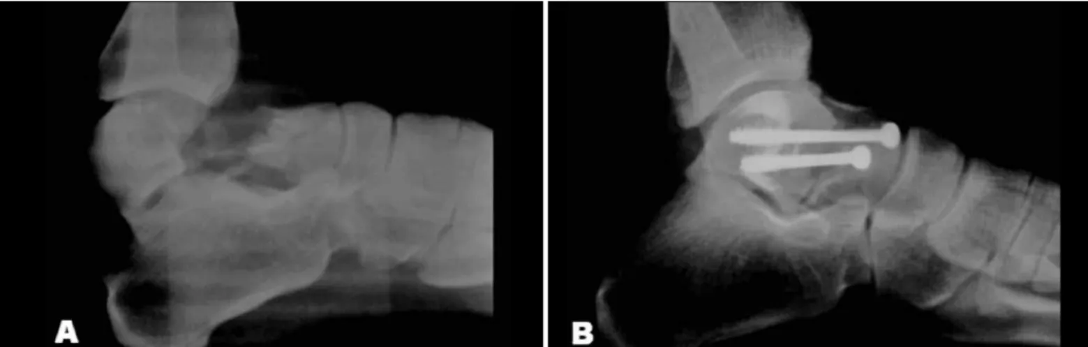
Figure 1 – A) Initial fracture of the talar neck, of Hawkins type II; B) Control in immediate postoperative period.
dity of the left ankle that had obliged him to use two forearm crutches over the last two years. He had the antecedent of an aviation accident four years earlier that resulted in a Hawkins type II fracture of the talar neck. That injury was treated surgically with open reduction and osteosynthesis, using two cancellous bone screws (6.5) by means of an anteromedial ap-proach. Figure 1 shows the initial radiograph and the postoperative control.
The clinical assessment revealed that the flexion--extension range of motion of the ankle was limited, with an extremely low score of 19 on the hindfoot and ankle scale of the American Orthopedic Foot and Ankle Society (AOFAS). The radiographic evaluation revealed signs of post-traumatic arthrosis of the left ankle and almost 100% collapse of the talar dome, secondary to avascular necrosis (Figure 2). Despite the radiographically visible degenerative abnormali-ties in the subtalar joint, the patient did not have any pain in inversion-eversion movements of the hindfoot.
Ankle arthroplasty was performed through an extensile anterior approach, in accordance with the original technique for the implant chosen. This con-sisted of a custom-made talar component prepared for cementation. A cutting guide for the talar dome was used to determine the cutting level, and we decided not to extract the lower cancellous bone screw, since this would not interfere with the positioning of the implant. Cement was used to fill in the bone defects, as well as to achieve fixation of the talar component. Both the surgical procedure and the postoperative pe-riod were free from intercurrences.
At the most recent follow-up consultation (12
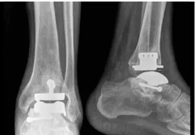
months after the operation), the patient presented an excellent clinical and radiological result. Currently, he is able to walk practically without pain, and without external supports, and he is extremely satisfied with the surgery. Radiological evaluations do not show any migration of components or signs of loosening, as can be seen from Figure 3. The score on the AOFAS hindfoot and ankle scale improved significantly and is now 87 (Table 1).
DISCUSSION
A high proportion of fractures of the body and neck of the talus lead to late post-traumatic arthrosis, which can cause significant functional disturbance(1-4). After
the initial disappointments from the initial designs, to-tal ankle arthroplasty has resurged recently as a valid alternative to arthrodesis for treating ankle arthrosis(7).
96
Figure 3 – Radiographs after 12 months of follow-up.
Table 1 – Clinical evolution observed after the treatment that was implemented.
Before the
operation After the operation
AOFAS 19 87
Pain Severe, almost always present occasionalSlight and
sagittal range of motion
(flexion and extension) 12o 33o
REFERÊNCIAs
1. Lindvall E, Haidukewych G, DiPasquale T, Herscovici D Jr, Sanders R. Open reduction and stable fixation of isolated, displaced talar neck and body fractures. J Bone Joint Surg Am. 2004;86(10):2229-34.
2. Vallier HA, Nork SE, Barei DP, Benirschke SK, Sangeorzan BJ. Talar neck frac-tures: results and outcomes. J Bone Joint Surg Am. 2004;86(8):1616-24. 3. Schulze W, Richter J, Russe O, Ingelfinger P, Muhr G. Surgical treatment of talus
fractures: a retrospective study of 80 cases followed for 1-15 years. Acta Orthop Scand. 2002 ;73(3):344-51.
4. Sousa R, Massada M, Pereira MA, Costa I, Costa e Castro J. Sequelas a longo prazo de fracturas do corpo e colo do astrágalo. Rev Bras Ortop. 2009;44(5):432-6. 5. Rammelt S, Zwipp H. Talar neck and body fractures. Injury. 2009;40(2):120-35.
6. Cracchiolo A 3rd, Deorio JK. Design features of current total ankle replacements: implants and instrumentation. J Am Acad Orthop Surg. 2008;16(9):530-40. 7. Hintermann B. Total ankle arthroplasty – historical overview, current concepts and
future perspectives. Wien-Austria: Springer-Werlag; 2005.
8. Bonnin M, Judet T, Colombier JA, Buscayret F, Graveleau N, Piriou P. Midterm re-sults of the Salto total ankle prosthesis. Clin Orthop Relat Res. 2004;(424):6-18. 9. Lee KB, Cho SG, Jung ST, Kim MS. Total ankle arthroplasty following revascu-larization of avascular necrosis of the talar body: two case reports and literature review. Foot Ankle Int. 2008;29(8):852-8.
10. Bullens P, de Waal Malefijt M, Louwerens JW. Conversion of failed ankle arthro-plasty to an arthrodesis. Technique using an arthrodesis nail and a cage filled with morsellized bone graft. Foot Ankle Surg. 2010 ;16(2):101-4.
Rev Bras Ortop. 2011;46(1):94-6
implant(8). Its porous surface, which is appropriate
for interdigitation, is a fundamental trait of all “latest generation” prosthesis models”(6). This characteristic
is one of the main reasons why avascular necrosis of the talus has been considered to be a contraindication. The potential for bone integration and uncemented fixation of the component is extremely low in areas of bone necrosis(9). Hintermann(7) even went as far
as stating that avascular necrosis affecting more than 25% of the body of the talus constitutes a relative contraindication, while necrosis affecting more than 50% constitutes an absolute contraindication for per-forming total ankle arthroplasty.
In order to surmount this difficulty, we decided to use bone cement to achieve fixation of the talar component. In addition, we used cement to fill in any bone defects in the talus that we found during the surgery. We decided not to extract the second screw, since it did not interfere with the cut that was made to correctly position the implant. In this way, we avoided further weakening the talus.
One year after the procedure, both the patient and the medical team are satisfied with the result achie-ved. Future complications, particularly loosening and/ or “sinking ” of the component/cement combination in relation to the remains of the talus or even the calcaneus cannot be ruled out. We are aware of the risks involved in this heterodox choice and they were properly explained to and discussed with the patient. Arthrodesis of the ankle was presented as an alterna-tive procedure, or as a rescue procedure in the event of failure of the arthroplasty(10). The patient chose to