Letters to the Editor
Radiol Bras. 2017 Mai/Jun;50(3):199–208
207
MRI, the fat seen within the lesion produces a hyperintense
sig-nal on T1-weighted images and sigsig-nal loss in fat-saturated
se-quences
(3,5,7). In rare cases, the presentation of MCT is atypical,
which can be a diagnostic challenge for radiologists
(2,6). Multiple
small floating spheres within a large cyst, as observed in the case
presented here, is one of those rare presentations, known as the
“floating ball” presentation
(4,6). Histologically, the spheres are
composed of keratin, fibrin, hemosiderin, sebaceous debris, hair,
and fat, in variable proportions
(2,6,13). Although the mechanism
of formation of these spheres has yet to be clarified, it is
specu-lated that it involves aggregation of sebaceous material around a
nidus
(2,4,14). The mobility of the spheres is due to their low density
relative to the other content of the cyst
(2,4,6). A finding of multiple
floating spheres within a single large cyst has not been reported
for other types of tumors and is therefore considered
pathogno-monic of MCT
(2,4,6,14–16).
REFERENCES
1. Rha SE, Byun JY, Jung SE, et al. Atypical CT and MRI manifestations of mature ovarian cystic teratomas. AJR Am J Roentgenol. 2004;183: 743–50.
2. Tandon A, Agarwal R, Tandon R, et al. Multiple intracystic floating balls: an unusual but unique sonographic pattern of mature cystic ter-atoma. BMJ Case Rep. 2011;2011:bcr0320113962.
3. Park SB, Kim JK, Kim KR, et al. Imaging findings of complications and unusual manifestations of ovarian teratomas. Radiographics. 2008;28: 969–83.
4. Gürel H, Gürel SA. Ovarian cystic teratoma with a pathognomonic ap-pearance of multiple floating balls: a case report and investigation of common characteristics of the cases in the literature. Fertil Steril. 2008;90:2008.e17–9.
5. Outwater EK, Siegelman ES, Hunt JL. Ovarian teratomas: tumor types and imaging characteristics. Radiographics. 2001;21:475–90. 6. Tongsong T, Wanapirak C, Khunamornpong S, et al. Numerous
intra-cystic floating balls as a sonographic feature of benign intra-cystic teratoma: report of 5 cases. J Ultrasound Med. 2006;25:1587–91.
7. Heo SH, Kim JW, Shin SS, et al. Review of ovarian tumors in children and adolescents: radiologic-pathologic correlation. Radiographics. 2014; 34:2039–55.
8. Salvadori PS, Bomfim LN, von Atzingen AC, et al. Spontaneous rupture of ovarian cystadenocarcinoma: pre- and post-rupture computed tomog-raphy evaluation. Radiol Bras. 2015:330–2.
9. Lima LLA, Parente RCM, Maestá I, et al. Clinical and radiological cor-relations in patients with gestational trophoblastic disease. Radiol Bras. 2016;49:241–50.
10. Guerra LFA, Pessanha LB, Oliveira GA, et al. Endometrial osseous metaplasia: sonographic, radiological and histopathological findings. Radiol Bras. 2016;49:62–3.
11. Queiroz RM, Costa PP, Oliveira NYF, et al. Female urethral diverticu-lum containing a urothelial carcinoma. Radiol Bras. 2016;49:406–7. 12. Manikkavasakar S, Ramachandram A, Ramalho M, et al. Malignant uterine disease with concurrent miometrial contraction at MRI: a pos-sible source of overstaging. Radiol Bras. 2016;49:342–3.
13. Donnadieu AC, Deffieux X, Le Ray C, et al. Unusual fast-growing ova-rian cystic teratoma during pregnancy presenting with intracystic fat “floating balls” appearance. Fertil Steril. 2006;86:1758–9. 14. Altinbas SK, Yalvac S, Kandemir O, et al. An unusual growth of ovarian
cystic teratoma with multiple floating balls during pregnancy: a case report. J Clin Ultrasound. 2010;38:325–7.
15. Rao JR, Shah Z, Patwardhan V, et al. Ovarian cystic teratoma: deter-mined phenotypic response of keratocytes and uncommon intracystic floating balls appearance on sonography and computed tomography. J Ultrasound Med. 2002;21:687–91.
16. Kawamoto S, Sato K, Matsumoto H, et al. Multiple mobile spherules in mature cystic teratoma of the ovary. AJR Am J Roentgenol. 2001;176: 1455–7.
Ana Paula Barroso Pazinatto Espindola1, Viviane
Brandão Amorim2, Hilton Augusto Koch1, Paulo Roberto
Valle Bahia2, Márcio V. P. Almeida2
1. Pontifícia Universidade Católica do Rio de Janeiro (PUC-Rio), Rio de Janeiro, RJ, Brazil. 2. Centro Estadual de Diagnóstico por Imagem do Rio de Janeiro (CEDI), Rio de Janeiro, RJ, Brazil. Mailing address: Dra. Ana Paula Barroso Pazinatto Espindola. Rua Vinícius de Moraes, 71, Ipanema. Rio de Janeiro, RJ, Brazil, 22411-010. E-mail: apazinatto@ yahoo.com.br.
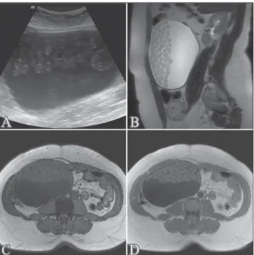
Figure 1. Ultrasound (A); sagittal T2-weighted MRI sequence (B); out-of-phase T1-weighted gradient-echo MRI sequence (C); and in-phase T1-weighted gradi-ent-echo MRI sequence (D). Note the expansile cystic lesion with heterogeneous content, containing numerous oval formations that were hyperechoic on the ultra-sound and showed intermediate signal intensity in the T1- and T2-weighted se-quences, with no evidence of signal loss in the out-of-phase T1-weighted gradi-ent-echo sequence.
Self-limiting thoracic aortic dissection during bronchial artery embolization
Dear Editor,
A 75-year-old woman presented with a 3-week history of
in-termittent hemoptysis related to a history of recurrent episodes of
pneumonia. Chest computed tomography (CT) showed
cylindri-cal bronchiectasis in the lingula, and bronchoscopy showed clots
in the left bronchial tree. Bronchial arteriography was requested
and revealed a shunt (Figure 1A) between the left bronchial
ar-tery and the left pulmonary arar-tery. During manual-injection
digi-tal subtraction angiography, enhancement and stagnation of the
contrast media were observed in a false lumen of the descending
thoracic aorta (Figures 1B and 1C), consistent with iatrogenic
aorta dissection. The iatrogenic aortic dissection extended to the
left bronchial artery, leading to obstruction of blood flow to the
shunt. However, there were no signs of hemodynamic instability,
and the patient therefore received conservative therapy with
clini-cal and radiologiclini-cal monitoring. A second CT scan, obtained 7
days later, showed that the iatrogenic aorta dissection was stable
Letters to the Editor
Radiol Bras. 2017 Mai/Jun;50(3):199–208
208
(Figure 1D), and a third scan, obtained 5 months later, showed
total resolution. During 7 months of follow-up, the patient reported
no pain or new episodes of bleeding.
During endovascular procedures, iatrogenic aortic dissection
can occur when the tip of the catheter is pushed into the vessel
wall during catheterization, as well as when high-pressure jets of
contrast media are directed toward the vessel wall. Although
un-common, iatrogenic aortic dissection accounts for approximately
5% of all thoracic aortic dissections
(1,2). In a review article, Ittrich
et al.
(3)showed rates of subintimal short segment dissection of
the aorta during bronchial arteriography ranging from 1% to
6.3%
(4,5), although there are virtually no images of such
dissec-tions in the literature. There is no standard for the management
of iatrogenic thoracic aorta dissection. Uncomplicated dissection
of the descending thoracic aorta is a relatively benign process, and
complete spontaneous resolution is observed in most cases.
Phar-macological treatment to control pain and blood pressure is
rec-ommended, as is short-term follow-up with CT
(6–8).
REFERENCES
1. Nienaber CA, Fattori R, Mehta RH, et al. Gender-related differences in acute aortic dissection. Circulation. 2004;109:3014–21.
2. Januzzi JL, Sabatine MS, Eagle KA, et al. Iatrogenic aortic dissection. Am J Cardiol. 2002;89:623–6.
3. Ittrich H, Klose H, Adam G. Radiologic management of haemoptysis:
D
Figure 1.A: Left bronchial arteriography showing a shunt (arrowheads) between the left bronchial artery (LBA) and the left pulmonary artery (LPA). B,C: Stagnation of the contrast media (arrowheads) can be seen at the false lumen of the descending thoracic aorta, indicating dissection. D: Coronal CT reconstruction at 7 days after bronchial arteriography showing persistence of the contrast media in the false lumen of the thoracic aorta (arrows), with no increase in the extent of the dissection.
Rafael Dahmer Rocha1, Joaquim Maurício da
Motta-Leal-Filho1, Francisco Leonardo Galastri1, Breno Boueri
Affonso1, Humberto Bogossian1, Felipe Nasser1
1. Department of Interventional Radiology and Pulmonology, Hospital Israelita Albert Einstein, São Paulo, SP, Brazil. Mailing address: Dr. Rafael Dahmer Rocha. Avenida Albert Einstein, 627/701, Jardim Leonor. São Paulo, SP, Brazil, 05652-900. E-mail: [email protected]. diagnostic and interventional bronchial arterial embolisation. Rofo. 2015; 187:248–59.
4. Uflacker R, Kaemmerer A, Picon PD, et al. Bronchial artery emboliza-tion in the management of hemoptysis: technical aspects and long-term results. Radiology. 1985;157:637–44.
5. Mal H, Rullon I, Mellot F, et al. Immediate and long-term results of bronchial artery embolization for life-threatening hemoptysis. Chest. 1999;115:996–1001.
6. Erbel R, Aboyans V, Boileau C, et al. 2014 ESC Guidelines on the diag-nosis and treatment of aortic diseases: document covering acute and chronic aortic diseases of the thoracic and abdominal aorta of the adult. The Task Force for the Diagnosis and Treatment of Aortic Diseases of the European Society of Cardiology (ESC). Eur Heart J. 2014;35:2873– 926.
7. LeMaire SA, Russell L. Epidemiology of thoracic aortic dissection. Nat Rev Cardiol. 2011;8:103–13.
8. Nienaber CA, Divchev D, Palisch H, et al. Early and late management of type B aortic dissection. Heart. 2014;100:1491–7.