Letters to the Editor
Radiol Bras. 2017 Mai/Jun;50(3):199–208
206
aorta (aortomesenteric compression)(1–5). In rare cases, the LRV is retroaortic. In such cases, compression occurring between the aorta and the spine is known as posterior NCS(2–4). The nutcracker phenomenon corresponds to these findings without clinical cor-relation(2–4). The prevalence of NCS is unknown, although it is known that it occurs predominantly in healthy, thin individuals between 20 and 40 years of age and in women(1–4). Clinically, hematuria is the most common finding, followed by pain on the left side, dyspareunia, dysmenorrhea, dysuria, varicoceles, and pelvic varices(1–5). In exceptionally rare cases, anatomic variations in the pancreas compress nearby vessels, including the LRV(2,6,7).
Renal vein thrombosis (RVT) is common in nephrotic syn-drome and in severely hypotensive neonates. Other causes: trau-mas, surgery, infections, neoplasias, vasculitis, venous compres-sions, contraceptives and myeloproliferative diseases. It’s infrequent in healthy adults, predominantly unilaterally(8,9). The clinical pre-sentation of RVT is much like that of NCS, with the added fea-tures of an acute increase in renal volume, late atrophy, and pro-gressive deterioration of renal function, as well as the complica-tion of pulmonary thromboembolism in up to 50% of cases(5,8,9). The pathophysiology of thromboses encompasses Virchow’s triad: endothelial lesions, stasis, and hypercoagulability. Gener-ally, thrombotic events involve at least two factors, although one may be sufficient(5,8,9).
One of the principal methods employed in the diagnosis of NCS is Doppler ultrasound, which is noninvasive and can be used in determining venous caliber and flow, the latter being sugges-tive of NCS when it exceeds 100 cm/s, with a sensitivity and speci-ficity of 78% and 100%, respectively, for the diagnosis(1–4). It shows high sensitivity in the investigation of RVT(8). Ultrasound, however, is operator-dependent and may not detect small thromboses(8,9). For the diagnosis of NCS and RVT, angiography has a sensitivity of 66.7–100% and a specificity of 55.6–100%(8). It is able to evalu-ate the aortomesenteric angle (compression); possible compres-sion and dilation of the LRV; filling defects; endoluminal blood clots; and signs of chronic thrombosis, such as thickening of the vessel walls and calcifications(1–4,9). However, it uses radiation and potentially nephrotoxic contrast agents(8,9). Retrograde venogra-phy is the gold standard examination in NCS(1–4) and RVT(8); it shows pressure gradients greater than 3 mmHg in the LRV, in
addition to the filling defects that represent thrombi(1–4,8). How-ever, it is invasive, potentially triggering thrombosis, and uses in-travenous iodine(8).
The therapeutic options are conservative treatment, reim-plantation/transposition of the LRV, the use of an external or in-ternal stent, renal autotransplantation, gonadocaval bypass, and nephrectomy(1–5). If RVT occurs, anticoagulation and thromboly-sis can also be employed(8–10).
REFERENCES
1. Eliahou R, Sosna J, Bloom AI. Between a rock and a hard place: clinical and imaging features of vascular compression syndromes. Radiographics. 2012;32:E33–49.
2. Lamba R, Tanner D, Sekhon S, et al. Multidetector CT of vascular compression syndromes in the abdomen and pelvis. Radiographics. 2014;34:93–115.
3. Butros SR, Liu R, Oliveira GR, et al. Venous compression syndromes: clinical features, imaging findings and management. Br J Radiol. 2013; 86:20130284.
4. Fong JK, Poh AC, Tan AG, et al. Imaging findings and clinical fea-tures of abdominal vascular compression syndromes. AJR Am J Roentgenol. 2014;203:29–36.
5. Mallat F, Hmida W, Jaidane M, et al. Nutcracker syndrome compli-cated by left renal vein thrombosis. Case Rep Urol. 2013;2013:168057. 6. Yun SJ, Nam DH, Ryu JK, et al. The roles of the liver and pancreas in
renal nutcracker syndrome. Eur J Radiol. 2014;83:1765–70. 7. Chauhan R, Roy TS, Chaudhury A, et al. Variant human pancreas:
aberrant uncinate process or an extended mesenteric process. Pancreas. 2003;27:267–9.
8. Asghar M, Ahmed K, Shah SS, et al. Renal vein thrombosis. Eur J Vasc Endovasc Surg. 2007;34:217–23.
9. Wang Y, Chen S, Wang W, et al. Renal vein thrombosis mimicking urinary calculus: a dilemma of diagnosis. BMC Urol. 2015;15:61. 10. Yoshida RA, Yoshida WB, Costa RF, et al. Nutcracker syndrome and
deep venous thrombosis in a patient with duplicated inferior vena cava. J Vasc Surg Venous Lymphat Disord. 2016;4:231–5.
Rodolfo Mendes Queiroz1, Daniel de Paula Garcia1, Mauro José Brandão da Costa1, Eduardo Miguel Febronio1
1. Documenta – Hospital São Francisco, Ribeirão Preto, SP, Brazil. Mailing address: Dr. Rodolfo Mendes Queiroz. Documenta – Centro Avançado de Diagnóstico por Imagem. Rua Bernardino de Campos, 980, Centro. Ribei-rão Preto, SP, Brazil, 14015-0130. E-mail: [email protected].
http://dx.doi.org/10.1590/0100-3984.2015.0192
Atypical presentation of mature cystic teratoma (“floating balls”)
Dear Editor,
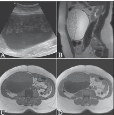
A 43-year-old female patient with no known diseases sought medical attention complaining of increased abdominal volume. The patient underwent ultrasound and subsequent magnetic reso-nance imaging (MRI) of the pelvis (Figure 1), which showed an expansile cystic lesion, with heterogeneous content, measuring 16.0 × 16.0 × 10.0 cm and containing numerous oval formations of various sizes. The lesion was hyperechoic on ultrasound and mobile upon a change in patient position. The oval formations showed intermediate signal intensity on T1- and T2-weighted MRI scans, with no evidence of signal loss in fat-saturated se-quences or signal drop on an out-of-phase T1-weighted gradient-echo sequence. These imaging findings, although uncommon, are pathognomonic of mature cystic teratoma (MCT). The pa-tient underwent surgery, and the diagnosis was confirmed by his-topathological analysis of the surgical specimen.
Also known as a dermoid cyst, MCT is the most common benign ovarian tumor, accounting for 10–25% of cases in adult
patients and 50% of those in pediatric patients(1–3). MCTs are typically asymptomatic and slow-growing(1,3). They are usually seen in women of reproductive age and are rarely diagnosed be-fore puberty. Its growth ceases at menopause(4–7). An MCT typi-cally contains well-differentiated tissues of the three germ lay-ers(1,5): the ectoderm, (derived from the skin and neural tissues); the mesoderm (osteomuscular and adipose tissues); and the en-doderm (ciliated and mucinous epithelium). The diversity of tis-sues in teratomas results in a wide variety of characteristics in imaging studies.
In most cases, pelvic tumors do not present imaging features that are considered diagnostic(8–12). However, MCTs often present typical imaging features, which facilitate the diagnosis. Among such features, one of the most common is that of a fatty tumor(3). In such cases, the most common ultrasound finding is that of a cystic mass with an echogenic tubercle (a Rokitansky nodule), presenting posterior acoustic shadowing secondary to calcifica-tions, strands of hair, or foci of fat(3,5,7).
Letters to the Editor
Radiol Bras. 2017 Mai/Jun;50(3):199–208
207
MRI, the fat seen within the lesion produces a hyperintense sig-nal on T1-weighted images and sigsig-nal loss in fat-saturated se-quences(3,5,7). In rare cases, the presentation of MCT is atypical, which can be a diagnostic challenge for radiologists(2,6). Multiple small floating spheres within a large cyst, as observed in the case presented here, is one of those rare presentations, known as the “floating ball” presentation(4,6). Histologically, the spheres are composed of keratin, fibrin, hemosiderin, sebaceous debris, hair, and fat, in variable proportions(2,6,13). Although the mechanism of formation of these spheres has yet to be clarified, it is specu-lated that it involves aggregation of sebaceous material around a nidus(2,4,14). The mobility of the spheres is due to their low density relative to the other content of the cyst(2,4,6). A finding of multiple floating spheres within a single large cyst has not been reported for other types of tumors and is therefore considered pathogno-monic of MCT(2,4,6,14–16).
REFERENCES
1. Rha SE, Byun JY, Jung SE, et al. Atypical CT and MRI manifestations of mature ovarian cystic teratomas. AJR Am J Roentgenol. 2004;183: 743–50.
2. Tandon A, Agarwal R, Tandon R, et al. Multiple intracystic floating balls: an unusual but unique sonographic pattern of mature cystic ter-atoma. BMJ Case Rep. 2011;2011:bcr0320113962.
3. Park SB, Kim JK, Kim KR, et al. Imaging findings of complications and unusual manifestations of ovarian teratomas. Radiographics. 2008;28: 969–83.
4. Gürel H, Gürel SA. Ovarian cystic teratoma with a pathognomonic ap-pearance of multiple floating balls: a case report and investigation of common characteristics of the cases in the literature. Fertil Steril. 2008;90:2008.e17–9.
5. Outwater EK, Siegelman ES, Hunt JL. Ovarian teratomas: tumor types and imaging characteristics. Radiographics. 2001;21:475–90. 6. Tongsong T, Wanapirak C, Khunamornpong S, et al. Numerous
intra-cystic floating balls as a sonographic feature of benign intra-cystic teratoma: report of 5 cases. J Ultrasound Med. 2006;25:1587–91.
7. Heo SH, Kim JW, Shin SS, et al. Review of ovarian tumors in children and adolescents: radiologic-pathologic correlation. Radiographics. 2014; 34:2039–55.
8. Salvadori PS, Bomfim LN, von Atzingen AC, et al. Spontaneous rupture of ovarian cystadenocarcinoma: pre- and post-rupture computed tomog-raphy evaluation. Radiol Bras. 2015:330–2.
9. Lima LLA, Parente RCM, Maestá I, et al. Clinical and radiological cor-relations in patients with gestational trophoblastic disease. Radiol Bras. 2016;49:241–50.
10. Guerra LFA, Pessanha LB, Oliveira GA, et al. Endometrial osseous metaplasia: sonographic, radiological and histopathological findings. Radiol Bras. 2016;49:62–3.
11. Queiroz RM, Costa PP, Oliveira NYF, et al. Female urethral diverticu-lum containing a urothelial carcinoma. Radiol Bras. 2016;49:406–7. 12. Manikkavasakar S, Ramachandram A, Ramalho M, et al. Malignant uterine disease with concurrent miometrial contraction at MRI: a pos-sible source of overstaging. Radiol Bras. 2016;49:342–3.
13. Donnadieu AC, Deffieux X, Le Ray C, et al. Unusual fast-growing ova-rian cystic teratoma during pregnancy presenting with intracystic fat “floating balls” appearance. Fertil Steril. 2006;86:1758–9. 14. Altinbas SK, Yalvac S, Kandemir O, et al. An unusual growth of ovarian
cystic teratoma with multiple floating balls during pregnancy: a case report. J Clin Ultrasound. 2010;38:325–7.
15. Rao JR, Shah Z, Patwardhan V, et al. Ovarian cystic teratoma: deter-mined phenotypic response of keratocytes and uncommon intracystic floating balls appearance on sonography and computed tomography. J Ultrasound Med. 2002;21:687–91.
16. Kawamoto S, Sato K, Matsumoto H, et al. Multiple mobile spherules in mature cystic teratoma of the ovary. AJR Am J Roentgenol. 2001;176: 1455–7.
Ana Paula Barroso Pazinatto Espindola1, Viviane Brandão Amorim2, Hilton Augusto Koch1, Paulo Roberto Valle Bahia2, Márcio V. P. Almeida2
1. Pontifícia Universidade Católica do Rio de Janeiro (PUC-Rio), Rio de Janeiro, RJ, Brazil. 2. Centro Estadual de Diagnóstico por Imagem do Rio de Janeiro (CEDI), Rio de Janeiro, RJ, Brazil. Mailing address: Dra. Ana Paula Barroso Pazinatto Espindola. Rua Vinícius de Moraes, 71, Ipanema. Rio de Janeiro, RJ, Brazil, 22411-010. E-mail: apazinatto@ yahoo.com.br.
Figure 1. Ultrasound (A); sagittal T2-weighted MRI sequence (B); out-of-phase T1-weighted gradient-echo MRI sequence (C); and in-phase T1-weighted gradi-ent-echo MRI sequence (D). Note the expansile cystic lesion with heterogeneous content, containing numerous oval formations that were hyperechoic on the ultra-sound and showed intermediate signal intensity in the T1- and T2-weighted se-quences, with no evidence of signal loss in the out-of-phase T1-weighted gradi-ent-echo sequence.
Self-limiting thoracic aortic dissection during bronchial artery embolization
Dear Editor,
A 75-year-old woman presented with a 3-week history of in-termittent hemoptysis related to a history of recurrent episodes of pneumonia. Chest computed tomography (CT) showed cylindri-cal bronchiectasis in the lingula, and bronchoscopy showed clots in the left bronchial tree. Bronchial arteriography was requested and revealed a shunt (Figure 1A) between the left bronchial