ISSN 1557-4989
© 2011 Science Publications
Corresponding Author: Martin Esqueda, Department of Food Research and Development, A.C. 0.6 Km Highway to La Victoria, 83304 Hermosillo, Sonora, Mexico Fax: +52 (662) 280-0422
Genetic Variability in Rhizoctonia solani Isolated from Vitis vinifera
Based on
Amplified Fragment Length Polymorphism
1
Amparo Meza-Moller,
1Martin Esqueda,
2Felipe Sanchez-Teyer,
1
Georgina Vargas-Rosales,
1Alfonso A. Gardea
and
1Martin Tiznado-Hernandez
1
Center for Food Research and Development, A.C.,
0.6 Km Highway to La Victoria, 83304 Hermosillo, Sonora,
2
Center for Scientific Research of Yucatan, A.C.,
Chuburna de Hidalgo Colony, 97200 Merida, Yucatan, Mexico
Abstract: Problem statement:Rhizoctonia solani is a potential grapevine pathogen. In order to develop effective methods of control, it is necessary to document its genetic diversity. Approach: The objective of this study was to analyze the genetic variability of R. solani isolated from the rhizosphere of ungrafted V. vinifera var. perlette seedless planted in Sonora, Mexico using Amplified Fragment Length Polymorphism (AFLP). Results: In the selective amplification using eight primer combinations we obtained a total of 446 AFLP markers with a 100% polymorphism. Out of 41 isolates, 36 different AFLP patterns were observed and five were replicates of the same pattern. The dendrogram shows inter- and intrapopulation similarity indexes of 0.26, 0.98 and 0.31, 0.98, respectively. Six groups emerged from the principal components analysis, five of which were clearly defined, while the other one was spread out. Conclusion: We conclude that R. solani growing in Sonoran vineyards shows a high degree of genetic variability, even under similar environmental conditions.
Key words: Rhizoctonia solani, thanatephorus cucumeris, molecular markers, vineyards, genetic variability, amplified fragment length polymorphism, grapevine pathogen, perlette seedless, similarity indexes, principal components, components analysis, sonoran vineyards
INTRODUCTION
As a pathogen, Rhizoctonia solani J.G. Kühn has a significant economic impact in the development and production of a wide variety of crops. It is considered a complex species because its physiological and pathogenic variability. Rarely it produces sexual structures (Teleomorph: Thanatephorus cucumeris A.B. Frank (Donk), thus its taxonomic classification is still unresolved (Gonzalez-Hernandez, 2002). This is the reason that the hyphal anastomosis reaction has been used to group relatively homogeneous groups; the fusion of hyphae indicates that both belong to the same anastomosis group. To date, 13 Anastomosis Groups (AGs) have been recognized (Tsror, 2010). Even though this classification system is one of the most widely used, it has been observed that some isolates occasionally fuse with others from a different AG, or else their lose their ability to fuse. Furthermore, the variability in the degree of hyphal fusion, morphology, pathogenicity and host range observed in AGs (AG-1, AG-13) has made it necessary to define intraspecific
groups, therefore reducing the usefulness of hyphal anastomosis for R. solani classification.
Genetic variability within and between AGs can be achieved by biochemical and molecular characters such as isozymes, soluble proteins, fatty acids, genomic DNA, variability in fragment length resulting from the enzyme decomposition of DNA using specific endonucleases (Restriction Fragment Length Polymorphism: RFLP), random amplification of variable length fragments (Random Amplification Of Polymorphic DNA: RAPDs), DNA sequences, Amplified Fragment Length Polymorphism (AFLP) And Internal Transcribed Spacer Region (ITS) (Sharon et al., 2006).
318 formation of subgroups within the AGs; e) detecting differences in variability among isolates derived from vegetative propagation relative to those resulting from sexual reproduction (Toda et al., 2004; Sharma et al., 2005). These results support the genetic and taxonomic relationships of R. solani and suggest the need for a system of classification that includes both its physiological and molecular characteristics.
Established that R. solani forms part of the rhizosphere of V. vinifera var. Perlette Seedless in two of the three most important grape growing regions in the state of Sonora, where 75% of Mexico’s grapes are grown. Several strains analyzed in this study were previously screened for pathogenicity by inoculating in V. vinifera, which caused a root rot with lesions larger than 5 mm, which suggests that this fungus represents a potential risk at replanting sites.
The aim of this study was to analyze the genetic variability of R. solani isolated from the rhizosphere of ungrafted V. vinifera var. Perlette Seedless.
MATERIALS AND METHODS
Abiotic factors: Using thematic and digital maps (INEGI, 2000), climate, geology, edaphology, physiography and hydrology of the sampling sites were obtained. The physical and chemical properties of the soil were also analyzed for six samples made up of 10 subsamples per quadrat. Following the methodology, determining: pH, electrical conductivity, nitrate phosphate and potassium concentration, as well as organic matter content and texture.
Strain isolation and culture: Three vineyards in La Costa de Hermosillo three in Pesqueira, Sonora, Mexico were selected. In each vineyard, three 5 Ha plots were randomly selected sampling was done in a zig-zag pattern, taking soil samples from the rhizosphere of ungrafted Vitis vinifera var. Perlette Seedless, from five plants at each point with 20-30 points per quadrat at a depth of 15-30 cm. The soil was processed using the Weinhold’s wet sieving method (Sneh et al., 1991). The residues obtained were placed in Petri dishes with 2% agar-water medium and incubated at 25°C for 24 h. Mycelial growth was observed with a light microscope hyphal tips were selected from individuals with characteristics similar to those of Rhizoctonia and then inoculated on nutritious medium (yeast-glucose extract) to develop pure strains.
Nuclei number and hyphal diameter: Four 1 cm2 samples were taken from the periphery of the mycelial growth in each of the colonies incubated at 25°C for 24 h and these were stained with 0.5% trypan blue in lactophenol. In each strain, a total of 50 hyphae were observed under a light microscope (400×), their diameters were measured and their nuclei counted (Sneh et al., 1991).
Anastomosis reaction: All of the group representative isolates were paired among themselves and with a control group isolates to determine their anastomosis reaction in a Petri dish with 2% agar-water with an 8 mm disc of the control group in the center surrounded by the multinucleate strain in three 8 mm discs. These were incubated at 25°C for 12,18 h until the two strains of hyphae had overlapped. Part of the overlapped area was removed and placed on a slide to determine the number and type of compatibility reactions (C0, C1,C2, C3) observed in three visual fields (400×) (Carling, 1996; Martin, 2000). A positive anastomosis reaction was recorded if, for each pairing there were five fusion points or a fusion frequency greater than 15% (Carling, 1996; Meyer et al., 1998). The anastomosis groups used as controls-selected considering regional environmental conditions and existing reports of the groups that affect the fruit crops were AG1-1C (CSKa), AG-6, AG-4 isolated from chili pepper plants (Capsicum annuum) in Sayula, Jalisco.
Vegetative compatibility: Based on mycelia similarity between each group isolates and isolates from AG-4, vegetative similarities were determined. An 8 mm disc of the control group was placed in the center of a Petri dish with a yeast-glucose culture medium and surrounded by another three 8 mm discs of the multinucleate strain. The dishes were incubated at 25°C for 72 h until the colonies from both strains overlapped. The results were recorded as: (a) vegetatively compatible isolates, if there was no dividing line at the overlap margin of the colonies; (b) weak vegetative incompatibility, if the margins of the colonies were delimited; and (c) strong vegetative incompatibility, if aerial hyphae were growing in the overlapping area (Ceresini et al., 2002).
8.0, 1% N-lauroylsarcosine and 150 µL of Ottawa sand. 50 µL of buffer were add to the tube and mixed for 2 min. Extracted DNA was resuspended on 50 µ L of TE and the integrity was evaluated by electrophoresis in agarose gel and the concentration of total genomic DNA was quantified by reading absorption at 260 nm on a Beckman DU6 spectrophotometer.
AFLP development: Genetic fingerprint (AFLP) of the R. solani isolated from the rhizosphere from the field was obtained according with Vos et al. (1995). Briefly, 100 ng of genomic DNA was double-digested with 5 U of EcoRI and MseI. Specific adapters were ligated and the mixture of fragment was diluted 1:10 and preamplified using primers containing a single selective nucleotides (Eco-A and Mse-C). After that, the preamplified fragments were diluted 1: 40 and then selectively amplified with a duplex PCR using a single Mse1 primer with two IRDye-labelled EcoR1 primers containing two selective nucleotides. In total 8 primer combinations were apply as follow: M-CC and M-CG with E-(AT, AG); M-CA and M-CG with E-(AA, AC). The reaction mixture contain 2.0 µL of preamplified DNA, buffer 1X, 1.5 mM MgCl2, 0.2 mM dNTPs, 2.5
units of Taq Polimerase 30 ng of MseI primer and 15 ng of each labeled-EcoR1 primer (700 EcoR1+2 (IRDye) and 800 EcoR1+2 (IRDye), in a final volume of 11 µL. Amplified fragments were separated using high resolution acrylamide gel. For this, 5 µ L of loading buffer solution was added to the amplified sample and denatured for 3 min at 94°C, after which it was immediately placed on ice. Then 1.0 µL of the molecular weight marker (standard 50-700 bp) was add to the reaction and fragments were separated by electrophoresis during 3 h. The labeled-amplified fragments were automatically captured and recorded during electrophoresis using an automatic IR2 sequencer (LiCor, Lincoln, Nebraska).
Data analysis: To obtain the genetic Similarity Index (SI) a binary matrix was generated in which 1 was assigned to the presence and 0 to the absence of a given band Nei and Li (1979) formula was applied. To generate the dendrogram, the Unweighted Pair Group Method with Arithmetic mean (UPGMA) was used (Hintze, 2007). Once data were generated, a resampling analysis with 1000 repetitions was done to determine the support value. Principal components analysis was carried out based on the similarity index to establish groups of isolates using Statgraphics ver 15.2.14. For the comparison of pairs of isolates, individuals with a
similarity index > 0.95 were considered clones since this represents less than 10% polymorphism.
RESULTS
Forty-one R. solani strains were isolated: 32 From Pesqueira and 9 from La Costa de Hermosillo. Table 1 shows the origin of the different isolates of R. solani studied. The 41 isolates belongs to anastomosis group AG-4 given by the morphological characteristics, such as hyphal diameter (5.2-6.8 µm), number of nuclei (3-5) and color (off white to dark brown), previously recorded for AG-4. There was no congruence between anastomosis and vegetative compatibility, since the isolates with higher reaction anastomosis (C2, C3) had a strong vegetative incompatibility.
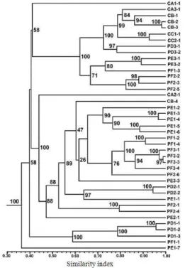
From the selective amplification using eight primer combinations in a duplex reactions, a total of 446 AFLP markers were obtained in a range of 50, 700 bp, of which 100% were polymorphic. Considering all of the 41 isolates analyzed, 36 differential AFLP patterns were identified and the following clones had a Similarity Index (SI) > 95%: CB-2 and CB-3; PD1-1 and PD1-2; PD3-1 and PD2-2; PE1-3 and PE1-4; PF3-2, PF3-3 and PF3-4. In every case, clones with the same band pattern came from the same vineyard (Table 1 and Fig. 1).
The similarity indexes for all the isolates ranged from 0.26,0.98, while isolates collected from the same field ranged from 0.31,0.98. According to the similarity dendrogram (Fig. 1) there are three major groups containing 98% of the isolates. In the first group there are two subgroups, one of which includes 7 of the 9 isolates from La Costa de Hermosillo, while the isolates from Pesqueira are distributed among the remaining groups and did not show a specific pattern with respect to their area of origin.
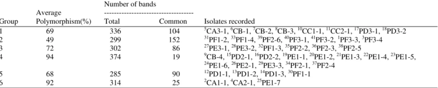
320 polymorphism (94%) was recorded for group 4 with 13 strains from both regions. 75% of the isolates of group 1 were from La Costa de Hermosillo and include all the isolates of vineyard C. In groups 2, 3 and 5 only groups from the Pesqueira region were found. All of those in group 2 were isolates from vineyard D; while those from groups 3 and 5 had organisms from vineyards E-F and D-E, respectively.
All localities considered on our sampling are characterized by very dry, very warm and warm subtype climate. The predominant soil in the three
Pesqueira vineyards is eutric regosol, while in La Costa de Hermosillo vineyards A, B C the soils are calcareous regosol, haplic xerosol luvic xerosol, respectively. The geology of all the vineyards goes back to the Quaternary period and is comprised of sedimentary rock. The nitrate content ranged from 20.2, 58.2 mg kg−1 and phosphates were 37.7, 98.7 mg kg−1. The pH was slightly alkaline, ranging from 7.2, 7.9. With the exception of vineyard F which had saline soil, all the vineyards had slightly saline soils between 2.7, 3.7 dS m−1. The texture was predominantly sandy with <2% humus Table 3.
Table 1: Sampling sites for isolates of Rhizoctonia solani from the regions of pesqueira and la costa de hermosillo in sonora, mexico
Region Vineyard Keya
La Costa de A CA1-1, CA2-1, CA3-1
Hermosillo B CB-1, CB-2, CB-3, CB-4
C CC1-1, CC2-1
Pesqueira D PD1-1, PD1-2, PD1-3, PD2-1, PD2-2, PD3-1, PD3-2
E PE1-1, PE1-2, PE1-3, PE1-4, PE1-5, PE1-6, PE1-7, PE2-1, PE3-1, PE3-2, PE3-3
F PF1-1, PF1-2, PF1-3, PF1-4, PF2-1, PF2-2, PF2-3, PF2-4, PF2-5, PF2-6, PF3-1, PF3-2, PF3-3, PF3-4 a: The first letter indicates vineyard location: Pesqueira (P) and La Costa de Hermosillo (C). Second letter indicates the vineyard (A-F). The first number indicates the plot in the vineyard and the last number indicates the sample replicate
Table 2: Description of the groups of Rhizoctonia solani obtained from the principal components analysis Number of bands
Average ---
Group Polymorphism(%) Total Common Isolates recorded
1 69 336 104 5CA3-1, 6CB-1, 7CB-2, 8CB-3, 10CC1-1, 11CC2-1, 17PD3-1, 18PD3-2
2 49 299 152 31PF1-2, 33PF1-4, 39PF2-6, 40PF3-1, 41PF3-2, 1PF3-3, 3PF3-4
3 72 302 86 27PE3-1, 28PE3-2, 32PF1-3, 35PF2-2, 36PF2-3, 38PF2-5
4 94 374 19 9CB-4, 15PD2-1, 16PD2-2, 19PE1-1, 20PE1-2, 21PE1-3, 22PE1-4, 23PE1-5,
24PE1-6, 26PE2-1, 29PE3-3, 34PF2-1, 37PF2-4
5 68 285 90 12PD1-1, 13PD1-2, 14PD1-3, 30PF1-1
6 92 314 25 2CA1-1, 4CA2-1, 25PE1-7
Superscript corresponds to the number assigned in Fig. 2
Table 3: Location, climate, edaphology, geology and soil chemistry of the vineyards in Sonora, Mexico
Vineyards in La Costa de Hermosillo Vineyards in Pesqueira
--- ---
A B C D E F
Latitude 28°57’42” 28°35’39” 28°55’31” 29°21’30” 29°20’47” 29°18’29”
Longitude 111°40’29” 111°35’13” 111°20’53” 110°54’25” 110°53’43” 110°55’25”
Climate BW(h’)hw(x’) BW(h’)hw(x’) BW(h’)hw(x’) BW(h’)hw(x’) BW(h’)hw(x’) BW(h’)hw(x’)
Edaphology Rc+Yh/1 Xh+Rc/1 Xl+Rc/2/s Re+Xh/1 Re+Xh/1 Re/1
Geology Q(al) Q(al) Q(al) Q(al) Q(al) Q(al)
NO3- (mg kg-1) d51.3a 20.2b 38.7ab 58.2a 56.0a 23.1 b
PO4≡(mg kg-1) 98.7a 53.4bc 82.2ab 55.2bc 37.7c 84.3ab
pH 7.9a 7.4a 7.7 a 7.5a 7.8a 7.2a
EC (dS m-1) 2.7a 3.2a 3.6 a 3.7a 3.5a 4.2a
Texture Ls Sl, Cl Sl Ls, Sl Ls, Sl Ls, Sl, S
Humus (%) 1.0bc 1.5ab 0.7 c 0. 9 bc 1.0bc 1.9a
CEC (cmol kg-1) 4.9b 12.7a 5.4 b 5.5b 5.6b 6.1ab
Fig. 1: Grouping of the AFLP phenotypes of Rhizoctonia solani based on similarity indexes Nodes numbers represent the resampling analysis support value. For each isolate, the first letter indicates vineyard location: Pesqueira (P) and La Costa de Hermosillo (C). Second letter indicates the vineyard (A-F). The first number indicates the plot in the vineyard and the last number indicates sample replicate
Fig. 2: Principal components analysis based on the similarity indexes of the AFLP patterns for Rhizoctonia solani. The numbers grouped together in the graph are: 1. isolate PF3-3…41. isolate PF3-2, described in Table 2. 68% of the variation was explained by the first two components
DISCUSSION
The 41 isolates had morphological characteristics, such as hyphal diameter, number of nuclei and color that have been recorded for anastomosis group AG-4 in previous studies by Sneh et al. (1991), Stevens and Jones (2001). The lack of congruence observed between anastomosis and vegetative compatibility could indicate that the points evaluated correspond to a C2 reaction and therefore the strains compared belong to the same anastomosis group but to a different vegetative compatibility group. The weak vegetative incompatibility reaction could indicate a closer relationship between the strains compared and would support the decision to classify these strains as AG-4 (Stevens and Jones, 2001; Ceresini et al., 2002). The genetic variability of the strains was not correlated with sampling site. Similarly, Arvayo-Ortiz et al. (2011) observed that morphological variability in Fusarium oxysporum f.sp. ciceris was high in the main chickpea producing regions of northwestern Mexico and was not a function of the physical or chemical properties of the soil; nor it was related to the geographic location of the cropfields.
For Rhizoctonia solani, of the AG-3 isolated from potato (AG-3 PT) and tobacco (AG-3 TB), Ceresini et al. (2002) recorded a total of 105 AFLP markers using two primer combinations. The 32 isolates of AG-3 PT had different band pattern and in the 36 AG-3 TB, 78% were polymorphic with four clones., studying 50 strains of R. solani isolated from chili pepper crops from different production regions in Mexico found 255 bands in total of these 91% were polymorphic, each of the isolates with different AFLP patterns. The greater polymorphism observed in our study could be the result of the great number of primer combinations used in those cases where a high similarity index in the AFLP was observed, could be due to the similar biotic and abiotic conditions of the places from which the isolates were collected.
322 exchange capacity were significantly different among vineyards owing to the different agricultural practices applied by each producer.
The genetic variability of a population is determined by gene migration and flow, sexual reproduction and somatic recombination processes (Burdon and Silk, 1997). Ceresini et al. (2002) and attributed the variability of R. solani to the processes of gene migration and flow caused by the movement of vegetative material inherent in the production of potato, tobacco and chili peppers. In our study, grapes are a perennial crop and the effect of transporting vegetative material on the variability of R. solani could be low. Nevertheless, own rooted vines are used when planting the vineyard and this could contribute to the dispersal of the fungus among the fields in the region sampled would explain the grouping of the isolates from different areas.
Although Rhizoctonia solani sometimes reproduces sexually, in Sonora vineyards its teleomorph Thanatephorus cucumeris is unknown. In etiologies where it is an underground pathogen, it is not thought to contribute significantly to the genetic variability of the species and, furthermore, its basidiospores viability is short-lived (Naito, 1996; Ceresini et al., 2002). Thirty single basidiospores obtained from fourfield isolates of T. cucumeris AG2-2 IV were examined for heterokaryon formation. AFLP banding patterns suggestedthat tuft mycelium was heterokaryotic and it is produced from betweenheterothallic and homothallic isolates. Results from these experimentsclarified that there were both homothallic and heterothallic isolates in the population of T. cucumeris AG2-2 IV that genetic exchange can occur between homothallic and heterothallic isolates (Toda and Hyakumachi, 2006).
Somatic recombination can be a source of variation through the nuclei exchange when hyphal anastomosis occurs, however for this process to allow high variation, it must occur between homokaryons and heterokaryons (Cubeta and Vilgalys, 1997). In all the groups of R. solani both the homokaryon and heterokaryon hyphae are multinucleate and have no fibulae. As such they lack the morphological characteristics necessary to identify outcrossing; similarly, the comparison of isolates does not always imply the formation of a heterokaryon. Although AG-4, the anastomosis group of our isolates, is heterothallic and its compatibility is controlled by a genetic factor with multiple alleles, it could be both homothallic and heterothallic this would lead to a heterokaryon-homokaryon recombination process (Cubeta and Vilgalys, 1997). Given the multinucleate characteristic of R. solani,the number of possible combinations is high (Burdon and Silk, 1997).
Managing Rhizoctonia diseases therefore remains a challenge, despite the recent useful knowledge developed on the biology and epidemiology of the pathogen (Tsror, 2010). Suppression of R. solani has been achieved with various fungi, bacteria and nematodes (Azadeh and Meon, 2009; Hernandez-Castillo et al., 2010; Suarez et al., 2011).
CONCLUSION
Although for the vineyards of Sonora the proportion of homokaryons and heterokaryons in the population is not known the sexual phase for R. solani has not been reported, the somatic recombination, added to the multinucleate condition of this phytopathogen, could provide enough genetic variability, even under similar environmental conditions. This would explain the high level of variability found in this study.
ACKNOWLEDGMENTS
We are grateful to R. Osuna (CESUES), A.C. Gallego-López (CESUES), A. Sanchez (CIAD) and A. Gutierrez (CIAD) for their assistance.
REFERENCES
Arvayo-Ortiz, M., M. Esqueda, E. Acedo-Felix, A. Sanchez and A. Gutierrez, 2011. Morphological variability and races of Fusarium oxysporum f.sp. ciceris associated with chickpea (Cicer arietinum) crops. Am. J. Agric. Biol. Sci., 6: 114-121. DOI: 10.3844/ajabssp.2011.110.113
Azadeh, B.F. and S. Meon, 2009. Molecular characterization of Pseudomonas aeruginosa UPM P3 from oil palm rhizosphere. Am. J. Appl. Sci., 6: 1915-1919. DOI: 10.3844/ajassp.2009.1915.1919 Burdon, J.J. and J. Silk, 1997. Sources and pattern of
diversity in plant-pathogenic fungi. Phytopathology, 87: 664-669. DOI: 10.1094/PHYTO.1997.87.7.664
Carling, D.E., 1996. Grouping in Rhizoctonia solani by Hyphal Anastomosis Reaction. In: Rhizoctonia Species: Taxonomy, Molecular Biology, Ecology, Pathology and Disease Control, Sneh, B., S. Jabaji-Hare, S. Neate and G. Dijst (Eds.). Kluwer Academic Publisher, Netherlands, pp: 37-47. ISBN: 0-7923-3644-5
Cubeta, M.A. and R. Vilgalys, 1997. Population biology of the Rhizoctonia solani complex. Phytopathology, 87: 480-484. DOI: 10.1094/PHYTO.1997.87.4.480
Gonzalez-Hernandez, D., 2002. Estado actual de la taxonomía de Rhizoctonia solani Kühn. Rev. Mex.
Fitopatol., 20: 200-205.
http://www.filogenetica.org/dolores_pdfs/Fitopatol ogia%202002.pdf
Hernandez-Castillo, F.D., F. Castillo-Reyes, G. Gallegos-Morales, R. Rodríguez-Herrera and C.N. Aguilar-Gonzalez, 2010. Lippia graveolens and Carya illinoensis organic extracts and there in vitro effect against Rhizoctonia solani Kuhn. Am. J. Agric. Biol. Sci., 5: 380-384. DOI: 10.3844/ajabssp.2010.380.384
Hintze, J., 2007. NCSS 2007. NCSS, LLC. Kaysville, Utah, USA. http://www.ncss.com
INEGI, 2000. Geographic information systems and digital mapping (Sonora) Scale 1:1000000. National Institute of Statistics, Geography and Informatics of Mexico, CD. Mexico. http://mapserver.inegi.org.mx/mgn2k
Martin, F.N., 2000. Rhizoctonia spp. recovered from strawberry roots in Central Coastal California. Phytopathology, 90: 345-353. DOI: 10.1094/PHYTO.2000.90.4.345
Meyer, L., F.C. Wehner, L.H. Nel and D.E. Carling, 1998. Characterization of the crater disease strain of Rhizoctonia solani. Phytopathology, 88: 366-371. DOI: 10.1094/PHYTO.1998.88.4.366
Naito, S., 1996. Basidiospore Dispersal and Survival. In: Rhizoctonia Species: Taxonomy, Molecular Biology, Ecology, Pathology and Disease Control, Sneh, B., S. Jabaji-Hare, S. Neate and G. Dijst (Eds.). Kluwer Academic Publisher, Netherlands, pp: 97-206. ISBN: 0-7923-3644-5
Nei, M. and W.H. Li, 1979. Mathematical model for studying genetic variation in terms of restriction endonucleases. P. Natl. Acad. Sci. 76: 5269-5273. DOI: 10.1073/pnas.76.10.5269
Raeder, U. and P. Broda, 1985. Rapid preparation of DNA from filamentous fungi. Lett. Appl. Microbiol., 1: 17-20. DOI: 10.1111/j.1472-765X.1985.tb01479.x
Sharma, M., S.K. Gupta and T.R. Sharma, 2005. Characterization of variability in Rhizoctonia solani by using morphological and molecular markers. Phytopathology, 153: 449-456. DOI: 10.1111/j.1439-0434.2005.01000.x
Sharon, M., S. Kuninaga, M. Hyakumachi and B. Sneh, 2006. The advancing identification and classification of Rhizoctonia spp. using molecular and biotechnological methods compared with the classical anastomosis grouping. Mycoscience, 47: 299-316. DOI: 10.1007/s10267-006-0320-x Sneh, B., L. Burpee and A. Ogoshi, 1991. Identification
of Rhizoctonia species. APS Press, St. Paul, Minnesota, ISBN: 978-0890541234, pp: 133. Stevens, J.J. and R.K. Jones, 2001. Differentiation of
three homogeneous groups of Rhizoctonia solani anastomosis group 4 by analysis of fatty acids. Phytopathology, 91: 821-830. DOI: 10.1094/PHYTO.2001.91.9.821
Suarez, M.H., F.D. Hernandez-Castillo, G. Gallegos-Morales, R.H. Lira-Saldival and R. Rodríguez-Herrera et al., 2011. Biocontrol of soil fungi in tomato with microencapsulates containing Bacillus subtilis. Am. J. Agric. Biol. Sci., 6: 189-195. DOI: 10.3844/ajabssp.2011.189.195
Toda, T., J.M. Mghalu, A. Priyatomojo and M. Hyakumachi, 2004. Comparison of sequences for the internal transcribed spacer region in Rhizoctonia solani AG -ID and other subgroups of AG 1. J. Gen. Plant Pathol., 70: 270-272. DOI: 10.1007/s10327-004-0123-x
Toda, T. and M. Hyakumachi, 2006. Heterokaryon formation in Thanatephorus cucumeris anastomosis group 2-2 IV. Mycologia, 98: 726-736. DOI: 10.3852/mycologia.98.5.726
Tsror, L., 2010. Biology, epidemiology and management of Rhizoctonia solani on potato. J. Phytopathol., 158: 649-658. DOI: 10.1111/j.1439-0434.2010.01671.x