IMMUNOGLOBULIN G PROTEOLYTIC ACTIVITY OF ACTINOBACILLUS ACTINOMYCETEMCOMITANS
Fernanda Akemi Nakanishi1; Mario Julio Avila-Campos2; Nádia Hizuru Kamiji3; Eiko Nakagawa Itano4*
1Coordenação de Aperfeiçoamento de Pessoal de Nível Superior - CAPES, São Paulo, SP, Brasil; 2Departamento de
Microbiologia, Instituto de Ciências Biomédicas, Universidade de São Paulo, São Paulo, SP, Brasil; 3IC-CPG, Universidade
Estadual de Londrina, Londrina, PR, Brasil; 4Departamento de Ciências Patológicas, Centro de Ciências Biológicas,
Universidade Estadual de Londrina, PR, Brasil
Submitted: December 20, 2004; Returned to authors for corrections: April 25, 2005; Approved: February 03, 2006
ABSTRACT
Actinobacillus actinomycetemcomitans produces a protease to human immunoglobulin G that is an important evasion mechanism. In this study, the proteolytic activity of A. actinomycetemcomitans strainATCC 43718on human immunoglobulin G associated with culture supernatant concentrations, the growth period and the period of incubation with immunoglobulin G were evaluated by an enzyme linked immunosorbent assay. The protease fraction was detected by Sephadex G 150 chromatography. The results showed that A. actinomycetemcomitans produced a protease to human immunoglobulin G in the culture supernatant, and the highest activity was achieved witen the concentration was 27.5 µg protein/mL, after culturing for 72 hours and incubating with IgG for 24 hours. The molecular mass of the protease active fraction was from 43 to 150 kDa.
Key words: immunoglobulin G, Actinobacillus actinomycetemcomitans, ELISA, protease
INTRODUCTION
Actinobacillus actinomycetemcomitans is a Gram-negative coccobacillus, frequently associated with localized aggressive periodontitis [LAP] which is characterized by rapid and marked bone loss around the incisor and molar teeth (1,9,14,20). This organism has been also associated with a variety of non-oral infectious diseases such as endocarditis, osteomyelitis, septicemia, pneumonia, arthritis and abscesses. It has been estimated that approximately 0.6% of infective endocarditis is caused by A. actinomycetemcomitans, and it has been detected in 18% of atherosclerotic plaque samples from patients (8).
A. actinomycetemcomitans produces several virulence factors, such as a leukotoxin belonging to the RTX family that destroys neutrophils and monocytes (3,8,18,20), a cytolethal distending toxin (CDT) that causes arrest of the mammalian cell cycle in G2 and a chaperonin 60 with potent leukocyte-activating
*Corresponding Author. Mailing address: Departamento de Ciências Patológicas, Centro de Ciências Biológicas, Campus Universitário, Universidade Estadual de Londrina. 86051-990, Londrina, PR, Brasil. Fax: (+5543) 3371-4207. E-mail: itanoeiko@hotmail.com
and bone resorbing activities (8). It has been shown that this bacteria also produces a protease to collagen type I and fibronectin characterized as serine or metallo protease with 50 kDa (19). The metallocollagenase produces multiple scissions in the collagen molecule, and seems able to degrade native Type I collagen, destroying the periodontal connective tissue (4). A. actinomycetemcomitans produces proteins inhibiting osteoblast proliferation and bone collagen synthesis, and a capsular-like polysaccharide that inhibits osteoblast proliferation by inducing apoptosis (8).
Considering the sparse data related to the Ig protease of A. actinomycetemcomitans, the aim of the present study was to partially characterize the protease produced by A. actinomycetemcomitans against human IgG.
MATERIALS AND METHODS Microorganism
A. actinomycetemcomitans [ATCC 43718] serotype b was grow at 37ºC for 24, 48 and 72 h on brain heart infusion broth [BHI] [Biobrás, Montes Claros, MG, Brazil], supplemented with 0.5% yeast extract [Biobrás, Montes Claros, MG, Brazil] under microaerophilic conditions [candle jar] as described by Avila-Campos et al. (2). The organisms were harvested by centrifugation [1x] [Hitachi himac CR21, Japan] [10.000 x g, 20 min, 4ºC] and the supernatant protein concentrations were determined by Folin method, using bovine serum albumin as standard (12). All samples were adjusted to the same protein concentration [880 mg/mL], and 1.0 mL aliquots were stored at -80ºC until use.
Human IgG purification
Human IgG was purified from serum in a Sepharose-protein G column [Sigma Chemical Co., St. Louis, MO, USA]. The IgG was eluted with 0.1 M glycine-HCl, pH 2.8, and immediately neutralized with 2 M Tris, pH 9.0. Fractions [1.0 mL] were collected and read in a spectrophotometer at 280 nm. The fractions with the highest absorbency were mixed. The resulting pool was dialysed against 0.15 M phosphate-buffered saline [PBS], and the protein concentration was determined using the Folin phenol method (12).
IgG proteolytic activity of A. actinomycetemcomitans ATCC 43718
The IgG proteolytic activity ofA. actinomycetemcomitans ATCC 43718 supernatants was performed according to Gregory et al. (5), with some modifications. Purified human IgG [250 ng well-1] was diluted in 0.1 M carbonate buffer, pH 9.6, and 96-well
flat polystyrene plates [Kartell S.P.A, Novigilio, Milan, Italy] were sensitized and incubated for 1 h, at 37ºC and overnight at 4ºC. The plates were washed 5 times with PBS-0.05% Tween 20 [PBS-T], blocked with PBS-T-5% skim milk [PBS-T-M] for 2 h, at 37ºC, and then incubated with A. actinomycetemcomitans ATCC 43718 supernatants [24, 48 and 72 h cultures]without dilutions and diluted 1/8, 1/16, 1/32, 1/64, 1/128, 1/256, 1/512, 1/1024 in a period of 4, 6 e 24 h of incubation. The BHI medium [Biobrás, Montes Claros, MG, Brazil] was used as control. The plates were washed with PBS-T [5x] and incubated with anti-human IgG peroxidase-labeled diluted 1:4000 [Sigma Chemical Co., St. Louis, MO, USA], washed 5 times, and a substrate solution [5mg orthophenylenediamine - Sigma Chemical Co., St. Louis, MO, USA, 10 mL of 0.1 M citrate buffer, pH 4.5 and 5 µL H2O2]
[100 µL well-1] was added. The reaction was halted with 50 µL of
4N H2SO4 and absorbency was read in a Titertek Multiskan EIA
reader [Labsystems, Helsinki, Finland] at 492 nm. The protease activity was determined in percentage of degradation, considering 100% non-degradation with BHI medium.
Sephadex G-150 chromatography of A. actinomycetemcomitans ATCC 43718 supernatants
A. actinomycetemcomitans ATCC 43718 supernatants [2 mL] were fractionated in a Sephadex G-150 [Sigma, St. Louis, Missouri, U.S.A.] column balanced with 0.15 M PBS pH 7.2. Fractions [1.0 mL] were collected and read in a spectrophotometer at 280 nm, and the active IgG protease fraction was determined by ELISA. For partial characterization, dextran blue [exclusion volume, more than 150 kDa] and 43 kDa glycoprotein from Paracoccidioides brasiliensis were applied to the same column.
Statistical analysis
Data were analyzed statistically by using ANOVA F and Tukey’s Test [variance analysis] and considered significant if the P value was lower than 0.05 [p<0.05].
RESULTS
IgG proteolytic activity of A. actinomycetemcomitans ATCC 43718
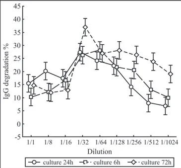
The mean value of three trials in triplicate of IgG proteolytic activityofA. actinomycetemcomitans ATCC 43718 supernatants determined in percentage of degradation was higher at 1/32 dilution [27.5 µg/mL] than pure and diluted by 1/8, 1/16, 1/64, 1/128, 1/256, 1/512, 1/1024 [p<0.05] and higher at 72 h than at 24 and 48 h of culture [p<0.05]. In relation to IgG incubation time, in a 1/32 dilution the higher activity was observed with 24 h of incubation [p<0.05] .However, in dilutions of 1/128, 1/ 256 and 1/512 the highest activity was observed after 6 h of incubation [p<0.05] [Figs. 1 and 2].
Analysis of IgG protease activity in A. actinomycetemcomitans ATCC 43718 fractions
The results of Sephadex G-150 chromatography of A. actinomycetemcomitans ATCC 43718 supernatants fraction by ELISA showed IgG protease activity in fractions 13 to 17 [Fig. 3]. The dextran blue eluted in a fraction 9 and 43 kDa glicoprotein in a fraction 19.
DISCUSSION
fluid, even in heatthy individuals. These antibodies may influence the oral microbiota by interfering with adherence or by inhibiting bacterial metabolism. Moreover, the IgG antibodies may enhance phagocytosis and death of oral microorganisms through activation of the complement or opsonization (13). However, Ig can be degraded by proteases produced by several bacterial species that colonize mucosal and tooth surfaces (13), including periodontal pathogens (4). Gregory et al.(5) demonstrated the presence of proteolytic enzymes in the supernatant of cultures of A. actinomycetemcomitans, and that these enzymes can cleave IgG, IgA and IgM in vitro.
This activity may be the factor which favors the growth of A. actinomycetemcomitans. Considering the bacteriocinogenic activity of A. actinomycetemcomitans (11), which can regulate intra- and interspecific microbiota, both of these virulence factors may play an important ecological role in the mouth.
Investigation of the factors that may modify the oral microbiota is important, considering the recent evidence suggesting that dental infection may be a predisposing factor Figure 1. The mean value of three trials in triplicate of human IgG incubated for 4, 6 and 24 h with pure, 1/8, 1/16, 1/32, 1/64, 1/128, 1/256, 1/512, 1/1024 diluted culture supernatant and then incubated with anti-human IgG peroxidase and the absorbancy read at 492 nm and the results determined in percentage of degradation, considering 100% non-degradation with BHI medium by ELISA.
1/1,1/8,1/16 x 1/32,1/64 p< 0.05;
1/32, 1/64 x 1/128, 1/256,1/512,1/1024 p<0.05; 4h x 6h = p>0.05;
4h x 24h [1/32] = p<0.05; 6h x 24h [1/32]= p<0.05.
Figure 2. Themean value of three trials in triplicate of human IgG incubated with pure, 1/8, 1/16, 1/32, 1/64, 1/128, 1/256, 1/512, 1/1024 diluted supernatant of 24, 48 and 72 h culture supernatant and then incubated with anti-human IgG peroxidase and the absorbency read at 492 nm and the results determined in percentage of degradation, considering 100% non-degradation with BHI medium by ELISA.
72h x 24h [1/32] = p<0.05; 72h x 48h [1/32] = p<0.05.
in systemic conditions such as coronary heart disease, diabetes and low birth weight (7).
In order to partially characterize the A. actinomycetemcomitans protease to IgG, supernatants of this organism were incubated with human IgG. The IgG degradation was revealed by using an anti-human IgG conjugated with peroxidase. The protease activity in A. actinomycetemcomitans strainATCC 43718 IgG associated with culture supernatant concentrations was dose-dependent; the highest activity was observed when supernatants were diluted 1/32 [27.5 µg protein/mL]. At high concentrations there was low activity, suggesting that some component not studied here may inhibit that activity. Additional studies are necessary to better understand the protease action. The highest protease activity was observed in the 72 hour culture of the supernatant. This could be explained either by high enzyme production or by accumulation, which may be growth-time-dependent.
and 1/512 the highest activity was observed, unexpectedly, after 6 hours of incubation. This result can be explained by the extensive IgG degradation after 24 hours, at these dilutions, and IgG conjugated with peroxidase binding in the ELISA plate, or by other variations including the supernatant culture period, or by some other factor. The use of purified IgG protease may eliminate the many components present in the culture supernatant that may influence the protease activity. This question will be the subject of future investigation.
In addition, to characterize the A. actinomycetemcomitans protease to IgG, the culture supernatant was fractionated by gel filtration chromatography and the protease activity was determined by ELISA. IgG protease activity was detected in fractions 13 to 17. The molecular masses [MM] of the components of these fractions were estimated as less than 150 kDa and more than 43 kDa, considering the later elution void volume [150 kDa] and prior to the reference 43 kDa glycoprotein. The protease activity observed in the five fractions may be due to the participation of proteases with different MMs or to the action of proteases at higher dilutions, as observed in this study. Gathering more precise data concerning the IgG protease MM requires further investigation.
In all of the culture supernatant concentrations, growth periods or incubation times investigated, the supernatant culture results were different from the BHI control, showing a protease activity in the supernatant, even in low degree [data not shown]. This A. actinomycetemcomitans IgG protease activity observed
in vitro may also act in vivo. Smith and Taubman (16) reported that IgG in dental plaque extracts was extensively degraded as a Fc fragment and Gregory et al.(5) observed severe degradation of IgG and IgA with crevice gingival samples from patients with localized aggressive periodontitis.
The A. actinomycetemcomitans protease in periodontal tissue may destroy IgG after 4-6 hours of contact, based on the in vitro results. Considering that this activity was present even in high supernatant dilutions, it is possible that this enzyme may act externally by diffusion through saliva, periodontal tissue, mucosa or even blood. However, further studies are necessary to confirm this hypothesis.
We concluded that A. actinomycetemcomitans produces a protease against human immunoglobulin G with high activity in BHI supernatant cultures at 27.5 µg protein/mL concentration, after 72 hours growth and incubation with IgG for 24 hours. The molecular mass of the active fraction of protease is between approximately 43 and 150 kDa.
ACKNOWLEDGEMENTS
The authors thank Mari Sumigawa Kaminami and Nilson de Jesus Carlos for their technical assistance, Dr Édio Vizoni for statistical analysis and Janet W. Reid for her careful correction of the English text.
RESUMO
Atividade proteolítica de Actinobacillus acitnomycetemcomitans sobre imunoglobulina G
Actinobacillus actinomycetemcomitans produz protease ativa sobre imunoglobulina G humana, sendo um dos mecanismos importantes de escape do microrganismo. No presente trabalho, foi analisada a atividade proteolítica de sobrenadante de cultivo de A. actinomycetemcomitans ATCC 43718sobre imunoglobulina G humana em função de concentração, tempo de cultivo do microrganismo e tempo de incubação com IgG, por ensaio imunoenzimático. Adicionalmente, foi determinada a fração com atividade de protease por meio de análise de eluatos de cromatografia em coluna de Sephadex G 150. Os resultados obtidos demonstraram que A. actinomycetemcomitans liberou protease ativa sobre imunoglobulina G humana em sobrenadante de cultivo, sendo a sua maior atividade evidenciada na concentração de 27,5 µg proteína/mL, com tempo de cultivo de 72 horas e com 24 horas de incubação com IgG.A massa molecular da fração ativa de protease foi compreendida entre 43 a 150 kDa.
Palavras-chave: imunoglobulina G, Actinobacillus actinomycetemcomitans, ELISA, protease
REFERENCES
1. Asikainen, S.; Jousimies-Somer, H.; Kanervo, A.; Summanen, P. Certain bacterial species and morphotypes in localized juvenile periodontitis and in matched controls. J. Periodontol., 58,
224-230, 1987.
2. Avila-Campos, M.J.; Farias, L.M.; Carvalho, M.A.R.; Damasceno, C.A.V.; Cisalpino, E.O.; Costa, J.E. Aspectos ecológicos de
Actinobacillus actinomycetemcomitans: aislamiento y caracterización de cepas. Rev. Latinoam. Microbiol., 30, 301-305, 1988.
3. Christersson, L.A.; Albini, B.; Zambon, J.J.; Wikesjö, U.M.; Genco R.J. Tissue localization of Actinobacillus actinomycetemcomitans
in human periodontitis. I Light immunofluoresce and electron microscopic studies. J. Periodontol., 58, 529-539, 1987.
4. Eley, B.M.; Cox, S.W. Proteolytic and hydrolytic enzymes from putative periodontal pathogens: characterization, molecular genetics, effects on host defenses and tissues and detection in gingival crevice fluid. Periodontol. 2000, 31, 105-124, 2003.
5. Gregory, R.L.; Kim, D.E.; Kindle, J.C.; Hobbs, L.C. Immunoglobulin-degrading enzymes in localized juvenile periodontitis. J. Periodont. Res., 27, 176-183, 1992.
6. Hammond, B.F.; Lillard, S.E.; Stevens, R.H. A bacteriocin of
Actinobacillus actinomycetemcomitans. Infect. Immun., 55,
686-691, 1986.
7. Henderson, B.; Wilson, M.; Sharp, L.; Ward, J.M. Actinobacillus actinomycetemcomitans. J. Med. Microbiol., 51, 1013-1020, 2002.
8. Henderson, B.; Nair, S.P.; Ward, J.M.; Wilson, M. Molecular pathogenicity of the oral opportunistic pathogen Actinobacillus actinomycetemcomitans. Annu. Rev. Microbiol., 57, 29-55, 2003.
9. Lang, N.; Bartold, P.M.; Cullian, M.; Jeffcoa, T.M.; Mombelli, A.; Murakami, S.; Page, R.; Papapanou, P.; Tonetti, M.; Van Dyke, T. Consensus report: Aggressive Periodontitis. Ann. Periodontol.,
4, 53, 1999.
10. Lima, F.L.; Farias, F.F.; Campos, P.C.; Totola, A.H.; Tavares, C.A.P.; Costa, J.E.; Farias, L.M.; Carvalho, M.A.R. Leukotoxic activity of
Actinobacillus actinomycetemcomitans isolated from human and
non-human primates. Braz. J. Microbiol., 32, 250-256, 2001. 11. Lima, F.L.; Farias, F.F.; Costa, J.E.; Carvalho, M.A.R.; Alviano,
C.S.; Farias, L.M. Bacteriocin production by Actinobacillus actinomycetemcomitans isolated from the oral cavity of humans
with periodontal disease, periodontally healthy subjects and marmosets. Res. Microbiol., 153, 45-52, 2002.
12. Lowry, O.H.; Rosobrough, M.J.; Farr, A.L.; Randall, R.J. Protein measurement with the Folin phenol reagent. J. Biol. Chem., 246, 1889-94, 1971.
13. Marcotte, H.; Lavoie, M.C. Oral microbial ecology and the role of salivary immunoglobulin A. Microbiol. Mol. Biol. Rev., 62, 71-109, 1998.
14. Page, R.C.; Vandesteen, G.E.; Ebersole, J.L.; Willians, B.L.; Dixon, I.L.; Altman L.C. Clinical and laboratory studies of a family with a high prevalence of juvenile periodontitis. J. Periodontol., 56, 602-609, 1985.
15. Shenker, B.J.; Vitale, L.A.; Welham, D.A. Immune suppression induced by Actinobacillus actinomycetemcomitans: Effects on immunoglobulin
production by human B cells. Infect. Immun., 58, 3856-3862, 1990. 16. Smith, D.J.; Taubman, M.A. Immune components in dental plaque.
J. Dent. Res., C55: 153-162, 1976.
17. Vale, C.H.B.; Fraga, L.A.O.; Costa, A.S.; Tavares, C.A.P.; Martins-Filho, O.A.; Farias, L.M.; Carvalho, M.A.R. Antiproliferative activity of Actinobacillus (Haemophilus) actinomycetemcomitans and
Fusobacterium nucleatum in peripheral blood mononuclear cells. Res. Microbiol., 155, 731-740, 2004.
18. Van-Sttenbergen, T.J.; Bosch-Tijhof, C.J.; Van Winkelhoff, A.J.; Gmür, R.; Graff, J. Comparison of six typing methods for
Actinobacillus actinomycetemcomitans. J. Clin. Microbiol., 32, 2769-2774, 1994.
19. Wang, P.L.; Shirasu, S.; Shinohara, M.; Daito, M.; Fujii, T.; Kowashi, Y.; Ohura, K. Purification and characterization of a tripsin- like protease from the culture supernatant of Actinobacillus actinomycetemcomitans Y4. Eur. J. Oral Sci., 107, 147-153, 1999. 20. Zambon, J.J. Actinobacillus actinomycetemcomitans in human
![Figure 3. Spectrophotometric profile at 280 nm of fractions obtained from A. actinomycetemcomitans ATCC 43718 culture supernatants chromatography in Sephadex G-150 [ ⎯ ⎯ ] and IgG proteolytic activity by ELISA [ ⎯ ⎯ ]](https://thumb-eu.123doks.com/thumbv2/123dok_br/15798913.648282/4.892.50.443.126.313/spectrophotometric-fractions-obtained-actinomycetemcomitans-supernatants-chromatography-sephadex-proteolytic.webp)
