Universidade de Trás-os-Montes e Alto Douro
Comprehending Inflammatory Bowel Diseases
in Dogs and Cats: Challenges in Diagnosis and
Dietary Management
Versão Definitiva
DISSERTAÇÃO DE MESTRADO EM MEDICINA
VETERINÁRIA
Ana Sara Almeida Leitão
Orientadora: Professora Doutora Ana Luísa Dias Guimarães
Lourenço
Co-orientador: Professor Doutor Ronald Jan Corbee
DECLARAÇÃO
NOME: Ana Sara Almeida Leitão
C.C.: 13946014
TELEMÓVEL: (+351) 932988342
CORREIO ELECTRÓNICO: [email protected]
DESIGNAÇÃO DO MESTRADO: Mestrado Integrado em Medicina
Veterinária
TÍTULO DA DISSERTAÇÃO DE MESTRADO EM MEDICINA
VETERINÁRIA:
Comprehending Inflammatory Bowel Diseases in Dogs and Cats:
Challenges in Diagnosis and Dietary Management
ORIENTADORES:
Professora Doutora Ana Luísa Dias Guimarães Lourenço
Professor Doutor Ronald Jan Corbee
ANO DE CONCLUSÃO: 2018
Declaro que esta Dissertação de Mestrado é resultado da minha pesquisa e
trabalho pessoal, e das orientações dos meus supervisores. O seu conteúdo é
original, e todas as fontes consultadas estão devidamente mencionadas no
texto e na bibliografia final. Declaro ainda que este trabalho não foi
apresentado em nenhuma outra instituição para obtenção de qualquer grau
académico.
“Let food be thy medicine and medicine be thy food”
Hippocrates, 460 BC
“When you rise in the morning, give thanks for the light, for your life, for
your strength. Give thanks for your food and for the joy of living. If you see
ACKNOWLEDGMENTS / AGRADECIMENTOS
À Universidade de Trás-os-Montes e Alto Douro, instituição de excelência que possibilitou a minha formação académica, onde cresci pessoalmente e tantas batalhas travei, ao longo destes 6 anos de curso.
À minha orientadora, Dra. Ana Luísa Lourenço, por ter aceite orientar esta dissertação de mestrado, pela disponibilidade e abertura manifestada desde o primeiro minuto desta jornada que começou em Vila Real, passou pela Holanda, e que culmina novamente agora em Vila Real. Grata por ter contribuído de forma tão importante para a realização do meu estágio curricular, pelo aconselhamento, pelo incentivo, pela hospitalidade e pelo contributo científico e humano, fundamental para a realização deste trabalho. Um muito obrigada.
I want to thank Dr. Ronald Jan Corbee, for having so kindly received me in his office for 17 weeks at the Universiteitskliniek voor Gezelschapsdieren, Utrecht. For all the combined efforts made to turn my traineeship in the Netherlands into a reality. It was without a doubt a period of intense learning. For his constant availability and valuable contribution for this dissertation, and for all the experiences and support provided during my time abroad, thank you so much.
À Maria João, por ser verdadeiramente o melhor que Vila Real e a Medicina Veterinária me trouxeram. Há 6 anos atrás começávamos a construir este “para o que der e vier”, e sei, se já me forem permitidas certezas nestes 24 anos de vida, que será para sempre assim. Obrigada por tudo, do fundo do coração.
Ao Christophe, por todos os momentos vividos no nosso Segundo Direito e que tantas saudades deixaram. Pela energia, pela palhaçada diária, pelas gargalhadas infinitas, por todas as viagens a bordo do Mini, pelo incentivo constante nessa maneira tão própria de ser, pela lufada de ar fresco que foi a meio deste percurso, obrigada.
Ao Marcos, pelo pilar que sempre foi na minha vida. Por ter sempre a palavra certa em todos os momentos difíceis, por todos os mimos e paciência infinita, pela motivação constante para terminar este percurso, por acreditar em mim em qualquer ocasião e desafio, um enorme obrigada.
À Carolina, pela tão boa surpresa que foi. Por todos os momentos partilhados em Vila Real, pelas conversas de mão cheia, pela admiração e carinho mútuo, obrigada.
Aos meus pais, por terem possibilitado e motivado o meu percurso académico do princípio ao fim.
À Rita Santos, um agradecimento especial pela ajuda, e por esta amizade de quase 20 anos que tanto estimo.
À Rita Sousa por todo o apoio, conselhos e por toda a amizade.
E por último, mas não menos importante, aos meus tios, irmã, avó, tias e avô pelo carinho e motivação demonstrados.
RESUMO
O termo “IBD”, do inglês “Inflammatory Bowel Disease” (mais corretamente traduzida para português como enterite idiopática) não representa uma só doença ou entidade clínica. É frequentemente aplicado a um grupo de doenças gastrointestinais com evidência histológica de inflamação intestinal sem causa definida, e caracterizadas clinicamente pela presença de manifestações gastrointestinais recorrentes ou persistentes ao longo do tempo. Ainda assim, a nomenclatura adotada não reflete o que ocorre com frequência na prática: a inflamação pode estar confinada ao intestino delgado, ou afetar todo o trato gastrointestinal, como uma gastroenterocolite difusa. Médicos veterinários usam com frequência o termo “IBD” para se referirem a vários tipos de enteropatias crónicas, não necessariamente idiopáticas. Nas publicações da área, o termo é, no entanto, usado para definir a enterite linfoplasmacítica e a enterite e gastroenterite eosinofílica idiopáticas, dado que as enterites granulomatosas e neutrofílicas são raras em animais de companhia, e estão geralmente, relacionadas com causas infeciosas. Devido ao fato de serem observadas e relatadas com frequência variações histológicas associadas às doenças gastrointestinais crónicas de etiologia idiopática, é possível observar-se em publicações mais recentes, uma tendência para classificar as enterites crónicas inflamatórias genericamente como Enteropatias Crónicas, classificando-as e dividindo-as posteriormente tendo em conta a resposta aos protocolos terapêuticos implementados, em: Food-Responsive Enteropathy (“FRE” ou “FRD” se a diarreia for o sinal clínico predominante), Antibiotic-Responsive Enteropathy (ARE) e Steroid-Responsive
Enteropathy (“SRE” ou “IRE” – Immunosuppressant-Responsive).
Ao longo desta dissertação, o termo “IBD” foi usado referindo-se a enterites responsivas à imunossupressão e enterites/gastroenterites linfoplasmacíticas e eosinofílicas idiopáticas.
O principal objetivo deste trabalho é rever os conceitos até à data relativos à “IBD” em cães e gatos, e compará-los com uma amostra de casos de enteropatias crónicas da
padrões que possam estar associados a diferentes fenótipos de enteropatias crónicas em pequenos animais. Adicionalmente será dado ênfase ao estudo dos padrões dietéticos e alimentares dos animais antes do diagnóstico e as recomendações nutricionais específicas prescritas.
Palavras-chave: enteropatias crónicas, enterite idiopática, IBD, Nutrição Clínica, cães, gatos.
ABSTRACT
Inflammatory bowel disease does not represent a clinical entity or a single disease in Veterinary Medicine. Instead, it is the term most commonly applied to a group of diseases that feature histological evidence of idiopathic intestinal inflammation and that are characterised by recurrent or persistent GI signs. However, the nomenclature does not reflect what is seen often in practice: inflammation can be confined to the small intestine (SI) or it can be spread as a diffuse gastroenterocolitis. Professionals often use the term IBD ambiguously to describe various forms of gastrointestinal disease that are associated with chronic inflammation, not truly idiopathic. On field publications, authors reserve the term to describe idiopathic lymphocytic-plasmacytic enteritis (LPE), eosinophilic enteritis (EE) and eosinophilic gastroenteritis (EGE). Due to the fact that histologic variations in chronic idiopathic gastrointestinal diseases are noticeable and widely reported, some authors in more recent publications generally nominate GI inflammatory conditions as Chronic Enteropathies (CE) and subdivide them according to the response to treatment in: Food-Responsive Enteropathy (FRE), Antibiotic-Responsive Enteropathy (ARE) and Steroid-Responsive Enteropathy (SRE or IRE – Immunosuppressant-Responsive). Throughout this dissertation, the term Inflammatory Bowel Disease “IBD” has been used to describe idiopathic LPE, EE and EGE, and Immunosuppressant-responsive enteritis. The main goal of this work is to review current knowledge about IBD in dogs and cats, and to compare it to a chronic enteropathy population of a Referral Small Animal Hospital, analysing retrospective and prospective clinical cases for that purpose. The focus will be to identify key clinical aspects of disease, steps to take to obtain a reliable diagnosis and trait marks of different chronic enteropathies in small animals. Furthermore, emphasis was given to the study of dietary and feeding patterns of the dogs and cats before and after diagnosis and to the specific nutritional recommendations prescribed.
Figures Index
Figures 1A, 1B: Ultrasonographic images of Jejunum………. 16
Figures 2A, 2B: Gastroduodenoscopy images of normal and abnormal duodenal mucosa……… 17
Figures 2C, 2D, 2E: Gastroduodenoscopy images of normal and abnormal duodenal mucosa………. 18
Figures 3A, 3B: Gastroscopy images of abnormal gastric mucosa………. 56
Figures 4A, 4B: Duodenoscopy images of abnormal duodenal mucosa……… 57
Tables Index
Table 1: Canine breed predispositions to GI disease……….... 4
Table 2: Clinical signs associated with Inflammatory Bowel Disease………. 5
Table 3: Cobalamin and Folate measurements on GI disease………... 14
Table 4:Clinical criteria for diagnosing IBD……… 19
Table 5: Activity Indices for IBD……….. 37
Table 6: Retrospective cases………. 41
Table 7: Prospective cases………. 42
Table 8: Diagnosis on the total population……… 43
Table 9: Summary characteristics of the 34 animals in the study………. 46
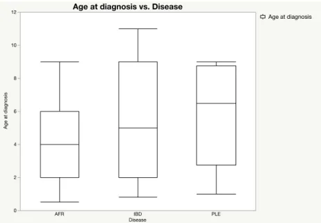
Table 10: Mean age at diagnosis for dogs according to disease groups……… 49
Table 11: Presenting clinical signs according to final diagnosis……… 50
Table 12: Relative and absolute frequencies of serum Cobalamin and Folate concentrations according to final diagnosis………... 53
Graphs Index
Graph 1: Diagnosis obtained on the Prospective study population……….. 44 Graph 2: Age distribution by disease 48 Graph 3: Duration of clinical signs in weeks before referral……… 51 Graph 4: Absolute frequencies of different patterns of disease development per
diagnosis group……….. 51
Graph 5: Ultranosonographic evidence for gastrointestinal disease according to final
diagnosis………. 54
Graph 6: Histopathology reports on IBD group 55 Graph 7: Histopathology reports on IBD group (severity of gastric inflammation) 55 Graph 8: Histopathology reports on IBD group (Helicobacter spp.)………... 56
Graph 9: Histopathology reports on IBD group (severity of small intestinal
inflammation)………. 56
Graph 10: Histopathology reports on PLE group………. 58
Graph 11: Histopathology reports on PLE group (severity of gastric inflammation).. 58 Graph 12: Histopathology reports on PLE group (Helicobacter spp.)………. 58
Graph 13: Histopathology reports on PLE group (severity of small intestinal
inflammation)………. 58
Graph 14: Histopathology reports on FRE group………. 59
Graph 15: Histopathology reports on FRE group (severity of gastric inflammation) 59 Graph 16: Histopathology reports on FRE group (Helicobacter spp.)………. 59
Graph 17: Histopathology reports on FRE group (severity of small intestinal
inflammation)………. 59
Graph 18: Identified dietary sources of protein of 31 dogs and cats on the sample
population………... 64
Graph 19: Identified dietary sources of carbohydrates of 31 dogs and cats on the
sample population……….. 64
Graph 20: Relative frequencies of novel protein sources in diets……… 65
List of Acronyms, Abbreviations and Symbols
AB – Antibiotic
ACTH – Adrenocorticotropic hormone AFR – Adverse food reactions
AGE – Advanced glycation end products ALP – Alkaline phosphatase
ALT – Alanine aminotransferase ANOVA – Analysis of variance
ARE – Antibiotic-responsive enteropathy BCS – Body condition score
BID – from Latin “bis in die”, twice a day BW – Body weight
CAFR – Cutaneous adverse food reaction
CCECAI – Canine Chronic Enteropathy Clinical Activity Index CD – Chron’s disease
CE – Chronic enteropathy
CGE – Chronic gastroenteropathy CH – Carbohydrates
CIBDAI – Canine IBD activity Index CKD – Chronic kidney disease
cPLI – Canine pancreatic lipase immunoreactivity cTLI – Canine trypsin-like immunoreactivity DM – Dry matter
DNA – Deoxyribonucleic acid E.coli – Escherichia coli EE – Eosinophilic enteritis
Felv - Feline Leukemia Virus
FIV – Feline Immunodeficiency Virus FOS – Fructooligosaccharides
fPLI – Feline pancreatic lipase immunoreactivity FRD – Food-responsive diarrhoea
FRE – Food-responsive enteropathy
fTLI – Feline trypsin-like immunoreactivity GI – Gastrointestinal
GSD – German Shepherd Dog IBD – Inflammatory Bowel Disease
IRE – Immunosuppressive-responsive enteropathy IU – International units
kD – kilo Daltons LI – Large intestine
LPE – Lymphocytic-plasmacytic enteritis
MAP – Mycobacterium avium subspecies paratuberculosis MCS – Muscle condition score
ME – Metabolizable energy MOS – Mannan-oligosaccharides MRP – Maillard Reaction products ND – No diagnosis
PARR – PCR for antigen-receptor rearrangements PCR – Polymerase Chain Reaction
PLE – Protein-losing enteropathy
PO – from Latin “per os”, oral administration PRR – Pattern recognition receptors
SCFA – Short-chain fatty acids
SCWT – Soft Coated Wheaton Terriers
SI – Small intestine
SIBO – Small intestine bacterial overgrowth SRE – Steroid-responsive enteropathy
TID – from Latin “ter in die”, three times a day TLR – Toll-like receptors
TSH – Thyroid stimulating hormone TT4 – Total Thyroxine
UC – Ulcerative colitis
UKG – from Dutch, “Universiteitskliniek voor Gezelschapsdieren”
UTAD – from Portuguese “Universidade de Trás-os-Montes e Alto Douro” WSAVA – World Small Animal Veterinary Association
Index
Acknowledgments ………..i
Figures Index ………...vii
Tables Index ………...viii
Graphs Index ………ix
List of Acronyms, Abbreviations and Symbols ………..x
Chapter I – Introduction 1. Chronic Gastroenteropathies in Household Animals ………..1
2. Idiopathic Inflammatory Bowel Disease – “IBD” ………..1
3. Clinical Nutrition ………....2
4. Objectives ………...3
Chapter II – State of the Art 1. Clinical presentation ………...………...3
2. Differential Diagnosis ………....6
3. Etiopathogenesis ………....6
i. Genetics and intestinal immunity ………...6
ii. Correlation between intestinal bacteria and IBD ………8
iii. Environmental Factors ………..10
iv. The undiagnosed infection hypothesis ………..11
4. Diagnosis ………..12
i. Laboratory evaluation ………...12
ii. Faecal examination ………...14
iii. Diagnostic Imaging ………...15
iv. Endoscopy and Intestinal biopsy ………..16
v. Histopathology ………..18
5. Treatment modalities ………...20
i. Parasiticides ………..20
ii. Exclusion diet ………...20
iii. Antibiotics ………....25
6. Dietary Management of Inflammatory Bowel Disease ………...29
i. Individual nutritional assessments ………29
ii. Diet selection ………....30
iii. Probiotics ………...33
iv. Feeding strategies ………...34
v. Managing Lymphangiectasia/Protein-Losing Enteropathy …...35
7. Disease Activity Indices ………..36
Chapter III – Practical section 1. Introduction ……….38
2. Objectives ………38
3. Materials and Methods ……….39
4. Results ………..41
i. Retrospective and prospective cases distribution………. 41
ii. Prospective cases final diagnosis ………..44
iii. Diagnostic Approach ………44
iv. Signalment ………46
v. History and presenting clinical signs ………49
vi. Clinicopathological data ………...52
vii. Ultrasonography ………54
viii. Histopathology ………..55
ix. Therapeutic Trials ……….60
x. Pharmacological Treatment ………..62
xi. Dietary History ……….62
xii. Clinical outcome ………...65
Chapter IV – Discussion ……….66
Chapter V – Conclusions ………78 Appendix ………..80 – 90
Chapter I
Introduction
1. Chronic Gastroenteropathies in Household Animals
The term chronic applies to the spectrum of disorders of the gastrointestinal (GI) tract that persist longer than 3 weeks time (Dandrieux, 2016). Typically these animals present for investigation of cardinal clinical signs such as vomiting and diarrhoea that do not fit the self-limiting or acute event category (Simpson & Jergens, 2011). When uncontrolled these can be life threatening per
se, but also represent a significant limitation to the animal’s well being, even in the most
moderate forms of disease, as well as a serious problem to the owner.
Different clinical manifestations can be present, usually related to the GI organs or segments involved (e.g. Large bowel: multiple defecations, slimy faeces with fresh blood; Small bowel: large amounts of stool, watery diarrhoea) and causality between the signs and extra-GI tract diseases should be excluded (Jergens et al., 1992). Infectious or parasitic diseases and gastrointestinal disease of other aetiology (mechanical obstruction from intussusception, foreign body or tumours) represent the first to investigate GI-tract related differentials that every clinician has to bare in mind (Jergens & Simpson, 2012). Other possible differentials for chronic gastroenteropathies comprise food-related disorders and gastrointestinal diseases showing inflammatory infiltrates with non-identifiable cause or idiopathic (Allenspach et al., 2007). Understanding chronic enteropathies clinically and obtaining a final diagnosis is often insidious.
2. Idiopathic Inflammatory Bowel Disease – “IBD”
Inflammatory bowel disease does not represent a clinical entity or a single disease in Veterinary Medicine. Instead, it is the term most commonly applied to a group of diseases that feature histological evidence of idiopathic intestinal inflammation and that are characterised by recurrent or persistent GI signs (Ettinger et al., 2017). However, the nomenclature does not reflect what is
inflammation, not truly idiopathic. On field publications, authors reserve the term to describe idiopathic lymphocytic-plasmacytic enteritis (LPE), eosinophilic enteritis (EE) and eosinophilic gastroenteritis (EGE), since granulomatous and neutrophilic enteritis are rarely seen in dogs and cats (Ettinger et al., 2017). Due to the fact that histologic variations in chronic idiopathic gastrointestinal diseases are noticeable and widely reported, some authors in more recent publications generally nominate GI inflammatory conditions as Chronic Enteropathies (CE) and subdivide them according to the response to treatment in: Food-Responsive Enteropathy (FRE or FRD if diarrhoea is the only clinical feature present), Antibiotic-Responsive Enteropathy (ARE) and Steroid-Responsive Enteropathy (SRE or IRE – Immunosuppressant-Responsive) (Allenspach et al., 2007; Walker et al., 2013; Allenspach et al., 2016; Dandrieux, 2016).
Throughout this dissertation, the term Inflammatory Bowel Disease “IBD” has been used to describe idiopathic LPE, EE and EGE, and Immunosuppressant-responsive Enteritis.
In humans, IBD represents two different chronic disorders characterized by inflammation of the intestinal wall: Crohn’s disease (CD) and ulcerative colitis (UC) (Lee et al., 2015) and the resemblances among species are limited both clinically and histologically. Nevertheless, science has been showing us over the years that inflammatory bowel diseases in humans and canine and feline populations might share the same aetiology. (Craven et al., 2004; Cerquetella, 2010; Khor
et al., 2011; Simpson et al., 2011; Lee et al., 2015).
3. Clinical Nutrition
The gastrointestinal tract is certainly the organ system most directly affected by nutrition. Dietary composition of macro and micronutrients has a deep impact not only on intestinal health but also through the influence it exerts on the microflora population. Although a dietary modification is believed to be a key element in managing chronic gastrointestinal diseases, there are not many controlled clinical trials that have evaluated specific dietary manipulation in preventing and managing canine and feline gastroenteric disorders (Cave, 2012). Despite this, a good nutritional approach to each patient can be useful to decrease pharmacological dosages used in medical therapy. Furthermore, the remission of clinical signs in some forms of chronic enteropathies can be achieved with dietary management as sole intervention (Allenspach et al., 2016).
4. Objectives
The main goal of this work will be to review current knowledge about IBD in dogs and cats, and to compare it to a chronic enteropathy population of a Referral Small Animal Hospital, by analysing retrospective and prospective clinical cases for that purpose. The focus will be to identify key clinical aspects of disease phenotypes, steps to take to obtain a reliable diagnosis and trait marks of different chronic enteropathies in small animals. Furthermore, emphasis will be given to the study of dietary and feeding patterns of the dogs and cats before and after diagnosis and specific nutritional recommendations prescribed.
Chapter II
State of the art
1. Clinical presentation
Idiopathic Inflammatory Bowel Disease is a widely discussed phenomenon throughout the veterinarian community. It is a diagnosis of exclusion, made when the cause(s) for GI mucosal inflammation in animals suffering from chronic gastroenteropathies fail to be documented (Ettinger et al., 2017). Its true incidence in dogs and cats is to this date unknown, but beliefs are that it is over diagnosed. No gender predisposition occurs both in dogs and cats, most affected animals are middle-aged, showing in some cases intermittent signs at an early age (Ettinger et al., 2017). The prevalence of Inflammatory Bowel Disease (IBD) in neutered and intact animals is not studied, but in a recently published study in dogs, gonadectomy in males and females was related with a greater risk for Atopic Dermatitis, Autoimmune Haemolytic Anaemia, Hypoadrenocorticism, Hypothyroidism, Immune-mediated Thrombocytopenia and IBD (Sundburg et al., 2016). This brings up the question whether sex steroids really play an important
inflammation (as shown in Table 1), which suggests that there might be some underlying genetic or immune triggers. Basenjis, Shar-Peis, German Shepherds, Boxers and Rottweilers are the breeds at higher risk of developing IBD (Washabau et al., 2010).
Table 1. Canine breed predispositions to GI disease
Breed Predisposition
Basenji Lymphocytic-plasmacytic Enteritis,
Hypertrophic Gastropathy
Beagle Selective Cobalamin Malabsorption,
Gastric Neoplasia
Border Collie Selective Cobalamin Malabsorption
Boxer Granulomatous Colitis
German Shepherd Idiopathic Antibiotic-responsive Diarrhoea, Lymphocytic-plasmacytic Enteritis
Giant Schnauzer Selective Cobalamin Malabsorption
Irish Setter Gluten-sensitive Enteropathy
Lundehund Lymphangiectasia, Atrophic Gastritis,
Gastric Neoplasia
Retrievers Dietary Allergy
Rottweiler Susceptibility to Parvovirus,
Lymphangiectasia
Soft-coated Wheaten Terrier Protein-losing Enteropathy
Shar-pei Lymphocytic-plasmacytic Enteritis,
Cobalamin Deficiency
Toy breeds Haemorrhagic Gastroenteritis
West Highland Terrier Dietary Allergy
Yorkshire Terrier Lymphangiectasia
Adapted from (Ettinger et al., 2017)
Concerning cats, also any breed can be affected, but there is a recognised predisposition for IBD in Siamese and other Asian breeds (Jergens, 2012). A finding often seen also in felines is
Triaditis - a term used by some authors to describe an association between three inflammatory
diseases in domestic felines: IBD (mostly lymphocytic-plasmacytic enteritis), lymphocytic cholangitis and chronic pancreatitis (Bailey et al., 2010; Jergens, Crandell, Evans, et al., 2010; Jergens, 2012; Fragkou et al., 2016).
Clinical signs are highly variable between species and individuals and usually correlate with the GI areas affected (Allenspach et al., 2007). Nevertheless, chronic small or large bowel diarrhoea (some patients show a mixed type) and/or vomiting are the most common complaints. Table 2 shows a list of clinical signs recognised to be associated with IBD.
Table 2. Clinical signs associated with Inflammatory Bowel Disease
Vomiting Common when gastric or upper SI inflammation is present; After food intake or on an empty stomach; Predominant sign in cats; Haematemesis in severe cases.
Small bowel diarrhoea Watery content, large volume of faeces and melena are often seen; Weight loss is a consequence.
Large bowel diarrhoea Increased frequency of defecation, haematochezia, tenesmus, smaller amounts of faeces and mucoid stools are features; Can be a consequence of prolonged SI-type diarrhoea or due to primary colonic inflammation.
Abdominal pain or discomfort Focal or diffuse. Excessive borborygmi Increased flatulence
Weight loss Due to SI-type diarrhoea or inappetence/anorexia. Changes in appetite and food intake Variable appetite, inappetence, anorexia,
polyphagia when severe weight loss occurs; Nauseated dogs frequently ingest grass to induce
where inflammation can cause lymphangiectasia and protein-losing enteropathy; Pleural effusion can also be seen.
Adapted from (Craven et al., 2004; Jergens, 2012; Ettinger et al., 2017)
2. Differential Diagnosis
Most-likely differentials for idiopathic Inflammatory Bowel Disease (Craven et al., 2004; Allenspach et al., 2007; Cerquetella, 2010; Dandrieux, 2016; Ettinger et al., 2017):
a) Chronic Infectious Disease – Parasites (Helminths, Protozoa such as Giardia spp. and
Tritrichomonas foetus), Fungi, Bacteria (Tuberculosis, Enteroadherent organisms such as E.colli and Streptococcus spp. in dogs, and Enterococcus hirae in cats);
b) Exocrine Pancreatic Deficiency (EPI);
c) Adverse Reactions to Food – Food Intolerance, Food Allergy;
d) Antibiotic-responsive Diarrhoea – previously nominated idiopathic SIBO; e) Lymphangiectasia;
f) Neoplasia – High and Low Grade Alimentary Lymphoma, Gastric Carcinoma, Intestinal Adenoma, Intestinal Adenocarcinoma, Mast cell tumours.
3. Etiopathogenesis
The exact mechanisms behind the development of IBD in small animals are poorly understood. In spite of this, comparisons are made with inflammatory bowel diseases in humans, which suggests their molecular pathogenesis might be the same. Three major components are thought to be the roots of disease: genetic predisposition, the intestinal microbiome and environmental aspects, in which dietary aspects are included (Cerquetella, 2010; Allenspach, 2011; Khor et al., 2011; Catchpole & Allenspach, 2012; D. Lee et al., 2015; Ettinger et al., 2017; Lewis & Abreu, 2017).
In humans, IBD is considered a heritable condition with familial history. In fact, almost a hundred genes and genetic loci that contribute to IBD susceptibility on man were already identified and the vast majority of them are directly involved with several metabolic pathways that are crucial to intestinal homeostasis. Moreover, 50% of these identified loci are involved in other inflammatory and autoimmune diseases in humans (Allenspach, 2011; Khor et al., 2011). There is currently no evidence for autoimmune mechanisms in IBD in dogs and cats (Ettinger et
al., 2017), but both innate and adaptive immune systems are thought to play important parts on
the pathogenesis.
Genetic studies in companion animals designed to identify polymorphisms linked to IBD are not many, but in German Shepherd Dogs (GSDs) at least two gene polymorphisms associated with this breed’s susceptibility to intestinal inflammation were identified (Allenspach et al., 2010). The same polymorphisms are present in affected animals from other breeds. One of the above referred genetic variations is present in genes encoding Toll-like-receptors (Cerquetella, 2010; Kathrani et al., 2010; Allenspach, 2011; Ettinger et al., 2017). Toll-like receptors (TLRs) are a class of cellular transmembrane pattern recognition receptors (PRRs), which play a key role on the induction of pro and anti-inflammatory genes, on the detection of microbial infection and on the induction of inflammatory and immune responses against microbial structures. These receptors allow cells from the innate immune system (macrophages and dendritic cells) and epithelial cells of the intestinal mucosa to recognise microbe-associated molecules (Burgener et
al., 2008). This is important not only for the recognition of pathogens and their products by
active antigen processing, but also for the cells to act as a barrier, and induce mucosal immune response through antigen presentation, secretion of cytokines, and recruitment of inflammatory cells (McMahon et al., 2010). Nevertheless, more than providing the adequate immune responses to pathogens, immune cell populations have to be able to maintain tolerance to harmless environmental antigens such as commensal bacteria and food, without triggering continuous inflammatory states (Kathrani et al., 2010). This is referred to as Oral Tolerance and it represents the induced hyporesponsiveness state that (mostly) dendritic cells mediate when “reading” commensal microbes through PRRs. So on an healthy individual, when identifying commensal
population, which can be the cause of the break on Oral Tolerance (Allenspach et al., 2010). This can be a strong element for the explanation of the pathogenesis of idiopathic inflammatory bowel diseases, supporting that an inflammatory response to commensal organisms can in fact be a reality. On the other hand, a different interpretation can also be highlighted. Increased expression of TLRs can arise as a consequence of a constant stimulus done by an altered intestinal microflora (Pierik et al., 2006; Burgener et al., 2008; Allenspach, 2011; Khor et al., 2011). Authors also mention structural intestinal abnormalities such as the disruption of the mucosal barrier to antigens due to a lack of production of epidermal growth factors, as another clue to understand IBD. If so, normal mucosal repair is inhibited and an abnormal intestinal permeability is expected in affected animals (Cerquetella, 2010; Ettinger et al., 2017).
In the face of the most recent findings, the inflammatory process seen on IBD mediated in many cases by lymphocytic-plasmacytic infiltrates can be explained by the adaptive immune response to an aberrant and continuous activation of Toll-like receptors on the enteric mucosa (Netea et al., 2008; Catchpole et al., 2012).
ii. Correlation between intestinal bacteria and IBD
The possibility for an intestinal dysbiosis or an altered microbiota on IBD patients is widely discussed in humans and animals. In spite of this, the actual host/bacteria interactions remain elusive. A major function of microbiota is to degrade dietary residues that were not digested, yielding fermentation products. An example of beneficial fermentation products is short-chain fatty acids (SCFA). On the other hand, putrefactive substances can be produced by fermentation of undigested ingesta. One study reports serum metabolite profiles in IBD dogs differ from serum metabolite profiles in healthy animals, which can be attributed to oxidative stress (Minamoto et
al., 2015). Oxidative stress in IBD dogs was in fact deeply investigated in a more recent study
(Rubio et al., 2017).
What is known so far from molecular studies in IBD dogs about their intestinal microbial communities, is that members of the families Enterobacteriaceae and Clostridiaceae appear to be overrepresented in some studies (Xenoulis et al., 2008; Suchodolski et al., 2010). Other study report decreases in Bacteroidetes and Firmicutes (where orders Bacillales, Lactobacillales and
Clostridiales are included) and concurrent increases in Proteobacteria (where family Enterobacteriaceae is included) (Minamoto et al., 2015). Clostridiales include family
Clostridiaceae, bacterial groups believed to be major producers of SCFA. The decrease of their
presence is therefore not beneficial for the host (Minamoto et al., 2015). The Enterobacteriaceae increases in the last mentioned study were mainly due to E. coli overrepresentation. Previous studies have reported an increased virulent potential of E. coli, such as adhesive capacity to cells, in human patients with IBD, and on granulomatous colitis of Boxer dogs (Rolhion & Darfeuille-Michaud, 2007; Craven et al., 2011). E.coli seems to be well adapted and in competitive advantage to survive on an intestinal environment of oxidative stress and lack of nutrients (Minamoto et al., 2015).
Interestingly, German Shepherd Dogs, a breed were many cases of antibiotic-responsive enteropathy are reported, indicate these animals possess a different microbiome when compared to a healthy dog population, characterised essentially by a higher representation of
Lactobacillales (Allenspach et al., 2010), usually regarded as beneficial bacteria. On the other
hand, works using faecal samples to study the microbiome profile of IBD patients found that the relative abundance of faecal Lactobacillus gradually decreases with disease severity, but no major bacterial group changes between the control and disease group were observed (Xu et al., 2016). However, it is still not clear whether using faecal samples is a reliable form to study the intestinal microbiota, but this finding is in accordance with the above mentioned molecular study (Minamoto et al., 2015), where decrease of Firmicutes is reported in IBD.
Many studies have been performed in several dog breeds and populations, and their results compiled and compared. These compilations conclude that there are in fact changes on the commensal bacterial and fungal communities of inflammatory bowel diseased dogs, (Suchodolski et al., 2012; Honneffer et al., 2014; Minamoto et al., 2015). In cats, few molecular studies were performed in IBD populations. A particularly relevant one analysed mucosal-adherent microbiota and revealed that Enterobacteriaceae are significantly increased in duodenal biopsies of cats with inflammatory bowel disease (Janeczko et al., 2008).
The composition of abnormal intestinal bacteria population seems to be variable among species and breeds of animals and the functional consequences of these alterations are yet to be understood. The fact that diet can influence the gut population since the very first days of every
effects on the host’s health status, namely the progressively recognised deleterious effect on immunity regulation.
Future focus lies on the possibility to manipulate the microbiome through faecal transplantation and the use of probiotics, hoping these measures can control/reduce GI inflammation (Honneffer
et al., 2014; Rossi et al., 2014; Sheehan & Shanahan, 2017). Further studies in coming years on
the microbiome profiles of IBD patients with a higher focus on environmental factors such as diet and living conditions are justified.
iii. Environmental factors – does the diet play a role on the pathogenesis?
The potential implication of diet-related factors on IBD is not clear and not many studies to this date in Veterinary Medicine focus on relating environmental factors and aetiology. The clinical benefit of dietary manipulation or restriction observed in many cases of eosinophilic and lymphocytic-plasmacytic enteritis suggests dietary antigens might be involved in the pathogenesis (Cave, 2012). In fact, one study examined pathological changes present in duodenal biopsies of 20 dogs diagnosed with Food-responsive enteropathy before and after a 6-week dietary trial (the animals were fed a hydrolysed soy protein-based commercial diet) using histopathological, immunohistochemical, and ultrastructural criteria. The results showed a lower density of the lamina propria’s mononuclear cell and eosinophil population. The study group also evidenced ultrastructural lesions of the mitochondria and brush border of enterocytes, which improved after the treatment (Walker et al., 2013). Despite the results, similarly with other studies, the control group used did not match breeds, ages, living conditions and diet. Once again this is a limitation to a more accurate comparison of results between groups.
It is widely accepted that the majority of household animals in Europe and North America are fed commercially formulated diets. Both dry and canned foods are processed and undergo essentially heat treatments that modify nutrient digestibility. Advanced Maillard-reaction products (MRPs) are formed during heat treatments in processed foods (a reducing sugar binds to a free reactive amino group of an amino acid) but also endogenously, occurring naturally on body tissues, and called Advanced Glycation End-products (AGEs) (van Rooijen et al., 2013). AGEs together with some ingested MRPs that are absorbed by the organism are accumulated on tissue proteins and related to several age-related diseases such as diabetes, cataracts and pro-inflammatory responses in humans, rats and dogs (van Rooijen et al., 2013). Authors point the average daily intake of
MRPs as being 122 times higher for dogs fed commercial diets, and 38 for cats, than the average daily intake for adult humans (van Rooijen et al., 2014). The real long-term implications of their ingestion, and if the amount of MRPs in commercial animal diets is in fact a reason for alarm needs to be deeper investigated. Nevertheless these conclusions are interesting and could stimulate further investigation to aid explain the mechanisms of some inflammatory and immune-mediated diseases in companion animals.
iv. The undiagnosed infection hypothesis
Granulomatous colitis of Boxers was considered in past years as a variation of idiopathic IBD, in such a fashion that most animals were diagnosed at a young age and prescribed life-long immunosuppressive therapy. The prognostic was not cheering for those who failed to respond to standard therapy and eventually would end up to be euthanised. But it was more than a decade ago that attaching and invasive E.coli colonies were identified as the aetiological agents of this disease, and the treatment use of Enrofloxacin related to long-term clinical remission (Simpson et
al., 2006; Craven et al., 2011). Considering this, it is plausible to hypothesize the presence of an
undiagnosed infectious agent causing chronic intestinal inflammation in alleged cases of idiopathic IBD. Toxoplasma gondii is an intracellular parasite thought to be involved in GI inflammation in genetically susceptible organisms, and is in fact used in mice to produce animal models for the study of IBD in humans (Egan et al., 2012). Furthermore, a recent study in 42 dogs with chronic gastroenteropathies identified Mycobacterium avium subspecies
Paratuberculosis (MAP) DNA by PCR in intestinal biopsies of 20% of the dogs. Of the 8
positive cases, 3 were diagnosed with food-responsive enteropathy, 2 with IBD and 3 with non-specific gastrointestinal changes, thus none with neoplasia. All positive dogs had access to rural areas (Glanemann et al., 2008). Nevertheless the clinical significance of this finding is not certain due to the fact that specific histologic evidence is not present. Also, a more recent study documents the identification of MAP in samples of dogs without evidence of clinical or histopathologic gastrointestinal disease (KuKanich et al., 2013).
4. Diagnosis
Intestinal biopsy is the necessary means to diagnose intestinal inflammation. However, the most important thing to retain before further advancing this matter is that not all animals presenting chronic gastrointestinal signs are candidates for intestinal biopsy. Performing endoscopy and collecting tissue samples to histologic examination is in fact the last step to be taken and, when done, histology reports should be carefully interpreted. Parasites, pathologic bacteria, anatomical GI problems and extra-GI diseases should be systematically ruled out before IBD can start to be prioritised on a list of differential diagnosis. Therefore, diagnostic means such as laboratory evaluation and diagnostic imaging should first be performed. The results of these tests are not used to prove that intestinal inflammation is idiopathic, but instead to eliminate common causal factors of GI diseases and the possibility for other organs’ involvement (Ettinger et al., 2017).
i. Laboratory evaluation
Even though no pathognomonic deviations are found in complete blood counts or in serum biochemistry profiles, both are useful to evaluate overall health status and possible consequences of malnutrition and electrolyte derangement due to chronic diarrhoea and vomiting.
Anaemia can be found as an indicator of chronic inflammation, chronic blood loss or severe iron-deficiency (Cave, 2012; Ettinger et al., 2017). Neutrophilia can also be present with or without a left shift, and in some cases the presence of eosinophilia in blood counts can be indicative for Eosinophilic Enteritis (EE). These changes are however highly variable, and one finding do not predict the other. Thrombocytopenia is rare in IBD. However, thrombocytosis can occur secondarily to chronic GI bleeding (Craven et al., 2004; Ettinger et al., 2017).
Malnutrition that occurs in prolonged gastrointestinal disease can have such consequences as hypocholesterolaemia and hypomagnesemia (Ettinger et al., 2017). Hypocalcaemia can be seen also, mainly in dogs with Protein-losing enteropathy (PLE). This feature has been in previous years explained as a result of low serum albumin concentrations, which resulted in the reduction of the protein-binding calcium fraction (Jergens et al., 1992). In more recent years, the possibility for measuring serum-ionised calcium became a reality, and PLE dogs were found to be equally susceptible to develop ionised hypocalcaemia. Calcium metabolism derangements in IBD animals are evidenced by hypoalbuminemia, low measures of ionised calcium and
hypovitaminosis D. Inappetence, malabsoption or intestinal loss of vitamin D could be implicated on the latter phenomenon (Gow et al., 2011). Protein-losing enteropathy associated with intestinal inflammation is more common in dogs, and losses of albumin and other proteins with different molecular weights are due to enhanced intestinal permeability (not always associated with Lymphangiectasis) and leakage, leading to hypoalbuminemia and in severe cases panhypoproteinaemia (Dossin & Lavoue, 2011). Hypoalbuminemia observed on serum biochemistry profiles can also be associated with protein-energy malnutrition in cases with prolonged deficient caloric intake, decreased intestinal absorption and increased catabolism (Cave, 2012). Fecal α-1-protease inhibitor is a plasma protein that is resistant to digestion and reaches the rectum intact. This protein is lost at the same rate as albumin in PLEs and is a useful diagnostic tool for non-panhypoproteinaemic animals. A fecal immunoassay was developed to measure fecal α-1-protease inhibitor (Melgarejo et al., 1998).
Measuring serum cobalamin and folate concentrations is especially important in cats and when animals present with excessive weight loss, decreased activity and anorexia or inappetence. Low measures of both parameters are associated with chronic gastrointestinal diseases in both dogs and cats (Table 3) and malabsorption in cats. Early supplementation is recommended, since delaying it can compromise the success of treatments applied as well as clinical remission of gastrointestinal signs (Simpson et al., 2001; Ruaux et al., 2005; Jergens, 2012). Furthermore, cats with low serum B12, and low B12 and folate together, are reported to have lower body condition scores than cats with normal values (Ruaux et al., 2005). In a more recent study, lower serum cobalamin concentrations in cats were more associated with alimentary lymphoma and IBD in comparison with other intestinal neoplasia (Jugan & August, 2017). In dogs, two large retrospective studies reported hypocobalaminaemia as an indicator for negative outcome in chronic enteropathies, highlighting the importance of a routine measurement and adequate supplementation, in chronic GI patients (Allenspach et al., 2007; Volkmann et al., 2017). Although parenteral supplementation is recommended, one recent study claims oral supplementation of hypocobalaminaemic dogs with chronic enteropathies is as effective in normalising serum cobalamin levels (Toresson et al., 2016).
Table 3. Cobalamin and Folate measurements on GI disease
Result Cobalamin Folate
Decreased Distal Small Intestine disease/dysbiosis; Exocrine pancreatic insufficiency
Proximal Small Intestine disease (impaired absorption)
Increased --- Bacterial overgrowth/ dysbiosis Adapted from (Reed et al., 2007; Toresson et al., 2016; Jugan et al., 2017)
The serum activity of liver enzymes can be found mildly increased in dogs with IBD, possibly a cause of a “reactive hepatopathy”, which is something unlikely in cats, since the half-life of these enzymes in domestic felines is shorter, and elevations observed in biochemistry profiles are more likely to be a result of primary hepatic disease (Ettinger et al., 2017).
ii. Faecal examination
Fecal examinations are fundamental for eliminating parasites as the cause for mucosal inflammation. Faecal flotations are commonly performed in veterinary laboratories to identify protozoa and protozoan cysts, and larvae of hookworms and whipworms. Giardia spp. infections are responsible for acute and chronic diarrhea in dogs, being cats less commonly affected (Ettinger et al., 2017). Oocysts can be identified in fecal flotation exams, and samples should be collected in 3 consecutive days, since shedding of oocysts is not regular. Fecal enzyme-linked immunosorbent assay (ELISA) delivers more accurate results for Giardia infestation (Rishniw et
al., 2010). Tritrichomonas foetus plays an important role in persistent diarrhea and chronic colitis
in cats (Ettinger et al., 2017). It can be diagnosed through fecal smears or by PCR which is a far more specific and sensitive procedure (Gunn-Moore et al., 2007).
A fecal marker (S100A12) used in human medicine to monitor IBD was recently found to be present in higher concentration in the faeces of dogs diagnosed with inflammatory bowel disease, and it correlates with disease severity (Heilmann et al., 2014). This can be potentially used as a non-invasive molecular marker of inflammation and disease activity by clinicians.
iii. Diagnostic Imaging
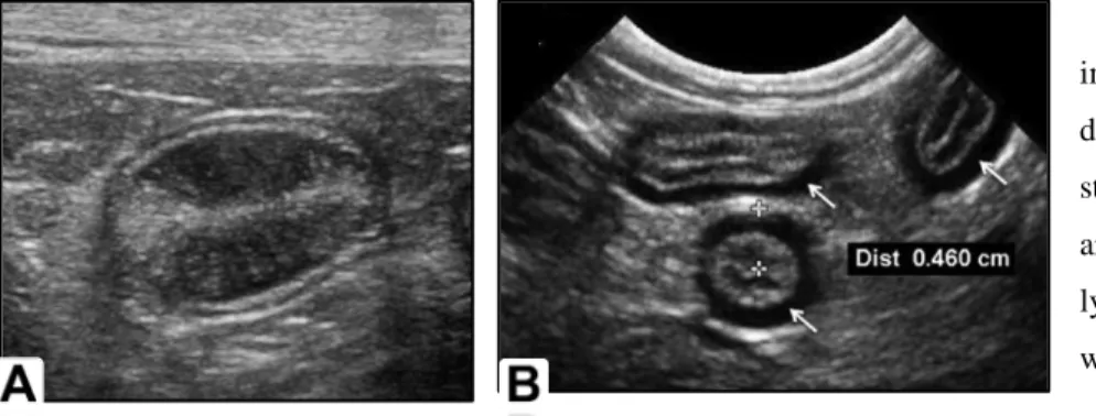
Radiographs are usually unremarkable and unhelpful to diagnose gastrointestinal inflammation. Information obtained is on the other hand useful to rule out anatomical gastrointestinal diseases. Moreover, ultrasonographic examination is preferable to detect abnormalities on gastrointestinal and abdominal organs. Unlike what happens in humans, ultrasonographic assessment of gut wall layering is an interesting tool to non-invasively detect intestinal changes in veterinary medicine (Le Roux et al., 2016). Distribution of disease can be visualised and changes classified in focal, multifocal or diffuse. Sonographic abnormalities observed in IBD patients are similar in dogs and cats: normal to mild (in some cases moderate) generalised thickening of the intestinal wall with maintenance of wall layering (Gaschen & Kircher, 2007; Jergens, 2012). Measuring wall thickness is not specific neither sensitive to diagnose IBD and the finding of normal mucosal layering does not rule out the presence of inflammation, though (Gaschen et al., 2007). Ultrasonography is a good tool to detect the presence of focal masses (which can be in some cases indicative of neoplastic disorders), to evaluate the presence of peristalsis movements, to search for the presence of abdominal free fluid and evidence of peri-intestinal hyperechoic mesentery, a sign of regional inflammation, neoplastic disorders and perforation. Corrugation of the intestines can also be seen in enteritis (Gaschen, 2011). In cases were Lymphangiectasia occurs, hyperechoic striations (representing dilated lacteals) and speckles, as well as increased mucosal echogenicity and mild small intestinal wall thickening can be observed (Gaschen et al., 2007; Dossin et al., 2011). In cats, ultrasonographic evidence of intestinal muscularis propria thickening is regarded as a consequence of cellular infiltration. A bigger prevalence of this finding is observed in cats diagnosed with Lymphoma rather than in those diagnosed with IBD (Zwingenberger et al., 2010). In spite of this, a pattern of muscularis propria thickening is recognised in both diseases and cannot be used to distinguish them (Zwingenberger et al., 2010; Daniaux et al., 2014). Mesenteric lymphadenomegaly can likewise be documented and it is associated with both Lymphoma and IBD (Zwingenberger et al., 2010).
Figure 1A. Transverse
image of a jejunal segment of a dog. Multiple parallel hyperechoic striations throughout the mucosa are visible. Lymphangiectasia and lymphocytic-plasmacytic enteritis were confirmed histologically.
Figure 1B. Transverse segments of the jejunum of a cat. The muscularis layer (arrows) is diffusely thickened
measuring 4.6mm. Final clinical and histopathological diagnosis was of cholangiohepatitis, pancreatitis and lymphocytic, plasmacytic and eosinophilic enteritis (triaditis). Adapted from (L. Gaschen, 2011).
A normal but hypoechoic gut mucosa at ultrasound evaluation is associated with 80% sensitivity and 81% specificity to diagnose food-responsive diarrohea in dogs. Authors suggest mucosal echogenicity is a better parameter than intestinal wall thickness to detect inflammatory disease in dogs with chronic diarrhea (Lorrie Gaschen et al., 2008).
Another paramount aspect of ultrasonography is that it enables clinicians to be on track for the best places to take biopsy samples.
iv. Endoscopy and Intestinal Biopsy
Since no other method can reliably confirm inflammation as tissue sampling and microscopic observation, intestinal biopsy is necessary to evidence inflammatory diseases. Biopsies are convenient also to rule out infectious agents such as parasites, bacteria and fungi and to document the type of inflammatory infiltrate and the extent of tissue damage and transformation. Collecting full-thickness intestinal biopsies during surgery is a procedure of more diagnostic value, since all intestinal layers can be analysed. Nevertheless, inclusion of deeper tissue is, according to some authors, essentially helpful to diagnose intestinal Lymphoma (Kleinschmidt et al., 2006). A more accurate distinction between inflammatory and neoplastic disease can be achieved this way. However, this procedure is far more invasive than endoscopy and surgical wound dehiscence can be a problem, especially when hypoproteinemia is present (Ettinger et al., 2017). Explorative laparotomy is not indicated as a routine procedure to collect biopsies to diagnose IBD. Instead, Endoscopy is a safer and less invasive method to collect tissue samples, even though it still
requires general anaesthesia to be performed. Endoscopy limitations are many, such as the quality of the samples obtained, which can be superficial (usually include mucosa and submucosa layers, leaving muscularis propria out of reach of endoscopic forceps), small (capture of villi only) and sometimes crushed, but also the inability of the majority of the endoscopes used in clinics to advance further inside the gastrointestinal tract, limiting sampling to the stomach and the proximal small intestinal region, or colon when Colonoscopy is performed (Washabau et al., 2010; Ettinger et al., 2017). Balloon Enteroscopy is an alternative to reach the Ileum, but quite expensive and not widespread in Veterinary Medicine. Limitations concerning Endoscopy are a reality but advantages do exist as well. The first one is that video endoscopy allows the operator to detect mucosal changes, unlike the serosal approach during surgery, and many samples can be collected at various points of the GI tract during the exam. This is especially important to assess diffuse disease and sample various lesions (Washabau et al., 2010). Clinical criteria for the decision whether to perform Endoscopy or not throughout the clinical course of a given animal should be guided according to its health status and diagnostic imaging results. Ultrasonographic evidence of substantial infiltrative disease, prolonged anorexia, hypoalbuminemia and poor body condition with progressive weight loss could guide the decision that endoscopy is appropriate, even before the institution of therapeutic antimicrobial and dietary trials (Washabau et al., 2010). Standardized report forms developed by WSAVA (The World Small Animal Veterinary Association) GI Standardization Group consider the following aspects to macroscopically assess the GI mucosa during Endoscopy: hyperaemia, oedema, discoloration, friability, haemorrhage, erosion/ulcer, lacteal dilation (Washabau et al., 2010).
Figures 2C, 2D and 2E. Gastroduodenoscopy images of the duodenal mucosa in the dog. C and D show increased
granularity and irregularity in mild and severe lymphoplasmacytic enteritis, respectively. E shows several dilated lacteals (glistening white patches) containing lymph: Lymphangiectasia. Adapted from (Ettinger et al., 2017).
v. Histopathology
Quality samples and an adequate number are essential for histopathological interpretation. The interpretation of mucosal inflammatory change has proved to be complex with long time recognised disparity among pathologist observations. Standard criteria for assessing small samples of GI tissue were developed as guidelines to avoid subjective evaluations of certain histopathological findings (Day et al., 2008). These guidelines contemplate 3 major important points to characterise samples: the dominant population of inflammatory cells, the severity of the changes and distinction between chronic mononuclear inflammation and lymphoid neoplasia. A normal histologic aspect of the GI tract was defined and pictorial and textual templates were developed as visual criteria for assessing and comparing inflammatory and morphological changes in the gastric body and antrum, duodenum and colon of the dog and cat.
Varying degrees of mucosal architectural disruption and minimal to pronounced inflammatory cells infiltration are common in IBD cats and dogs. Typical changes of duodenal mucosa include villus stunting, epithelial injury, crypt distention, lacteal dilation and mucosal fibrosis (Day et al., 2008; Washabau et al., 2010; Jergens, 2012). Concerning inflammatory infiltrates associated with IBD, lymphocytic, lymphoplasmacytic, eosinophilic and mixed inflammation, represent the types observed in companion animals (Jergens et al., 1992; German et al., 2001; Craven et al., 2004; Garcia-Sancho et al., 2007; Jergens, 2012). Lymphoplasmacytic gastroenteritis represents the most common finding in IBD cats (Jergens, 2012). In dogs, mixed inflammation is often found
along with lymphoplasmacytic disease. Lower gastrointestinal disease (colitis) is observed more in IBD dogs (Craven et al., 2004).
Even tough lymphoid neoplasia usually exhibits predominantly lymphocytic infiltrates (mononuclear cell population), chronic mucosal lymphoplasmacytic inflammation is regarded as a precursor for alimentary lymphoma in humans, dogs and cats (Washabau et al., 2010). Differentiation of severe IBD from well-differentiated lymphoma may be especially problematic in cats since alimentary lymphoma is this species’ most common presentation of lymphoid neoplasia (Jergens, 2012). Specific tests to detect monoclonality and differentiate both types of diseases are required. Immunohistochemical phenotyping of the infiltrate population is one option, performing PARR (PCR for antigen receptor rearrangements), which is a clonality test to identify T-cell receptor genetic rearrangements (Moore et al., 2005; Pohlman et al., 2009), is the other. The latter is superior in sensitivity but not available as a routine test in most veterinary laboratories, and some cases of IBD evidence some degree of clonal rearrangements (Ettinger et
al., 2017).
Finally, histopathological evidence of GI inflammation alone does not have diagnostic value, and histologic scores are reportedly not associated with disease outcome (Allenspach et al., 2007). Further criteria should be applied to institute a clinical diagnosis of IBD as represented in Table
4.
Table 4. Clinical criteria for diagnosing IBD
Chronic gastrointestinal signs >3 weeks vomiting, diarrhoea, anorexia, weight loss, haematochezia, mucoid faeces (not all signs need to be present)
Failed antibiotic and dietary trials Clinical signs persist after intervention
Thorough diagnostic evaluation not conclusive Inability to document the cause of inflammation
Intestinal biopsies Histopathological evidence of mucosal inflammation
5. Treatment modalities
As mentioned before in this work, Idiopathic Inflammatory Bowel Disease is a diagnosis of exclusion. A logical and stepwise diagnostic approach to every chronic gastroenteropathy (CGE) case is required, especially since a presumptive diagnosis is still difficult to obtain after performing a thorough clinical evaluation. Even though its often empirical nature, dietary and antimicrobial trials are treatment modalities used as clinical criteria for diagnosing IBD, as well as indicators of prognosis and predictors of the need for further treatments in CGE patients. In clinically stable cases where a more aggressive therapy is not a priority, the use of parasiticides, an exclusion diet and antibiotics should be encouraged in this exact sequence before immunosuppression treatment.
i. Parasiticides
The presence of intestinal parasites can be confirmed by a correctly performed faecal examination or by the cessation of clinical signs in response to parasiticide treatment.
Tritrichomonas foetus infection is linked to persistent diarrhoea in cats, causing chronic colitis
even in adult animals (Gookin et al., 2004). False negatives can arise from faecal examination if antibiotics were administered in the previous 7 days, therefore detection by PCR is preferable, and treatment protocols include the only effective agent against the pathogen – Ronidazole at 30 to 50mg/kg PO q 12h for 2 weeks (Gookin et al., 2006; Ettinger et al., 2017). Other intestinal pathogens such as Helminths and Giardia sp. usually cause acute infections in younger animals. Nevertheless, when suspicions persist in chronic diarrhoeas, Benzimidazoles such as Fenbendazole can be empirically used to effectively treat both infections at 50mg/kg PO, once or twice daily, 3 to 5 days) or Metronidazol if only Giardiasis is present at 25mg/kg PO q 12h for 5 days (Ettinger et al., 2017).
ii. Exclusion diet
Implementing an exclusion diet is a paramount step to help diagnose and manage chronic enteropathies in companion animals. Studies in dogs indicate that about 2/3 of the animals presented for consultation due to chronic gastrointestinal disorders in secondary to tertiary referral centres are diagnosed with food-responsive diarrhoea, or as having adverse food
reactions (AFR), successfully treated with dietary manipulation after well conducted elimination
diet trials (Craven et al., 2004; Allenspach et al., 2007; Allenspach et al., 2016; Volkmann et al., 2017). Similarly, the feline referral population diagnosed with adverse food reactions in another study is 1/3 of the cases referred for chronic idiopathic gastrointestinal disorders (Guilford et al., 2001).
Adverse reactions to food are detrimental and repeatable responses to a certain dietary component (Ettinger et al., 2017). Underlying immunologic mechanisms are a feature of true food allergies (hypersensitivity) and non-immunologic reactions are associated with food intolerances. They share the same or very similar clinical signs, although a different pathogenesis (Gaschen & Merchant, 2011; Ettinger et al., 2017). Hypersensitivity can be linked to the breakdown of Oral Tolerance and intestinal aberrant immune responses. Contrastingly, Food Intolerance is associated with idiosyncratic phenomena thought to have a genetic basis. Assumptions are that problems with enzyme activities, intestinal permeability, microbiome and individual metabolism together or individually play a role (Ettinger et al., 2017). Interconnections between these pathogenetic mechanisms are so intricately involved in chronic intestinal diseases that food sensitivities can cause gastrointestinal problems, or gastrointestinal disease itself can induce food sensitivities (Guilford et al., 2001; Mandigers et al., 2010). Despite the above mentioned differences, clinical distinction between types of adverse food reactions cannot be made using exclusion diets because dietary manipulation is known to benefit and to improve several gastrointestinal conditions with various aetiologies (Cave, 2012). Yet close monitoring of responses to dietary trials can be very elucidative for establishing the prognosis.
The prevalence of Food Allergy in dogs and cats is unknown. Animals are thought to react to a specific food and related foods, most often proteins (Gaschen et al., 2011; Ettinger et al., 2017). Food allergen sources in dogs and cats have been reviewed mostly in cases of cutaneous adverse food reactions and are beef, dairy products, chicken, wheat and lamb for dogs, and beef, fish and chicken for cats (Mueller et al., 2016). Non-seasonal pruritus is in fact a common clinical sign that follows GI signs in true food allergies, but exclusive intestinal allergy is difficult to prove, since no reliable diagnostic tests are available in Veterinary Medicine (Guilford et al., 2001;
case, since discrepancies are observed between laboratories and results do not correlate with exclusion diet responses (Guilford et al., 2001; Gaschen et al., 2011; Ettinger et al., 2017). In the absence of a specific protein allergic reaction other hypotheses apply. Behind non-immunological adverse food reactions can be the composition and quality of the diet. Diets rich in poorly digestible nutrients prone to fermentation and consequent osmotic-diarrhoea, lactase deficiency, nutrient deficiencies that cause intestinal mucosal dysfunction (hypocobalaminemia) and idiosyncratic reactions to food additives are a few (Cave, 2012). The latter hypothesis is the main reason why exclusion diets should have as few ingredients as possible.
The fundaments to start an elimination diet in companion animals suffering from long-term gastrointestinal manifestations lay on the introduction of dietary components that the animal has not previously been in contact with. By removing potential sources of allergy or adverse reaction on frequently ingested foods and using one preparation as a single source of feeding, attempts are made to exclude dietary factors as the cause for the malaise (Cave, 2006; Gaschen et al., 2011; Cave, 2012; Ettinger et al., 2017). There is currently no standardised protocol for elimination trials in dogs or cats, but collecting a complete dietary history is important and should be transversely achieved before its design and prescription.
Nutritional recommendations for an elimination diet concern evidently a change in diet, and 3 possible options are available: hydrolysed-protein commercial preparations, single-source novel protein commercial preparations or home-cooked diets (Jergens et al., 1992; Guilford et al., 2001; Craven et al., 2004; Cave, 2006; Gaschen et al., 2011; Cave, 2012; Jergens, 2012; Dandrieux, 2016; Ettinger et al., 2017).
Novel-protein commercial diets include a protein source from venison, rabbit, duck, kangaroo, moose, elk, goose, goat, ostrich, or emu and are combined with a carbohydrate source such as potatoes, sweet potatoes, rutabagas, oats, barley (for dogs), and green peas (for cats). There are already commercially formulated diets using also insect proteins. Cross-reactivity among meats of various proveniences has not been studied yet in allergic cases of companion animals (Gaschen et al., 2011).
In regard to hydrolysed-protein formulas, the disruption of protein structures done by hydrolysis, results in peptides with lower molecular weight, which in turn prevents reactions in patients sensitised to the intact protein (Cave, 2006). Therefore, the principle for the use of hydrolysed diets as exclusion diets is that antigenicity is substantially reduced in these formulations and