PKD2-DEPENDENT SENSING MECHANISM IN KUPFFER’S
VESICLE: HOW IT AFFECTS THE LEFT-RIGHT AXIS
ESTABLISHMENT IN ZEBRAFISH
ANA RAQUEL MARQUES JACINTO
Tese para obtenção do grau de Doutor em Mecanismos de Doença e Medicina
Regenerativa
na NOVA Medical School | Faculdade de Ciências Médicas
PKD2-DEPENDENT SENSING MECHANISM IN KUPFFER’S
VESICLE: HOW IT AFFECTS THE LEFT-RIGHT AXIS
ESTABLISHMENT IN ZEBRAFISH
Ana Raquel Marques Jacinto
Orientador: Susana Santos Lopes
,
PhD
Tese para obtenção do grau de Doutor em Mecanismos de Doença e Medicina
Regenerativa
Agradecimentos i
Abstract vii
Resumo ix
Resumo Alargado xi
List of abbreviations xvi
CHAPTER 1:
Introduction1
1. Introduction 3
1.1 What we need to know about cilia (for this study) 3
1.2 What we need to know about Nodal Signalling (for this study) 6
1.3 Left-Right Axis Establishment 12
1.3.1 Mice 12
Left-Right Hypothesis 12
Nodal – the left-side determinant 17
1.3.2 Zebrafish 20
Left-Right establishment 20
Pkd2-mediated sensing and Nodal pathway 26
1.4 Cilia, flow and LRO architecture – Mechanosensory vs Chemosensory 31
1.5 Zebrafish as a disease model 34
2. Aims 36
CHAPTER 2:
Notch/Her12 signalling modulates motile/immotile cilia ratio downstream of Foxj1a in zebrafish left-right organizer57
CHAPTER 3:
No fluid flow and different levels of Pkd2-mediated sensing affect left-right axis establishment in the same manner95
125
CHAPTER 5:
Dand5 and Nicalin1: two Nodal signalling inhibitors compared de novo in theLeft-Right context
157
CHAPTER 6:
Usefulness of zebrafish larvae to evaluate drug-induced functional and morphological renal tubular alterations187
CHAPTER 7: Discussion 213
To move or not to move 217
Downstream of calcium 221
Two inhibitors, One pathway (or maybe two?) 223
Zebrafish as a good model for Human Diseases 225
To my family and friends!
“Serpenteio, mas não me desvio”
Em primeiro lugar, gostaria de agradecer à Susana pela oportunidade que me deu. Tu
aceitaste-me no teu laboratório numa altura crítica da minha vida em que provavelmente
muitos outros PIs talvez não o fizessem. Espero ter estado à altura das expectativas e dos
desafios que tu tinhas para mim e espero que saibas que sempre dei o meu melhor em tudo
o que fiz. Quero que saibas que estarei sempre grata pela tua aposta e desejo-te continuado
sucesso em tudo o que faças! Obrigado por tudo!
De seguida, gostaria de agradecer a todos os membros do grupo que nestes 4 anos me
acompanharam. Quero começar com as duas posdocs, pois se houve alguma coisa que estes
4 anos me ensinaram é que um laboratório sem posdocs é um laboratório mais pobre.
Bárbara, tu és uma fonte de inspiração e boa disposição. A tua visão da vida e do mundo e a
tua atitude definem-te como uma pessoa com uma forte conduta moral e grande código de
trabalho. Posso dizer à vontade que aprendi contigo uma metodologia e disciplina de trabalho
que realmente funciona e dá frutos. Adorei trabalhar e conviver contigo, fosse a discutir ciência
ou a contar cílios imóveis em 3D ou a discutir a atualidade política. Obrigada por tudo. Mónica,
a tua boa disposição e visão clara das coisas iluminou-me o caminho em várias ocasiões. Vou
para sempre guardar as nossas conversas e as tuas palavras sábias e de conselho. Sim, foste
tu que sempre me disseste que tudo ia correr bem e que eu ia terminar com algo do qual me
ia orgulhar. E não é que tinhas mesmo razão? O teu percurso científico e de vida é algo que
vou guardar como uma inspiração para os dias difíceis. E nessas ocasiões quero parar e
pensar: o que será que a Mónica faria nesta situação? Obrigada por todas as discussões
científicas, por me obrigares a parar e a ver melhor as coisas, por todos os puxões de orelhas
e por todos os sorrisos e gargalhadas! Não podia deixar de agradecer à pessoa que primeiro
me mostrou onde estava tudo no laboratório quando cheguei. A estudante de doutoramento
que já lá estava quando comecei: Petra. A nossa relação cresceu e ultrapassou as barreiras
do laboratório. Eu vi-te crescer de estudante de doutoramento com dúvidas e incertezas, para
doutorada com uma confiança redobrada nas suas capacidades, para mulher feliz e realizada
em vários aspetos da vida. Poder ver isto tudo foi uma bênção e algo que sempre me deixou
feliz. Poder ajudar neste processo deixou-me também realizada. És uma grande e boa amiga
que vou estimar para a vida! Obrigada por todas as confidências no telhado do IGC. Quero
também agradecer ao meu parceiro de flow: Pedro. A tua energia calma e racional, o teu
sentido de humor e os teus comentários certeiros tornam-te alguém com quem é
pela piada na altura certa e pelo comentário científico certeiro. No tempo em que tive no
laboratório da Susana, tive a oportunidade de ajudar a formar novas gerações de cientistas.
De todos, gostaria de primeiramente destacar aquela com quem acabei por ter uma
convivência que ultrapassou o iniciar de alguém num laboratório: Sara. É difícil para mim
descrever o orgulho que senti quando te vi a defender a tua tese. Tu és uma pessoa especial,
que me ensinou muita coisa e com quem tenho um prazer imenso de conviver e chamar
amiga. Quero que nunca te esqueças do teu valor como pessoa e como cientista. Tens
capacidade para ir longe mas, mais que tudo, tens coragem para isso também! Pedro e Sara,
obrigada pela ajuda, apoio e camaradagem e nunca se esqueçam: cuidado com as
leguminosas ácidas! Finalmente, quero agradecer a todos os outros estudantes que
partilharam a vida do ‘lab’ comigo: Diana, Hanna, Rita, Inês, Catarina Bota, Margarida Rasteiro
e Filipe Tiago. Espero não vos ter aborrecido com as conversas “de velha” e que vos tenha
conseguido ajudar. Quero agradecer a todos os técnicos de Fish Facility com quem tive o
privilégio de trabalhar, particularmente ao mais recente: Fábio Valério. Obrigada pela
animação, pela ajuda e camaradagem.
Gostava de deixar um agradecimento especial às pessoas do CEDOC que sempre se
disponibilizaram para me ajudar e encorajar, particularmente à Ana Roberto, Sílvia Costa,
Marta Santos e Rita Teodoro, mas também a toda a comunidade científica que está em franco
crescimento. Quero também agradecer aos membros do Programa Doutoral ProRegeM, pela
oportunidade e pela convivência.
Sou uma sortuda, pois tenho uma lista de bons amigos a quem devo a manutenção da
sanidade mental. Quero agradecer a: Patrícia Gomes, Lucas Coito, Diana Espadinha, Andreia
Nunes, Raquel Silva, Tiago Marinho, Inês Fragata, Carlos Almeida, Bruno Franco, Mariana
Liberato, Joana Silva, Sofia Carvalho, Margarida Bárbaro, Alexandra Santos, Inês
Albuquerque, Catarina Nabais, Eduardo Marabuto, Inês Pereira, Sofia Henriques, Tiago
Mendes e Daniela Grácio. Inês, Sofia e Daniela, as originais do Team Banana, e Tiago,
Banana Honorário: a vida tratou de nos afastar a todos e é difícil combater isso ao ritmo em
que vivemos. No entanto, a minha dívida para convosco é grande e por isso quero
agradecer-vos. Gosto de saber que sempre que conseguimos nos encontrar todos, é como se o tempo
não tivesse passado. Joana, Sofia, Margarida, Alexandra, Inês, Catarina e Eduardo: os nossos
encontros são sempre mais divertidos quando estamos todos juntos. São momentos como
vocês – sinto que saio sempre uma pessoa mais rica e feliz ao fim de cada encontro nosso.
Obrigada por me fazerem sentir sempre incluída! Bruno e Mariana: a vossa adição aos nossos
convívios tornam-nos imensamente mais ricos. Vocês são impecáveis e agradeço-vos a
amizade e o carinho com que sempre me trataram. Tiago: o meu informático favorito.
Relativamente à tese, obrigada pela ajuda quando o meu PC decidiu apagar todas as fontes
no dia 2 da escrita da tese. Nunca me tinha posto na Malveira tão depressa. Sem ser
relativamente à tese, obrigada por me relembrares sempre que mão direita é penalti e pela
diversão que é sempre garantida. A forma como vês a vida é uma inspiração. Raquel Silva:
adoro o facto de seres um doce e uma fonte de conforto. As nossas parelhas para o jantar
têm sido fantásticas e creio que temos o dever moral de as continuar. A tua presença e energia
calma faz tudo parecer naturalmente melhor! Andreia Nunes: oh well…. Tu és a minha
definição de cientista. Persistente, inteligente, trabalhadora e focada; és uma força da
natureza e nada te consegue parar! Por todas as vezes que disseste que conseguia e que ia
dar, o meu muito obrigado! O teu entusiasmo é sempre contagiante e apenas desejo que sejas
sempre feliz! Di: amiga desde os primeiros dias da licenciatura, ainda não nos tornamos
jardineiras nos jardins da Gulbenkian! Mas ainda vamos a tempo! És o meu mori e quero
manter-te na minha vida para sempre. Espero que nunca nada se ponha entre a nossa
amizade! Obrigada pelo apoio sempre e pelas palavras de conforto. Lucas: és impecável.
Adoro conversar contigo, os teus conselhos são sempre bem-vindos, e a tua fantástica “sorte”
em SW é algo digno de nota! Obrigada por tudo! Patrícia: simplesmente, obrigada! Tu estás
sempre presente, mesmo quando o que mais precisas é de não estar. Tu preocupas-te
sempre e dás sempre mais, mesmo quando o esforço já é inumano. É algo que aprecio em ti
e agradeço imensamente que me aches digna de tal. Obrigada!
Finalmente, quero agradecer à minha família. Aos meus pais, Modesto e Olga Jacinto. Por
terem apoiado a decisão da filha deles que sempre disse que queria ser médica e um dia
decidiu que afinal queria ser cientista. Por continuarem a apoiar quando essa decisão levou a
filha deles para longe e que tem sempre o potencial de continuar a levar. A nossa amizade é
tão forte que não há distância que a desfaça. Eu amo-vos e agradeço-vos e espero ter sido
sempre digna do vosso orgulho. Aos meus tios, Ana e Victor Jacinto, por estarem sempre por
perto e positivos, pelas sobremesas e pelos filmes ao sábado à tarde e pelas gargalhadas.
Aos meus avós, Mário e Irene Jacinto. Porque nem sempre perceberam o que a neta deles
estava a fazer, mas que mesmo assim sempre manifestaram apoio, à maneira deles. Á minha
Hugo Murta (e respectivos!). A vossa boa disposição é contagiante e as idas a Coimbra são
sempre entusiasmantes! Á minha nova família, que sempre demonstra interesse no que faço.
Ao meu primogénito: Luke. Por sempre me lembrar que qualquer hora do dia é hora de parar
e dar festas ao gatinho. Ao meu adotado: Sora. Porque o adoro como se fosse meu e é
claramente o mais fotogénico! Ao meu mais novo: Rocky. Por me lembrar que a paciência é
uma virtude e que o amor está nas pequenas coisas. E a ti, Nelson, o meu companheiro de
luta, que tornas tudo isto mais fácil a cada gargalhada dada e momento partilhado. Porque
tens estado incansável na linha da frente e na linha de trás, a segurar as pontas e a fazer com
que eu ultrapassasse a reta final com sucesso. Por tudo isso e muito mais, muito Obrigado!
Left-Right (LR) axis establishment is a complex process that happens early in development. It
requires the interplay of several genetic pathways like TGF-β, Notch, Wnt and Calcium
signalling. It also involves the integration of fluid dynamics, morphogen diffusion and cilium
biosynthesis to correctly position the internal organs in their final destinations. Problems in LR
axis establishment are often associated with chronic diseases. The first asymmetric decision
commonly happens in a small transient structure, the Left-Right Organizer (LRO), a ciliated
structure present in many vertebrates. Motile cilia generate an asymmetric fluid flow that is
perceived differently between the left and the right side, which generates a calcium response
and asymmetric gene expression. These signals are then transferred to the Lateral Plate
Mesoderm, the tissue that will later give rise to the heart and influence the endoderm derived
organs such as the liver and pancreas.
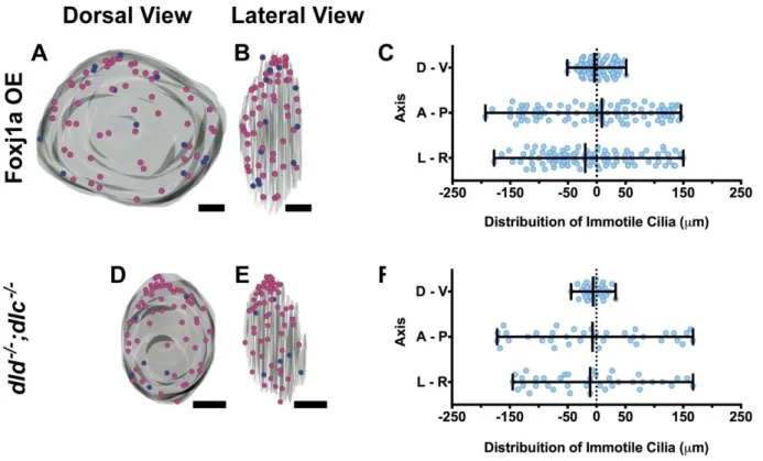
In Chapter 2, we focused in understand the pathways behind deciding between being a motile
vs immotile cilium in the LRO. Although all cilia are made motile in terms of ultrastructure due
to Foxj1a expression, the decision to move or not is dependent on Notch signalling alone.
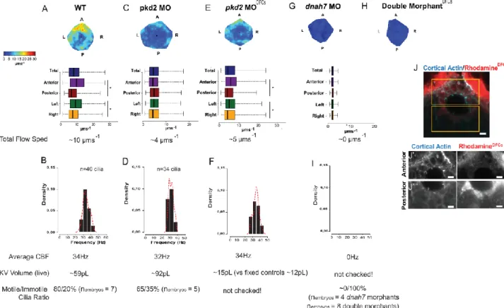
Then, we focused on further characterization of an important calcium channel, Pkd2, in the LR.
This channel is thought to partner with Pkd1l1 and sense flow, an important feature in LR. In
Chapter 3, we asked if having no flow had the same impact as having no Pkd2-mediated
sensing. The only manipulation that did not affect the LRO architecture was to target Pkd2 on
the LRO cells only, which still left visible Pkd2 protein and a slower flow. Still, all our
manipulations had the same phenotype: randomization of organ situs. In Chapter 4, we
performed a LRO-specific microarray between WT and pkd2 morphant embryos in order to
find other asymmetric genes present in the LRO. Although we did not find any gene expressed
asymmetrically between left and right side, we indeed find four new genes with minor roles in
LR: cacybp, frzb, pvalb6 and ncl1. In Chapter 5, we further focused in manipulating ncl1, a new
impacting on mesendoderm patterning. Indeed, we found that it has an impact on LR, probably
by influencing the secretion of some TGF- β signalling player. In Chapter 6, we set to establish
zebrafish as a good model to study kidney toxicity when metabolizing drugs.
Together, the results presented in this thesis provide new clues for LR axis establishment, from
cilia motility to new downstream genes of Pkd2 and calcium. It also highlights the zebrafish as
a good model to study human disease.
Key-words: Left-Right axis establishment, Pkd2, Cilia motility, TGF-
β signalling, Notch
Estabelecimento do eixo Esquerda-Direita (ED) é um processo complexo que ocorre cedo
durante o desenvolvimento. Exige a integração de várias vias de sinalização, tais como
TGF-β, Notch, Wnt e Cálcio. Também envolve a coordenação de dinâmica de fluídos, difusão de
morfogénios e movimento ciliar. Tudo junto e coordenado no tempo leva à correta localização
dos órgãos internos. Problemas no estabelecimento deste eixo estão normalmente
associados a doenças crónicas. As primeiras decisões assimétricas acontecem numa
pequena e transiente estrutura chamada o Organizador Esquerda Direita, uma estrutura ciliar
que existe em vários vertebrados. Cílios móveis geram um fluxo de fluido assimétrico que se
traduz numa expressão génica assimétrica entre o lado esquerdo e o lado direito. Estes sinais
são depois transferidos à Mesoderme Lateral, o tecido que mais tarde dá origem ao coração
e influencia a endoderme que dá origem a órgãos como o fígado e o pâncreas.
No Capítulo 2, focámo-nos em tentar compreender como é que as vias de sinalização se
coordenavam para decidir entre um cílio móvel e imóvel no Organizador. Embora a expressão
de Foxj1a em todas as células do Organizador produza cílios com ultra-estrutura compatível
com motilidade, a decisão entre mover ou não é exclusiva da via de sinalização Notch. A
seguir, focámo-nos em caracterizar Pkd2, um canal de cálcio importante no estabelecimento
do eixo ED. Este canal está associado ao Pkd1l1, uma molécula com domínios capazes de
sentir fluido e responder com entrada de cálcio na célula. No Capítulo 3, questionámos se não
ter fluxo no Organizador tinha o mesmo impacto que não ter um mecanismo para sentir esse
fluxo através da ausência do Pkd2. A única manipulação que não afetou a arquitetura do
Organizador foi remover o Pkd2 apenas nas células do Organizador, o que não é muito
eficiente e tem impacto na velocidade do fluxo dentro do Organizador. Ainda assim, todas as
manipulações efetuadas deram o mesmo fenótipo: randomização da posição dos órgãos. No
Capítulo 4, fizemos um estudo de transcritómica usando apenas as células do Organizador
encontrado genes com expressão assimétrica à volta do Organizador, encontramos quatro
novos genes que influenciam o estabelecimento do eixo: cacybp, frzb, pvalb6 e ncl1. No
Capítulo 5, focámo-nos na ncl1, um gene nunca antes associado ao estabelecimento do eixo
e que atua como antagonista da via de sinalização TGF-β ao influenciar a secreção de Lefty
e influenciando a padronização da mesoderme-endoderme. Nós descobrimos que este gene
impactua no estabelecimento do eixo, provavelmente ao influenciar a secreção de algum
elemento da via de sinalização TGF-β. No Capítulo 6, focámo-nos em estabelecer o
peixe-zebra como um bom modelo para estudar toxicidade no rim em resposta a fármacos.
Em suma, os resultados apresentados nesta tese providenciam novas pistas para o
estabelecimento do eixo ED, desde o movimento do cílio a novos genes a jusante do Pkd2 e
do cálcio. Também reforçámos a ideia de que o peixe-zebra pode ser um bom modelo para
estudar doenças humanas.
Palavras-Chave: Estabelecimento do eixo Esquerda-Direita, Pkd2, Motilidade ciliar, Via de
O correto estabelecimento dos órgãos internos no seu devido lugar dentro do organismo é
importante para o correto funcionamento dos mesmos. É um processo comum a vertebrados
e invertebrados, embora a forma como o atingem nem sempre é a mesma. Algo em comum
é a expressão assimétrica de genes da via de sinalização TGF-β nos tecidos embrionários
que vão dar origem a estes órgãos mais tarde durante o desenvolvimento. Em alguns
vertebrados, incluindo nós seres humanos, as primeiras decisões assimétricas ocorrem numa
pequena e transiente estrutura denominada Organizador Esquerda Direita (ED). Esta
estrutura é composta por células que têm à sua superfície um cílio inclinado. Este tem a
capacidade de mexer e a combinação de vários cílios móveis faz com que se gere um fluxo
de líquido assimétrico da direita para a esquerda. Este fluxo gera uma resposta assimétrica
nas células do Organizador: as células da esquerda têm uma subida da concentração de
cálcio intracelular e expressão de nodal enquanto as células do lado direito expressam
cerl2/dand5, o primeiro sendo um ativador e o segundo um inibidor da família TFG-
β. Esta
primeira assimetria é propagada pelos tecidos até chegar à Mesoderme Lateral, o tecido que
origina o coração e influencia a endoderme, que dá origem aos órgãos viscerais.
Muitas questões permanecem em aberto neste processo, particularmente como é que o fluxo
é interpretado pelas células do Organizador. Tipicamente, o Organizador é composto por
células com cílios móveis, responsáveis por gerar o fluxo, e células com cílios imóveis,
potenciais sensores desse mesmo fluxo. Há duas grandes teorias sobre isto: o fluxo
transposta um morfogénio em vesículas que embatem do lado esquerdo do Organizador e
libertam aí o seu conteúdo, dando assim origem à expressão assimétrica de genes; ou a força
e direção do fluxo são de alguma forma percebidos pelos cílios imóveis. A descoberta de um
complexo proteico composto por polycysteins (Pkd2, um canal de cálcio e Pkd1l1, um recetor)
nos cílios do Organizador deu força redobrada a esta segunda hipótese. Este complexo
mecânica do fluxo de urina, respondendo com uma entrada de cálcio nas células. A ausência
deste complexo dá origem à doença dos rins policísticos e em ratinho original também
problemas de lateralidade – perde-se a expressão assimétrica de genes da família TFG-β na
proximidade do Organizador e na mesoderme lateral e tem impacto na posição dos órgãos.
Com esta tese, começámos por tentar compreender melhor a base molecular por detrás da
decisão entre ser um cílio móvel ou imóvel no Organizador. Depois, tentámos averiguar se há
diferenças entre abolir completamente o fluxo ou não ter uma forma de sentir esse fluxo atrás
do knockdown do Pkd2 em termos de fenótipos ao nível da expressão génica e da posição
final dos órgãos. Através de um estudo de transcritómica, averiguamos quais os genes que
estão a jusante do Pkd2 e debruçámo-nos sobre 4 em particular: parvalbumin6, calcyclin
binding protein, frizzled-related protein e nicalin1. Por fim, fizemos uma comparação mais
exaustiva entre dois inibidores pertencentes à via de sinalização TFG-β: Dand5, que já era
conhecido por influenciar o estabelecimento ED, e Nicalin1, que nunca tinha sido descrito
neste processo. Todos estes trabalhos usaram o peixe-zebra como animal modelo. É possível
encontrar 70% de genes humanos neste vertebrado, o que o torna bastante atrativo para
investigar processos que ocorram de forma semelhante em humanos. O facto de os embriões
terem propriedades óticas que os tornam atrativos para microscopia e gerar um grande
número de embriões são algumas das características mais apreciadas pelos cientistas. Por
isso, terminámos com uma avaliação do peixe-zebra como um bom modelo para testar
toxicidade renal à exposição de fármacos.
Todas as células do Organizador expressam foxj1a, um gene necessário e suficiente para
especificar cílios móveis. No entanto, os resultados apresentados no segundo capítulo desta
tese mostraram que a via de sinalização Notch é responsável pela decisão móvel vs imóvel.
Recorrendo a estudos transcricionais, microscopia eletrónica com amostragem exaustiva do
todos os cílios expressarem foxj1a e os axonemas ciliares terem dynein arms e radial spokes
responsáveis pela motilidade, alguns cílios se mantém imóveis durante todo o processo. Esta
decisão é tomada cedo e parece ser a jusante da transcrição de her12, um elemento da via
de sinalização Notch. A sobrexpressão de her12 aumenta o número de cílios imóveis em troca
de cílios móveis, o que leva à perturbação do fluxo normal dentro do Organizador e tem
impacto na posição dos órgãos.
A importância do canal de cálcio Pkd2 no estabelecimento do eixo ED está bem fundamentada
no número de trabalhos feitos com este gene em vários animais modelo. Há várias evidencias
que apontam para esta proteína fazer parte do mecanismo capaz de sentir o fluxo e iniciar a
onda de cálcio observada no lado esquerdo. No terceiro capítulo desta tese perguntámos se
abolir o fluxo, que se sabe estar a montante da expressão assimétrica de dand5, originava
defeitos tão graves como abolir a forma de sentir este fluxo. Através deste trabalho
percebemos que tirar diferentes níveis de Pkd2 em todo o peixe-zebra levam a diferentes
respostas em termos de volume e arquitetura do Organizador. A única abordagem que
minimiza a interferência com a arquitetura do Organizador é quando se tira o Pkd2
exclusivamente das células do Organizador. No entanto, esta abordagem não remove
completamente o Pkd2 e resulta numa velocidade de fluxo mais baixa. De qualquer forma,
todas as manipulações deram o mesmo resultado em termos de dand5 (maioritariamente
simétrico) e randomização da posição dos órgãos.
Embora haja evidencias que suportam a importância da onda de cálcio observada à esquerda
do Organizador, pouco se sabe sobre o que é ativado a jusante da mesma. Por isso, no
Capítulo 4, fizemos um estudo de transcritómica usando apenas as células do Organizador
de embriões normais e embriões sem Pkd2. Da lista de genes obtida, escolhemos 4 para
manipular individualmente e perceber o seu impacto no eixo ED. Primeiro avaliou-se o padrão
dand5, o primeiro gene que se sabe ser assimétrico no Organizador do peixe-zebra. A seguir
avaliámos o impacto da sua manipulação na assimetria de dand5 e na posição dos órgãos.
Nenhum revelou ter impacto tão forte como manipular Pkd2, mas todos parecem ter algum
papel no estabelecimento do eixo ED. Em suma e para futura verificação, parvalbumin6 pode
estar a controlar a cálcio do lado esquerdo, frizzled-related protein pode afetar o tamanho
ciliar, calcycling binding protein pode influenciar a degradação de dand5 através de β-catenin
e nicalin1 pode influenciar a secreção de outros elementos da via TGF-β como Dand5 ou
Lefty.
Uma vez que Ncl1 está descrito na literatura como inibidor de TGF-β e nunca foi associado
ao estabelecimento do eixo ED, no Capítulo 5 decidimos explorar o impacto deste gene.
Knockdown deste gene leva a um aumento de defeitos de posição dos órgãos com um efeito
dependente da dose de morfolino usado. Afeta a expressão de dand5 e spaw, randomizando
o padrão e levando a uma menor expressão dos mesmos. Experiências onde se fez o
knockdown de ambos dand5 e ncl1 mostraram mais defeitos em termos de posição de órgãos,
apontando para uma potencial epistasia dos dois genes. Sobrexpressar dand5 em embriões
com knockdown de ncl1 não consegue melhorar os efeitos da ausência de ncl1, o que aponta
para potenciais vias separadas. Dados preliminares do mutante de ncl1 suportam os fenótipos
observados com o morfolino.
Finalmente, no Capítulo 6, confirmámos que o peixe-zebra pode ser um bom modelo para
testar a metabolização de fármacos como anti-retrovirais usados no tratamento da SIDA. Os
prónefros presentes nas larvas do peixe-zebra apresenta elevada homologia com o que se
encontra no rim humano. Neste estudo, através de várias técnicas como espectrometria de
massa para avaliar metabolismo, estudos de filtração de inolina para testar a função renal,
microscopia com multifotão e eletrónica de transmissão, avaliámos os danos que a exposição
estão em concordância com o tipicamente se observa nos rins humanos expostos aos
mesmos fármacos, confirmando o peixe-zebra um animal modelo bastante atrativo para este
tipo de estudos.
Os capítulos desta tese ilustram a versatilidade do peixe-zebra para testar o estabelecimento
do eixo ED nos vários níveis que influenciam este processo. Podemos estudar a formação
dos cílios no Organizador e podemos estudar em detalhe a expressão génica dos vários
intervenientes das diferentes vias de sinalização ao nível do Organizador e ao nível dos
tecidos que vão dar origem aos órgãos. Descobrimos novos interveniente nunca antes
descritos neste processo, adicionando novos níveis de complexidade a um processo já de si
fascinante.
BMP
– Bone Morphogenetic Protein
Cacybp – Calcyclin Binding Protein
Cerl2
– Cerberus-like 2
Dand5
– DAN family member 5
DFCs – Dorsal Forerunner Cells
DNA
– Deoxyribonucleic Acid
Dnah – Dynein axonemal heavy chain
ECM
– Extracellular Matrix
ER – Endoplasmic Reticulum
Fgf
– Fibroblast Growth Factor
Foxj1a – Forkhead box J1a
Frzb
– Frizzled related protein
ICOs – Intracilliary calcium oscillations
Her12 – Hairy-related 12
HPF
– Hours Post Fertilization
KV
– Kupffer’s Vesicle
LPM
– Lateral Plate Mesoderm
LR
– Left-Right
LRO
– Left-Right Organizer
mRNA
– Messenger Ribonucleic Acid
Ncl1 – Nicalin1
Pkd – Polycystin kidney disease
PSM
– Presomitic Mesoderm
Pvalb6 – Parvalbumin 6
SHH
– Sonic Hedgehog
SS
– Somite Stage
TGFβ – Transforming Growth Factor Beta
Wnt
– Wingless/Integrated Family Members
CHAPTER 1
Introduction
“It always seems impossible until it’s done”
1. INTRODUCTION
Correct Left-Right (LR) axis establishment comes from a combination of cell morphology,
ciliogenesis, fluid flow physical dynamics, several signalling pathways (calcium, Nodal, Notch
and Wnt), signal transduction and organogenesis. Although a lot has been done do address
all these steps and we already have a good idea how these steps coordinate to give rise to
correct organ position, still a lot is missing. One of the biggest questions that remains highly
debated in the field is how the biophysical properties of fluid flow are interpreted from the
Left-Right Organizer (LRO) cells. Is there a morphogen being released in the LRO space or
is it the mechanical force that is being somehow interpreted? Other related question regards
the two cilia types present in the LRO; what are the relevant differences between motile and
immotile cilia and whether these immotile cilia are the predicted sensors by McGrath et al. in
2003. What is also still unknown is what are the downstream targets of the asymmetric
calcium signalling on the left, and its relationship to the complementary asymmetry observed
for dand5. This thesis aims to answer to some of these questions and add more knowledge
to the field.
1.1 WHAT WE NEED TO KNOW ABOUT CILIA (for this study)
For many vertebrates, LR is decided downstream of the beating motion of cilia. So, in order
to understand LR, one needs to acquire some notions about this organelle. Cilia are hair-like
structures that occur on the surface of most cells. There are several subtypes of cilia but,
typically, cells have a primary, sensory cilium. These tend to be very dynamic in terms of
morphology and molecular composition, so they can sense fluid flow, light, odorants or
signalling molecules. Primary cilia are seen like a hub for signalling pathways like sonic
hedgehog (Huangfu et al., 2003), Wnt signalling (Corbit et al., 2008), Notch signalling
(Ezratty et al., 2011; Leitch et al., 2014) and others further reviewed in Pala et al., (2017).
The ultrastructure of a primary cilium is typically 9+0, which means that it has only 9 doublets
of microtubules and lacks motility components. Conventional motile cilia on the other hand
have a 9+2 organization, presenting an extra central pair of microtubules connected to the 9
outer doublets by radial spokes (Figure 1C). They also present dynein motors arranged in
outer and inner arms that allow for movement (as reviewed in Choksi et al., 2014). This kind
of cilia generally beat in a wavelike or corkscrew fashion to generate fluid movement or to
allow cells to move through fluid (Kramer-Zucker et al., 2005a) (Figure 1D). These 9+2 motile
cilia are what is typically observed in the LRO of zebrafish and Xenopus. The mouse LRO
presents motile cilia with a 9+0 configuration, with dynein arms but without central pair
(Hirokawa et al., 2006; Nonaka et al., 1998; Takeda et al., 1999) (Figure 1A). Odate et al.
(2016) observed very few 9+2 cilia in the mouse LRO, these being randomly distributed
(Odate et al., 2016). The absence of central pair and radial spokes makes the cilia
ultrastructurally more fragile than 9+2 cilia, but allows stable unidirectional rotation of node
cilia (Shinohara et al., 2015) (Figure 1B). In the mouse model, the direction of the flow is
Figure 1 – Cilia in Left-Right axis establishment(A) Motile cilium 9+0 typically found in the mice LRO. It has 9 pairs of microtubule doublets, with inner (yellow) and outer (blue) dynein arms. (B) Rotational movement of a 9+0 cilium. Scheme from (Huang et al., 2009). (C) Motile cilium 9+2 typically found in the zebrafish LRO. It has 9 pairs of microtubule doublets plus a central pair, with inner (yellow) and outer (blue) dynein arms and radial spokes (black). (D) Typical movement of a 9+2 cilium. Scheme from Pintado et al. (2017)
determined by two features: posterior tilt and clockwise rotation. Due to these two features,
mice cilia can generate a leftward effective stroke and a rightward recovery stroke on the cell
surface (Cartwright et al., 2004; Nonaka et al., 2005; Okada et al., 2005). Cilia tilt is given by
the basal body position in the cell. The basal body is initially positioned centrally but then
gradually shifts toward the posterior side of the node cells. Positioning of the basal body and
unidirectional flow were found to be impaired in mice lacking Dishevelled (Hashimoto et al.,
2010), Prickle (Antic et al., 2010) or Vangl (Antic et al., 2010; Song et al., 2010). All these are
components of the noncanonical Wnt signalling planar cell polarity (PCP) pathway. While
Dishevelled protein is localized to the posterior side of the apical membrane of mice LRO
cells (Hashimoto et al., 2010), Vangl and Prickle localized to the anterior side (Antic et al.,
2010). Vangl is also critical for LR determination in zebrafish and Xenopus (Antic et al., 2010;
Borovina et al., 2010). Also at play are opposing gradients of Wnt5a/b posteriorly and
secreted Frizzled-related proteins (Sfrp) inhibitors anteriorly that help polarize node cells
along the anterior-posterior axis (Minegishi et al., 2017).
The transcription factor Forkhead box J1 (Foxj1) is important to regulate the motile
ciliogenesis. Loss of Foxj1 in mice disrupts 9+2 motile cilia, leading to defective ciliogenesis
in airway epithelial cells and Left-Right axis establishment defects due to abnormal centriole
migration and/or apical membrane docking (Brody et al., 2000). Foxj1 is also important to
make motile monocila, including the 9+0 subtype found in the mice LRO (Alten et al., 2012;
Chen et al., 1998). Knockdown of Foxj1 in both Xenopus and zebrafish can cause loss of all
motile cilia (Stubbs et al., 2008; Yu et al., 2008) revealing that Foxj1a has a larger role than
motility per se. Foxj1 is considered a master regulator of motile cilia since it is responsible for
the regulation of a cohort of ciliary genes required for different structural and functional
aspects of motile cilia, such as the ones used to make, assemble, transport and dock the
inner and outer dynein arms, radial spokes and central pair (Didon et al., 2013; Jacquet et
al., 2009; Newton et al., 2012; Stubbs et al., 2008; Yu et al., 2008). In zebrafish, fibroblast
growth factor (FGF) signalling induces foxj1 expression in LRO (Neugebauer et al., 2009)
and Wnt signalling seems to act downstream of FGF to directly control foxj1 expression
through TCF/LEF transcription factor-binding sites within the foxj1 promoter (Caron et al.,
2012). This relationship between Wnt signalling and foxj1 expression is conserved in
Xenopus GRP (Walentek et al., 2012). Notch signalling has also been linked to motile cilia
through length and motility control. In zebrafish LRO, Notch signalling is required for proper
foxj1 expression, deltaD homozygous mutants showed significantly shorter cilia and
abnormal motile/immotile ratio, making more motile cilia at the expense of the immotile.
Overactivation of Notch signalling through Notch Intracellular Domain (NICD) lead to longer
and more immotile cilia in the zebrafish LRO (Lopes et al., 2010; Tavares et al., 2017). In
terms of length, two other studies reported opposing results. While, overactivation of Notch
signalling led to shorter cilia in CL4 cells (Jurisch-Yaksi et al., 2013), it led to neural tube
longer primary cilia in vitro and in vivo. In terms of motility, data from Xenopus LRO had
suggested a similar role for Notch signalling, through Galnt11 and NICD manipulation,
regarding motile/immotile cilia ratio (Boskovski et al., 2013). Although decreasing cilia length
has a high impact on left-right axis establishment, increasing length seems to cause milder
left-right defects in zebrafish. Arl13b overexpression, which increases cilium length without
affecting cilia beat frequency, results in reduced beat amplitude and similar flow strengths as
control embryos (Pintado et al., 2017).
1.2 WHAT WE NEED TO KNOW ABOUT NODAL SIGNALING (for this study)
LR axis establishment requires TGF-β family ligands to function at many different levels: from
LRO establishment, to interpretation of symmetry breaking fluid flow, to transducing and
propagating this asymmetric information into the presumptive tissues that will give rise to
organ positioning, the lateral plate mesoderm and the endoderm. The main players so far
studied in LR development of mouse, xenopus and fish are Nodal/Southspaw (Spaw) and its
auto-induction capabilities (Long et al., 2003; Ohi and Wright, 2007; Oki et al., 2007; Osada
et al., 2000; Saijoh et al., 2000; Wang and Yost, 2008), the inhibitors Cerberus/Dan and Lefty
(Branford et al., 2000; Hashimoto et al., 2004; Katsu et al., 2012; Marques et al., 2004; Meno
et al., 1996; Schweickert et al., 2010; Vonica and Brivanlou, 2007; Wang and Yost, 2008;
Yokouchi et al., 1999) and the transcription factor Pitx2 (St. Amand et al., 1998; Campione et
al., 1999; Essner et al., 2000; Logan et al., 1998; Piedra et al., 1998; Yoshioka et al., 1998).
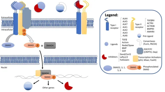
Figure 2 – TGF-β signalling pathway overview
There are many ligands in TGF-β signalling. Focusing on Nodal protein, it has to be processed in the extracellular space by Convertases to become fully mature. Mature Nodal binds to Type I and Type II receptors and the co-receptor from the EGF/CFC family. When activated, the receptors phosphorylate SMAD, which can bind to SMAD4. This complex can enter the nucleus and activate transcription by binding to transcription factors like FoxHI. The ligand Nodal and its repressor Lefty are both expressed in response to Nodal signalling. The interaction of Nodal with inhibitors like Lefty or Cerl2 in the outside of the cells affects its ability to bind to the receptors and activate the pathway. Scheme adapted from Schmierer and Hill, (2007).
Nodals are synthesized as proproteins that are proteolytically processed extracellularly by
conversates (subtilisin-like proprotein 1 and 4, also known as Furin and Pace4) (Beck et al.,
2002; Le Good et al., 2005). Several experiments led to the understanding that Nodal acts
via activin receptors, Smad transcription factors and associated transcription factors such as
FoxH1 (as reviewed in Shen and Schier, 2000; Whitman, 2001). Nodal/Spaw bind to a
receptor complex composed of type I and type II receptors that can function as
serine/threonine kinases (Attisano and Wrana, 2002; Shi and Massague, 2003).
Ligand/receptor assembly results in phosphorylation and activation of type I receptor through
the type II receptor, which in turn starts the phosphorylation of a downstream Smad cascade.
Nodal signalling acts by regulating the phosphorylation of Smad2 (Kumar et al., 2001; Lee et
al., 2001; Yeo and Whitman, 2001), which forms a complex with Smad4 and enter the
nucleus. Then, it binds to transcription factors like FoxH1 (previously known as Fast2) and
activates transcription of lefty1/2, nodal itself and pitx2, as well as other genes (reviewed in
Whitman, 2001). Multiple receptors/ligands can diversify the output of Nodal signalling.
Members of the epidermal growth factor (EGF-CFC) family are extracellular and GPI-linked
proteins that include zebrafish one-eyed pinhead (Oep), frog FRL-1, chick CFC and
mouse/human Cripto/Cryptic (reviewed in Shen and Schier, 2000). These are important
components of the Nodal signalling pathway by acting as coreceptors for Nodal ligands. Its
absence leads to defects in germ layer formation, organizer development and positioning of
the anterior-posterior axis (Gritsman et al., 1999). Lefty action is also EGF-CFC-dependent,
by interacting with EGF-CFC proteins and competing with Nodal for binding to these
coreceptors (Cheng et al., 2004). In the LR context, EGF-CFC is expressed at both right and
left Lateral Plate Mesoderm (LPM), where it is thought to interact with Nodal and induce
Nodal signalling, and at the notochord, where it physically binds to Lefty1 to antagonize
Nodal signalling (Shen et al., 1997; Yan et al., 1999). LR nodal antagonists Lefty proteins
are secreted and can compete with Nodals by binding to common receptors and by
physically binding to Nodal (Chen and Schier, 2002; Cheng et al., 2004). Nodal signalling
induces the expression of both nodal and of its antagonists (Hamada et al., 2002).
In
zebrafish, lefty1 is expressed first in the posterior notochord, prior to asymmetric gene
expression, where it might be inhibiting Nodal signalling from happening before the
asymmetric cues from the LRO. An oep-dependent signalling event in the midline weakens
the midline lefty1, alleviating the repression and allowing for the asymmetric left cue from the
LRO to start Nodal signalling in the left LPM (Burdine and Grimes, 2016). Another important
inhibitor is Dand5, a member of the differential screening-selected gene in neuroblastoma
(DAN) family of predicted secreted proteins, that exhibits a finger-wrist-finger architecture
with the cystine-knot motif, which makes the overall architecture similar to other cystine-knot
containing proteins including BMP and Noggin (further reviewed in Nolan and Thompson,
2014). Cerberus2 in mouse, Coco in xenopus and Charon in zebrafish were all renamed as
Dand5 and are potent secreted inhibitors of Nodals, impacting on mesendoderm patterning
and left-right establishment (Bell et al., 2003; Belo et al., 2000; Hashimoto et al., 2004;
Pearce et al., 1999; Piccolo et al., 1999). Most of TGF-β signalling interactions are illustrated
in Figure 2.
Since Nodal act as a morphogen, it is important to understand how gradients are formed.
Alan Turing, in his paper entitled “The Chemical Basis of Morphogenesis” in 1952 predicted a
chemical mechanism for biological gradient pattern formation (Turing, 1952). He suggested a
reaction-diffusion system where chemicals react to each other and diffuse across the space.
This
concept was redefined by Lewis Wolpert in his “French Flag Model” (Wolpert, 1969),
where morphogens affect a field of cells in a concentration-dependent fashion
– more
morphogen activate certain genes while less morphogen activates other genes. Only
decades later, it was formally demonstrated by Christiane Nüsslein-Volhard in Drosophila,
with the identification of bicoid as a morphogen (Berleth et al., 1988). There are many models
for morphogen gradient formation based in complex mathematical equations, it can be put in
simplified terms (thoroughly reviewed in Müller et al., 2013). It is possible to describe five
major models: free diffusion, tortuosity and binding-mediated hindered diffusion, facilitated
diffusion, transcytosis and directed transport via filopodial extensions known as cytonemes
(Figure 3). Free diffusion is the simplest case, where molecules move freely from the source
to the target tissue. However, this would not produce a gradient since this would result in
uniform distribution of morphogen in the target tissue. Gradient formation happens when
either the morphogen is degraded or is permanently trapped by a cell, producing a graded
distribution of morphogens on the target tissue. Hindered diffusion assumes that the target
tissue is densely packed with cells, which can cause hindrance by making the morphogens
going around them (tortuosity-mediated hindrance), and/or by transiently binding
morphogens through receptors or extracellular matrix components (binding-mediated
Figure 3 - Theories of Morphogen Transport
There are 5 major models to explain gradient formation of a morphogen: free diffusion, hindered diffusion by tortuosity or binding, facilitated diffusion, transcytosis and cytonemes. Scheme adapted from Müller et al. (2013).
hindrance). This would allow a faster gradient formation. Facilitated diffusion and shuttling
assumes
that a morphogen is largely immobile until a “positive” diffusion regulator comes
and release the hindrance exerted by the “negative” diffusion regulators. In this scenario,
morphogens and “negative” diffusion regulators are uniformly distributed while the “positive”
diffusion regulators have a localized source and help to generate the morphogen gradient.
These diffusion-based models have four potential weaknesses: length of patterning fields,
solubility of morphogens, reliability of patterning and geometry of patterning fields. Because
of that, two additional models were proposed. Transcytosis, where morphogens bind to cell
surface and are endocytosed, only to be exocytosed again and endocytosed by other cells.
This process leads to a slow shaped gradient over time. The final model is through
cytonemes, where long dynamic filopodia-like structures project from target cells and contact
morphogen-producing cells. Therefore, morphogens are
‘handed over’ to and transporter
along cytonemes, forming a concentration gradient (reviewed by Müller et al., 2013).
Nodal and Lefty have signal peptide sequences required for secretion (Beck et al., 2002;
Blanchet et al., 2008; Le Good et al., 2005; Jing et al., 2006; Marjoram and Wright, 2011;
Meno et al., 1996; Müller et al., 2012; Sakuma et al., 2002; Tian et al., 2008; Zhou et al.,
1993) and experiments with fused GFP support an extracellular localization (Müller et al.,
2012). This supports the idea that these two proteins move through tortuosity-mediated
diffusion, where cells increase the path length of molecules diffusion in the extracellular
space. Therefore, global effective diffusion coefficient of molecules moving through a tissue
should be lower than the local diffusivity. Lefty and Nodal have similar local diffusivity (which
could explain by having similar molecular weight) (Müller et al., 2013). However, while Lefty
showed a local diffusivity double of its effective diffusivity, Nodal had a local diffusivity 90%
higher than its effective diffusivity. This suggests that Lefty is mostly hindered by cell packing,
while Nodal is hindered by cell packing and extracellular diffusion regulators (Müller et al.,
2013), explaining why Lefty is long range while Nodal is short range (Chen and Schier, 2002;
further reviewed in Schier, 2003). Diffusional movement can be altered by transient binding
to other molecules such as receptors or components of the extracellular matrix (Baeg et al.,
2004; Belenkaya et al., 2004; Wang and Yost, 2008). Also, different affinities for receptors
might account for different ranges of distribution (Müller et al., 2012). Another mechanism
might be rapid clearance of molecules during diffusion (Kicheva et al., 2007). All data seems
to indicate that Nodal gradient is dependent of diffusion, binding and degradation of the
morphogen (Wang et al., 2016).
1.3 LEFT-RIGHT AXIS ESTABLISHMENT
1.3.1 MICE
Left-Right Hypothesis
Immotile cilia syndromes in Humans, such as Kartagener syndrome, are typically
accompanied with LR axis abnormalities of organ situs which were thought to be associated
with ciliary dysfunction (Afzelius, 1976; Kosaki and Casey, 1998). Finding asymmetric gene
expression like lefty (Meno et al., 1996) and nodal (Collignon et al., 1996; Lowe et al., 1996)
and motile cilia (Nonaka et al., 1998) in the embryonic node linked both fluid flow dynamics
and gene expression. Two mice models were extremely important for the progress of LR
studies: situs inversus viscerum (iv) and inversion of embryonic turning (inv). While the first
was a mouse with a spontaneous mutation in the axonemal dynein heavy-chain 11 gene
(also known as left/right-dynein) that led to random organ situs (Hummel and Chapman,
1959; Supp et al., 1997), the second was an insertion mutation that affected inversin gene
and always resulted in situs inversus (Mochizuki et al., 2002; Morgan et al., 2002; Yokoyama
et al., 1993). These mutants presented problems in nodal expression around the node and in
the Lateral Plate Mesoderm (LPM), becoming bilateral or reversed (Lowe et al., 1996). Both
iv and inv mutants had flow abnormalities, but while iv had all cilia immotile and no flow, inv
mutant had motile cilia that produced a very weak and disorganized leftward flow. Besides,
inv mutants had a misshaped node (Okada et al., 1999) which confirmed the shape of node
was also important for proper flow. A third mutant, a KIF3B knockout mice, was done around
the same time and also showed LR randomization problems. Disruption of KIF3B, a
microtubule-dependent motor, was shown to be important for cilia assembly and
maintenance. Without it, the node lacked cilia and therefore showed randomization of organ
position and randomization of lefty2, a nodal antagonist (Nonaka et al., 1998). Mutations in
KIF3A, that heterotrimers with KIF3B (Kondo et al., 1994; Yamazaki et al., 1995), showed
similar phenotypes (Takeda et al., 1999). In fact, Nonaka et al. (1998) proposed the first
morphogen model for LR axis establishment, where a secreted factor X would be transported
by the fluid flow. Moreover, this model can explain why cultured embryos frequently reverse
their body situs. A mature node comprises approximately 250 cells, with 50-60um wide and
70-90um long, and up to 50um deep (Sulik et al., 1994; Bellomo et al., 1996; Yamanaka et
al., 2007). It is a concave, tear-drop-shaped epithelial field of cells located distally on the
ventral surface of the embryo, covered by the Reichert’s membrane (Figure 4A). The removal
of this membrane during the culture procedure exposes the node to the outer environment,
increasing the external turbulence and allowing diffusion of the factor X.
Further confirmation that motile cilia were crucial in LR came from targeted deletion of the
ATP binding domain of lrd, which also resulted in randomization of laterality due to immotility
of node cilia (Supp et al., 1999). All this body of information indicates that flow occupies an
upstream position in this process, elegantly backed up by the manipulative flow experiments
done by Nonaka et al. (2002). This work showed that artificially provided strong or weak
leftward flow did not affect pitx2 or heart looping, while strong (but not weak) rightward flow
could reverse organ situs in cultures of WT mice submitted to a fluid peristaltic chamber. The
same experiments were repeated with iv mutant embryos, that lack fluid flow due to immotile
cilia. When iv mutant embryos were submitted to strong or weak leftward flow, they showed
left sided pitx2 and normal heart looping, while strong or weak rightward flow induced
reversal LR situs (Nonaka et al., 2002).
Figure 4 – Left-Right axis establishment in Mice
(A) Schematic of a mice LRO, with immotile cilia in the periphery and motile cilia in the pit. The flow generated has two main directions: from right to left in the middle plane and from left to right ventrally, near the Reichert’s membrane and dorsally on the LRO cell surface. NVPs are depicted in yellow spheres with blue content, that are released on the right side by microvilli (in yellow) and break on the left side, releasing there the content and leading to an intracellular calcium rise (orange end). Scheme adapted from Tanaka et al. (2005). (B) Schematics of Nodal signalling pathway in LR. The node has a tear-drop shape with motile cilia (black dots). Nodal is expressed on the left side of the LRO (yellow crescent moon) where is also observed an intracellular calcium rise (black dots on the yellow crescent moon). The antagonist cerl2 is expressed on the right side (light blue crescent moon). Another antagonist, lefty is expressed in the midline. Since there is stronger Nodal signalling on the left side versus the right side, Nodal protein can travel to the left Lateral Plate Mesoderm (LPM) and start Nodal signalling there (big crescent moon in orange). Blue contours around the LPM and midline means EGF/CFC expression. A – Anterior, P – Posterior, D – Dorsal, V – Ventral, L – Left, R - Right