Evaluation of different cell disruption processes on encysted cells of
Haematococcus pluvialis:
effects on astaxanthin recovery and implications
for bio-availability
M.M. Mendes-Pinto, M.F.J. Raposo, J. Bowen
1, A.J. Young
1& R. Morais
∗Escola Superior de Biotecnologia, Universidade Cat´olica Portuguesa, Rua Dr. Ant´onio Bernardino de Almeida, 4200-072 Porto, Portugal
1Carotenoid Research Group, School of Biological & Earth Sciences, Liverpool John Moores University, Byrom
St., Liverpool L3 3AF, UK
(∗Author for correspondence; fax +351-225090351; e-mail [email protected])
Key words: astaxanthin, bio-availability, Haematococcus, microalgae, processing
Abstract
Although Haematococcus pluvialis is one of the most important natural sources of the carotenoid astaxanthin as a pigmentor for the aquaculture industry, the thick sporopollenin cell wall in the cysts hinders astaxanthin extraction and its subsequent bio-availability to fish. A range of physical and chemical processes were tested to promote the disruption of the encysted cells. The efficacy of these processes was evaluated in terms of astaxanthin recovery, which was assessed by determining the extent of leaching of astaxanthin into an organic solvent. The processes tested were: autoclave 30 min, 121◦C, 1 atm; HCl 0.1 M, 15 min and 30 min; NaOH 0.1 M, 15 min and 30 min; enzymatic treatment with a mixture of 0.1% protease K and 0.5% driselase in a phosphate buffer, pH 5.8, 30◦C, for one hour; spray drying, inlet 180◦C, outlet 115◦C; and mechanical disruption, with a cell homogeniser developed for this purpose. The mechanical (homogenisation) and autoclave treatments were the most effective in terms of extraction and availability.
Introduction
Haematococcus pluvialis Flotow (Chlorophyceae) has
a complex life-cycle involving several stages from motile flagellated zooids through to palmella and en-cysted stages (Elliot, 1934). This alga is one of the most important natural sources of the carotenoid pig-ment astaxanthin (3,30-dihydroxy-β,β’-carotene-4,40 -dione) and is cultivated commercially by several com-panies. Under optimum conditions, astaxanthin is completely absent from the cells. Upon exposure of the cells to growth-limiting conditions (typically as a result of nutrient – especially nitrogen – limitation or exposure to very high irradiance or high temperature: Droop, 1954; Fan et al., 1994; Harker et al., 1996a), astaxanthin is synthesised and accumulated within oil droplets.
Haematococcus can accumulate up to 8% dry
weight of astaxanthin, in the form of 3S,30S
enan-tiometer, as a mixture of mono and di-esters (Harker et al., 1996b). The composition of the aplanospores is somewhat dependent upon the age of the culture so that the ratio of mono-esters: di-esters decreases with time (Harker et al., 1996b).
Astaxanthin biosynthesis in this alga is gener-ally, but not exclusively (A. Hartley & A.J. Young, unpublished data), linked to the generation of apla-nospores and the associated formation of a thick sporopollenin wall around the cell. The subsequent bio-availability of the astaxanthin is limited due to this wall in the astaxanthin-rich cysts (Good & Chapman, 1979). Intact astaxanthin-rich cysts of
et al., 1991). Thus there is a need to extract or rup-ture these cysts prior to use in order to achieve a desired level of pigmentation in the target organism. Whilst it is possible to obtain a ‘cell-free’ extract of carotenoids, this is not practical on a large scale. Nor is it feasible to extract and purify ‘free’ astaxanthin from Haematococcus, in part due to the sensitivity of astaxanthin to oxidation. A range of alternative pro-cesses have been suggested for processing algal cells. For example, Bubrick (1991) described a process in-corporating cryogenic (–170 ◦C) grinding of dried
Haematococcus biomass in the presence of butylated
hydroxytoluene.
The main aim of this study was to evaluate a range of different physical and chemical treatments on the disruption of encysted cells of H. pluvialis, in terms of astaxanthin recovery and the effect on subsequent ‘po-tential bioavailability’ of pigment from the processed biomass.
Materials and methods
Haematococcus pluvialis (34/7 strain of CCAP –
Culture Collection of Algae and Protozoa, Win-dermere,UK) was cultivated in modified Bold’s Basal Medium (Nichols & Bold, 1964). The culture was grown at 21 ± 1 ◦C for 14 days in plastic sleeves of 10-L volume into which ambient air was used to agitate the culture. Illumination was provided by cool white fluorescent lamps (L36 W/21 Hellweiss Luminux Coolwhite, Osram, Lisbon, Portugal) at an irradiance of 46 µmol photon m−2s−1. After 14 days, the irradiance was increased to 80 µmol photon m−2 s−1 in order to stimulate astaxanthin biosynthesis in the alga.
Processing of algal biomass
After sedimentation and centrifugation (10 min,
1000 × g) of the encysted cells, the de-watered
astaxanthin-rich biomass was divided into aliquots and submitted to the following processes: (i) autoclaved 30 min, 121◦C, 1 atm; (ii) HCl 0.1 M, 15 min and, (iii) 30 min; (iv) NaOH 0.1 M, 15 min and, (v) 30 min; (vi) enzymatic treatment for 1 hour with a mixture of 0.1% protease K and 0.5% driselase in a phosphate buffer, pH 5.8, 30◦C; (vii) spray drying, inlet 180◦C, outlet 115◦C; and, (viii) mechanical disruption using a cell homogeniser developed for this purpose (patent pending). A steady stream of nitrogen was applied
during the biomass mechanical processing in order to prevent or minimise oxidation reactions that might oc-cur. All the treatments were carried out in triplicate. Following treatment, the astaxanthin content and com-position of the processed biomass was determined by HPLC (see below).
Astaxanthin determination
After the addition of 2 mL ethanol and glass beads to the test tubes, triplicates (30 mg) were submit-ted to a combination of vortex mixing (1 min) and ultrasound treatment (15 min). Samples were centri-fuged (5 min at 1000× g), the supernatant removed and the pellet re-suspended in 2 ml diethyl ether / ethanol (1:1, v/v). After repeating all the steps the pellet was extracted with 2 ml diethyl ether and finally with 3 ml n-hexane. All supernatants were pooled and the sample was evaporated to dryness under a steady stream of oxygen-free nitrogen.
Extraction into acetone
Pigment availability in processed algal biomass was assessed by extraction of astaxanthin into acetone (2 mL), from 30 mg cells (centrifuged as above). The material was gently mixed using a magnetic stir-rer and left for 16 hours at room temperature. The sample was filtered prior to analysis to remove any cell debris. These conditions were previously found to permit maximum extraction of cellular astaxanthin from astaxanthin-rich materials, without any degrada-tion of carotenoid. This assay was used to compare the effects of different processing methods on subsequent astaxanthin availability from cells of Haematococcus.
Spectrophotometry
Astaxanthin content was determined using a double beam UV-VIS spectrophotometer (Shimadzu 1601) within a wavelength range 400–700 nm. For astax-anthin quantification, the extracts originated from the solvent mixture extraction were re-suspended in n-hexane; those obtained from the contact with acetone were analysed directly in acetone. The extinction coef-ficient of astaxanthin in both solvents is 2100 and the
λmax is 470 nm in n-hexane and 476 nm in acetone
Thin-Layer Chromatography (TLC) – one dimensional separation of carotenoids
Pre-coated silica gel H60 thin layer chromatography plates 20 × 20 cm (Merck, Darmstadt, FRG) were used for the separation of carotenoids. Acetone, n-hexane, methanol (all from Merck, Darmstadt, FRG) and citric acid (Sigma) were laboratory grade. The carotenoid standards, β-carotene and astaxanthin (pur-chased from Sigma) were dissolved in a small volume of n-hexane at 1mg/ml; 2.5 µg was applied to the TLC plate. Samples of 30 mg from all treatments were evaporated to dryness and reconstituted individually in 500µl n-hexane, from which 1.2 mg was applied to the TLC plate. All samples and standards were kept dried, under oxygen-free nitrogen, in the dark at –4◦C until used.
All separations were carried out in developing chambers presaturated with the appropriate solvent system, at room temperature. Carotenoids were sep-arated using a solvent system of acetone: n-hexane, 3:7 (v/v) (Kobayashi et al., 1991). Before immersing the plate into the solvent system, the plate was pre-run in a 2.5% (w/v) solution of citric acid in methanol. This prevented the tailing of carotenoids that posses a 3-hydroxy, 4-keto end-group such as astaxanthin and astacene. The TLC plates were then dried at 40◦C for 10 min.
Analysis of chromatograms was made using an imaging densitometer (Model GS-700, Bio-Rad) op-erating with a reflectance resolution of 56 µm (450 dpi), with a blue filter at a pixel depth of 8.
High Performance Liquid Chromatography (HPLC)
The HPLC analyses were carried out using a mod-ified method based on that described by Vecchi et al. (1987). Samples were analysed by normal phase HPLC using a diode array detector (HP1100, Hewlett Packard). A Lichrosorb column (12.5× 4.6 cm, 5µm particles) was pre-coated with 1% H3PO4in methanol
(v/v) before used, to prevent tailing of astaxanthin. The mobile phase was n-hexane: acetone, 86:14 (v/v; Merck, Darmstadt FRG) operating at a flow rate of 1.2 ml/min. In order to improve separation efficiency, the polarity of the mobile phase was changed during the run (run time 25 min); the gradient elution was 1:1 (v/v) n-hexane: acetone. The sample volume applied was 20 µl following solubilisation of the carotenoid extract in 100 µl n-hexane.
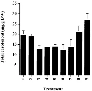
Figure 1. Total carotenoid content of Haematococcus cysts follow-ing processfollow-ing: 1. Control (intact); 2. Autoclave; 3. HCl 15 min; 4. HCl 30 min; 5. NaOH 15 min; 6. NaOH 30 min; 7. Enzyme; 8. Mechanical disruption; 9. Spray drying. See Materials and Methods for details.
Scanning Electron Microscopy (SEM)
The Haematococcus processed biomass was examined by scanning electron microscopy using a JEOL JSM-840 microscope at an accelerating voltage of 1– 15 KV. The samples were lyophilised (CHRIS AL-PHA 1–4,B-Braun Biotech International) at –52 ◦C, and coated with a thin gold-palladium film (10–15nm) using a Polaron E5000 sputter unit.
Results
The carotenoid composition of the Haematococcus biomass used in this study was 70.5% astaxanthin mono-esters, 24.7% astaxanthin di-esters and 4.8% lutein. Traces of β-carotene were observed but were not quantified. The astaxanthin content of the algal biomass was approximately 2% (w/dw) which is com-parable to the commercial source of this material currently available (NatuRose, Cyanotech Corp.). This pigment composition is typical of encysted cells of this alga, with high levels of astaxanthin mono-esters predominating (Grung et al., 1992; Harker et al., 1996b).
The effects of processing of the algal biomass on the carotenoid content of the cells is shown in
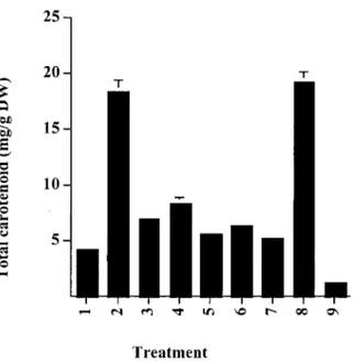
Figure 2. Extraction of total carotenoids into acetone from pro-cessed Haematococcus biomass. 1. Control (intact); 2. Autoclave; 3. HCl 15 min; 4. HCl 30 min; 5. NaOH 15 min; 6. NaOH 30 min; 7. Enzyme; 8. Mechanical disruption; 9. Spray drying. See Materials and Methods for details.
Figure 1. This shows that, compared to the control, the highest recovery of astaxanthin was obtained in the autoclaved, spray-dried and mechanical disrupted cells. None of these processes has a detrimental effect on the carotenoid content and composition of the algal biomass. However, enzymatic treatment of
Haemato-coccus cells or exposure to alkali or acid, resulted
in a significant loss of carotenoid (20–35% of total carotenoid) as a direct result of processing. Caroten-oids such as astaxanthin which possess the 3-hydroxy, 4-keto end-group are unstable, especially in the pres-ence of alkali. However, the prespres-ence of oxidative products of astaxanthin, namely astacene and semi-astacene, could not be confirmed by HPLC analysis. Chromatographic analysis of these processed materi-als revealed that selective degradation of carotenoids did not occur and that losses were seen for all caroten-oids. The high yield of carotenoid from the biomass obtained by spray-drying may be explained by the re-duced water content facilitating better extraction by the organic solvents.
The use of algal astaxanthin as a pigmentor in the aquaculture industry is dependent upon the bio-availability of the carotenoid from the algal biomass. The physical barrier of the sporopollenin cell wall in the cysts of Haematococcus is thought to be a key factor in limiting carotenoid bio-availability in the
gastro-intestinal tract of salmonids. The leaching of carotenoids, under gentle conditions, from biological materials into an organic solvent provides a simple, rapid estimation of the extent to which the cell wall is disrupted. Intact cysts of Haematococcus only re-lease∼20% of astaxanthin into acetone over a 16 hour period (Figure 2). Similar low yields were observed for cells that were subject to enzymatic treatment, spray-drying or exposure to acid or alkali. High re-coveries of carotenoid (>85%) were only observed for cells that had been autoclaved or mechanically processed. This suggests that these two methods of processing algal biomass may be most effective in terms of pigmentation as the cell contents are readily accessible.
In general, the leaching of carotenoids into acet-one did not result in a high recovery of lutein and
β-carotene (data not shown). This may be due to
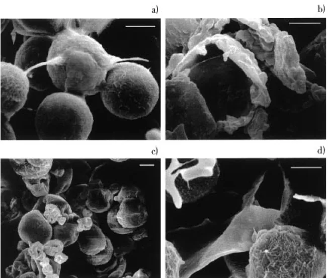
their binding to the various pigment-protein com-plexes within the chloroplast, whilst the astaxanthin esters are accumulated in extra-chloroplastic globules. In addition to using solvent extractability as a means of assessing the effectiveness of processing on the algal biomass the direct effect that processing had on the cell wall of the alga can be observed by scanning electron microscopy (Figure 3 a-d). The un-processed cysts of Haematococcus (Figure 3a) are fully intact with no signs of pitting or damage to the cell wall. In contrast, the cells subjected to mechanical processing (Figure 3b) and autoclaving (Figure 3d) are clearly disrupted. A noticeable effect of spray-drying the algal biomass (Figure 3c) was the clumping of cells, although the cells are not damaged by this pro-cess. Exposure to NaOH did result in some distortion and collapse of some cells, especially those exposed to alkali for a longer period. Some pitting of the cell wall was observed with treatment by HCl, whilst enzymatic treatment had no visible effect on cell morphology (data not shown).
Discussion
The bio-absorption or bio-availability of carotenoids in animals and humans is a critical aspect concern-ing the use of these natural pigments / anti-oxidants in the food and feedstuff industries (Castenmiller & West, 1997). In aquaculture, the relatively poor re-tention (typically only a few percent; Torrissen et al., 1989) of astaxanthin and canthaxanthin in salmonids has significant implications in terms of cost. Nat-ural sources of astaxanthin include krill waste, the
Figure 3. Scanning electron micrographs of Haematococcus pluvialis (× 2000), bar size 10 µm: (a) control (intact) cells, (b) mechanical disrupted cells, (c) spray-dried cells, and (d) cells autoclaved.
yeast Phaffia rhodozyma, flowers of Adonis annua and a number of microalgae, notably Haematococcus
pluvialis (Bernhard, 1990).
Salmonids appear to lack the digestive enzymes necessary to break down the sporopollenin wall found in cysts of Haematococcus. A number of factors may be responsible for limiting the bio-availability of astaxanthin from Haematococcus cells. The use of astaxanthin-rich cells of this alga in pigmentation tri-als on rainbow trout has shown that intact cysts do not permit pigmentation and that cellular astaxanthin only becomes bio-available once the cyst is broken or disrupted (Johnson & An, 1991; Sommer et al., 1991; J. Bowen, S. Davies, S. Lagocki, A.J. Young, unpublished). In addition, astaxanthin accumulated in algae such as Haematococcus is esterified, principally
as mono-esters, whilst the synthetic products are free carotenoid. A study by Foss et al. (1987) demonstrated that racemic astaxanthin di-palmitate did not pigment salmonids as effectively as free astaxanthin. A more recent study in rainbow trout has however demon-strated that both the natural mono- and di-esters of astaxanthin from Haematococcus, when administered to fish in an isolated form, pigment the muscle tis-sues as effectively as synthetic astaxanthin (J. Bowen J, S. Davies, R. Serwata, & A.J. Young, unpublished data). Another factor that distinguishes algal astax-anthin from the synthetic form is the configuration of the carotenoid. In algae, astaxanthin occurs as the (3S, 30S) form (Renstrøm et al., 1981) whilst the synthetic
product is a racemic mixture of the (3R, 30R), (3S, 30S)
et al., (1987) demonstrated that all three epimers of astaxanthin are equally utilised by salmonids, which suggests that the algal form of the carotenoid would not be discriminated against by the fish.
The cracking or disruption of the algal cell there-fore appears to be the single most important factor in utilisation of algal astaxanthin. It is, however, import-ant to note that astaximport-anthin accumulation in
Haemato-coccus is not restricted to the formation of cysts and
that this carotenoid can be biosynthesised at a number of stages of the alga’s complex life cycle (see Intro-duction). Similarly the formation of the sporopollenin cell wall, and hence the apparent ‘toughness’ of the cell, alters with the age of the cyst (Burczyk, 1987).
Several methods have been proposed to disrupt algal cells (Farrow & Tabenkin, 1966; Ruane, 1977; Nonomura, 1987) although most methods are not very efficient for disrupting the sporopollenin wall of Haematococcus cysts. These cysts are similar to those found in many microalgae and in pollen from higher plants (VanWinkle, Swift & Rickoll, 1997). They are particularly resistant to chemical attack, in-cluding KOH and acetolysis but can be attacked by chromic acid. Indeed, resistance to acetolysis is used in part to characterise such cell walls. According to the results obtained from this study it can be observed that both mechanical disruption and autoclaving of the astaxanthin-rich algal biomass are effective treat-ments. Both processes result in a very high yield of astaxanthin (Figure 1; i.e. no detrimental effects were observed due to processing per se). In addition, the extractability of the astaxanthin (Figure 2) from these processed cells was very efficient suggesting effective cracking of the cyst wall. This was confirmed by SEM for both products (Figure 3).
Acknowledgements
This work was supported by a Framework IV Project FAIR CT-97-1518.
References
Bernhard K (1990) Synthetic astaxanthin. The route of a carotenoid from research to commercialisation. In Krinsky NI, Mathews-Roth MM, Taylor RF (eds), Carotenoids. Chemistry and Biology. Plenum Press, New York, pp. 337–364.
Britton G (1995) UV/Visible spectroscopy. In Britton G, Liaaen-Jensen S, Pfander H (eds), Carotenoids Vol. 1B Spectroscopy. Birkhaüser Verlag, Basel, pp. 13–62.
Bubrick P (1991) Production of astaxanthin from Haematococcus. Biores. Technol. 38: 237–239.
Burczyk J (1987) Cell wall carotenoids in green algae that form sporopollenin. Phytochemistry 26: 121–128.
Castenmiller JJM, West CE (1997) Bioavailability of carotenoids. Pure appl. Chem. 69: 2145–1250.
Choubert G, Heinrich O (1993) Carotenoid pigments of the green alga Haematococcus pluvialis: assay on rainbow trout, Onco-rhynchus mykiss, pigmentation in comparison with synthetic astaxanthin and canthaxanthin. Aquaculture 112: 217–226. Droop MR (1954) Conditions governing haematochrome formation
and loss in the alga Haematococcus pluvialis. Arch. Mikrobiol. 20: 391–397.
Elliot AM (1934) Morphology and life history of Haematococcus pluvialis. Arch. Protistenk. 82: 250–272.
Fan L, Vonshak A, Boussiba S (1994) Effect of temperature and irra-diance on growth of Haematococcus pluvialis (Chlorophyceae). J. Phycol. 30: 829–833.
Farrow WM, Tabenkin B (1966) Process for the preparation of lutein. U.S. Patent n◦3 280 502.
Foss P, Storebakken T, Austreng E, Liaaen-Jensen S (1987) Caroten-oids in diets for salmonids V. Pigmentation of rainbow trout and sea trout with astaxanthin and astaxanthin di-palmitate in comparison to canthaxanthin. Aquaculture 65: 293–305. Good BH, Chapman RL (1979) A comparison between the walls of
Haematococcus cysts and the loricas of Dysmorphococcus and the possible presence of sporopollenin. J. Phycol. 15: 17. Grung M, D’Souza MCD, Borowitzka M, Liaaen-Jensen S (1992)
Algal carotenoids 51. Secondary carotenoids 2. Haematococcus pluvialis aplanospores as a source of (3S,30S) astaxanthin esters. J. appl. Phycol. 4: 165–168.
Harker M, Tsavalos AJ, Young AJ (1996a) Factors responsible for astaxanthin formation in the chlorophyte Haematococcus pluvialis. Biores. Technol. 55: 207–214.
Harker M, Tsavalos AJ,Young AJ (1996b) Autotrophic growth and carotenoid production of Haematococcus pluvialis in a 30 liter air-lift photobioreactor. J. Ferment. Bioengng 82: 113–118. Johnson EA, An G-H (1991) Astaxanthin from microbial sources.
Crit. Rev. Biotechnol. 11: 297–326.
Kobayashi M, Kakizono T, Nagai S (1991) Astaxanthin production by a green alga, Haematococcus pluvialis accompanied with morphological changes in acetate media. J. Ferment. Bioengng 71: 335–339.
Nichols HW, Bold HC (1964) Trichsarcina polymorpha gen. et nov. J. Phycol. 1: 34–38.
Nonomura AM (1987) Process for producing a naturally-derived carotene/oil composition by direct extraction from algae. U.S. Patent no. 4 680 314.
Renstrøm B, Borch G, Skulberg OM, Liaansen-Jensen S (1981) Optical purity of (3S,30S)-astaxanthin from Haematococcus plu-vialis. Phytochemistry 20: 2561–2564.
Ruane M (1977) Extraction of caroteniferous materials from algae. Australian Patent n◦72 395 74.
Sommer TR, Potts WT, Morrisey NM (1991) Utilization of mi-croalgae astaxanthin by rainbow trout (Oncorhynchus mykiss). Aquaculture 94: 79–88.
Torrissen OJ, Shearer KD (1989) Pigmentation in salmonids – carotenoid deposition and metabolism. CRC Crit. Rev. Aqua. Sci. 1: 209–225.
VanWinkle, Swift KP, Rickoll WL (1997) The zygospore wall of Chlamydomonas monoica (Chlorophyceae): Morphogenesis and evidence for the presence of sporopollenin. J. Phycol. 33: 655– 665.
Vecchi M, Glinz E, Meduna V, Schiedt K (1987) HPLC Separation and determination of astacene, semiastacene, astaxanthin, and other keto-carotenoids. J. High Res. Chromatogr. Commun. 10: 348–351.