Effect of the
Nanohydroxyapatite
Formulation NanoXIM•HAp102
®
on the Proliferation and
Osteogenic Differentiation of
Human Bone Mesenchymal
Faculdade de Engenharia da Universidade do Porto
Effect of the Nanohydroxyapatite Formulation
NanoXIM•HAp102
®
on the Proliferation and
Osteogenic Differentiation of Human Bone
Mesenchymal Stem Cells
Elisabete Marina Nunes Pires
Dissertation performed under the
Integrated Master in Bioengineering- Biomedical Engineering
Supervisor: Prof. Doutora Maria Helena Fernandes (FMDUP)
Co-supervisor: Doutor Paulo Quadros (Fluidinova,S.A)
ii
iii
Abstract
Tissue engineering approaches that are inspired in the hierarchical nanostructure of the bone have shown that this structure presents a higher reactivity and functional improvement when compared to microsized materials. Nanophase ceramics, especially nano-hydroxyapatite (nano-HA), are widely used for bone regeneration and/or replacement in many biomedical applications due to their great chemical similarity with bone and teeth and their documented ability to promote mineralization. Therefore, there is a strong interest for the syntheses of synthetic extra pure and well defined hydroxyapatite nanocrystals.
Fluidinova, Engenharia de Fluidos S.A, has developed and patented a novel continuous reactor, NETmix®, for nano-hydroxyapatite manufacture. This Netmix® technology allows the production of the nanoXIM•Hap102®. This product is a highly pure nanocrystalline hydroxyapatite in paste form, at 15% (weight/volume) in pure water, with promising features for bone regeneration.
This work aims to study the biological profile of the commercially available preparation nanoXIM•HAp102® regarding relevant cellular and molecular events involved in the proliferation/differentiation of human Mesenchymal Stem Cells (hMSCs).
Initially, a preliminary experiment was performed in order to select the appropriate concentration range of nano-HA to be tested in mesenchymal stem cells. Thereby, the dose-dependent effects of nano-HA were investigated in MG63 cells cultured for 7 days in the presence of nano-HA in the concentration range 1-1000 μg/mL, by evaluating the metabolic activity/cell proliferation by the MTT assay. Results of this preliminary experiment allowed to select the concentrations 1, 10, 50 and 100 µg/mL to evaluate the effect of nano-HA in hMSCs.
The response of hMSCs to nano-HA was characterized at days 7, 14 and 21 for cell viability/proliferation, histochemical staining of alkaline phosphatase (ALP) and collagen, apoptosis and expression of osteoblastic genes. In addition, cultures were observed by scanning electron microscope (SEM), confocal laser scanning microscopy (CLSM) and transmission electron microscopy (TEM).
The cell culture experiments showed negligible cytotoxicity of nano-HA in the range 1 - 50 µg/mL. In addition, the presence of nano-HA at 10 µg/mL caused an increase in the cell viability/proliferation, accompanied by the synthesis of ALP and collagen and a normal F-actin cytoskeleton organization, without any increase in the apoptosis rate. Moreover, hMSCs were able to increase the expression of BMP-2. The presence of higher nano-HA levels (50 and 100 g/mL) were associated with slight deleterious effects in the measured cell response
iv
parameters. TEM analyses revealed that nano-HA was readily internalized by hMSCs by endocytosis. Nano-HA aggregates were seen in varying size lysosomes and showed low intracellular dissolution rate.
In conclusion, these data contribute to further understanding of the functional properties of the nano-HA formulation NanoXIM•HAp102®. Accordingly, nano-HA was able to modulate the proliferation and osteoblastic differentiation of hMSCs. At selected conditions, nano-HA exhibits an interesting profile for bone tissue applications.
v
Resumo
Abordagens de engenharia de tecidos que são inspiradas na nanoestrutura hierárquica do osso têm mostrado que esta estrutura apresenta uma maior reatividade e desempenho funcional quando comparada com materiais micrométricos. Cerâmicos nanofásicos, especialmente a nano-hidroxiapatite (nano-HA), são amplamente usados na regeneração e /ou substituição óssea em muitas aplicações biomédicas, devido à sua grande similaridade química com o osso e os dentes; e devido à sua capacidade para promover a mineralização. Neste contexto, existe grande interesse em sintetizar hidroxiapatite sintética extra pura e com nanocristais bem definidos.
A “Fluidinova, Engenharia de Fluidos S.A”, desenvolveu e patenteou um novo reator contínuo, NetMix®, para a fabricação de nano-hidroxiapatite. Esta tecnologia, NetMix®, permite a produção da nanoXIM•Hap102®. Este produto é uma hidroxiapatite nanocristalina de elevada pureza, comercializada na forma de pasta aquosa (15% peso/volume), com características promissoras para regeneração óssea.
Este trabalho tem como objetivo estudar o perfil biológico da preparação comercial nanoXIM•HAp102® relativamente a eventos celulares e moleculares pertinentes, envolvidos na proliferação e diferenciação osteoblástica de células estaminais mesenquimais humanas (hMSCs).
Inicialmente, foi realizada uma experiência preliminar com o objetivo de selecionar a gama de concentração apropriada de nano-HA a ser testada nas hMSCs. Desse modo, os efeitos dose-dependentes da nano-HA foram investigados em células osteoblásticas MG63, cultivadas durante 7 dias na presença de nano-HA na gama de concentrações de 1-1000 µg/mL, por avaliação da actividade metabólica/proliferação celular através do ensaio de MTT. Os resultados desta experiência preliminar permitiram determinar as concentrações de 1, 10, 50 e 100 µg/mL para avaliar o efeito da nano-HA em hMSCs.
A resposta das hMSCs à nano-HA foi caracterizada nos dias 7, 14 e 21 para a viabilidade celular/proliferação, coloração histoquímica da fosfatase alcalina e do colagénio, apoptose e expressão de genes osteoblásticos. Além disso, as culturas foram observadas por microscópia electrónica de varrimento, microscopia confocal de varrimento a laser e microscopia electrónica de transmissão.
Os resultados dos estudos celulares mostraram uma toxicidade mínima da nano-HA na gama 1 - 50 µg/mL. Além disso, a presença de 10 µg/mL nano-HA causou um aumento na viabilidade/proliferação celular, acompanhado pela síntese de fosfatase alcalina e de
vi
colagénio, e por uma organização normal da actina-F do citoesqueleto, sem qualquer aumento na taxa de apoptose. Observou-se ainda um aumento na expressão de BMP-2. A presença de níveis mais elevados (50 and 100 g/mL) causou um ligeiro efeito citotóxico, considerando os parâmetros avaliados. A observação das culturas por microscopia eletrónica de transmissão mostrou que a nano-HA foi rapidamente internalizada por um processo de endocitose. Observou-se a presença de agregados de nano-HA nos lisossomas, que mostraram uma taxa baixa de dissolução intracelular.
Em conclusão, este estudo contribui para uma melhor compreensão das propriedades funcionais da formulação NanoXIM•HAp102®. Assim, a nano-HA apresentou capacidade de modular a proliferação e a diferenciação osteoblástica de células mesenquimais humanas. Em condições apropriadas, a nano-HA exibe um perfil interessante para aplicações no tecido ósseo.
vii
Acknowledgements
First of all I would like to express my sincere gratitude to Prof. Doutora Maria Helena Fernandes, my supervisor, for giving me opportunity to undertake this work, for all her support, advices and encouragement. Her contributions, detailed comments and insight have been of great value to me.
I thank “Fluidinova, Engenharia de Fluidos S.A”, especially Dr.Doutor Paulo Quadros, by had provideding the productnanohydroxyapatite particles for his this study, and for the support he provided at different levelsduring this work.I also would like to thank to Prof. Doutor Fernando Jorge Monteiro for helping me to look for a project.
My sincere thanks to Prof. Doutor Pedro Gomes and Doutora Mónica Garcia, at “Laboratory for Bone Metabolism and Regeneration”, for their support, all the comments and working suggestions; it helped me being a better student.
Prof José Duarte of Faculdade de Desporto, Universidade do Porto, for scientific support in the characterization of the samples for TEM analysis.
To my friends and colleagues, Diogo Constante, Diogo Neto and Joana Santos, my genuine recognition for the friendship, encouragement and really good moments of laugh.
I express many thanks and affection to my parents, brother and sister to whom I own what I am.
Finally, I wish to thank my boyfriend, Nuno Machado, for his true love, patience and understanding in this moment of my life. Moreover, I thank you for the never-ending support, words of advice and for always being there for me, during this long term journey.
ix
Contents
Abstract ... iii
Resumo ... v
Acknowledgements ... vii
Contents ... ix
List of Figures ... xi
List of Abbreviations ... xiii
Chapter 1 ... 1
Introduction ... 1
1.1 - Thesis Objectives and Layout ... 2
Chapter 2 ... 3
Literature Review ... 3 2.1 - Tissue engineering ... 3 2.2 - Bone ... 4 2.2.1 - Bone function ... 4 2.2.2 - Bone composition ... 4 2.2.3 - Bone Structure ... 4 2.2.4 - Bone cells ... 5 2.2.4.1 - Osteoblast ... 52.2.4.2 - Bone lining cells ... 6
2.2.4.3 - Osteocytes ... 7
2.2.4.4 – Osteoclasts ... 7
2.2.5 - Bone remodelling, healing and repair ... 7
2.3 - Bone marrow and stromal cells ... 8
2.3.1 - Mesenchymal Stem Cells ... 9
2.3.2 - Osteogenic differentiation potential of MSCs ... 9
2.3.2.1 - Gene expression of MSCs during osteogenic differentiation ... 9
2.4 - Nanomaterials in bone regeneration ... 11
2.4.1 - Hydroxyapatite ... 12
x
2.4.1.2 - Synthesis Methods ... 13
2.4.1.3 - NETmix® Technology ... 14
2.4.1.4 - Applications ... 15
2.5 - Interactions cells – Nanostructured materials ... 16
2.5.1 - Apoptosis ... 17
Chapter 3 ... 19
Materials and Methods ... 19
3.1 - NanoXIM•HAp102® ... 19
3.2 - Pre-test: dose-dependent effect of nano-HA in MG63 osteoblast-like cells ... 19
3.3 - Culture of hMSCs ... 20
3.4 - Characterization of the cell cultures ... 20
3.5. Statistical analysis ... 23
Chapter 4 ... 25
Results ... 25
4.1 - Pre-test: dose-dependent effect of nano-HA in MG63 osteoblast-like cells ... 25
4.2 - Behaviour of hMSCs in the presence of nano-HA ... 26
4.2.1 - Metabolic activity/cell proliferation... 26
4.2.2 - Alkaline phosphatase and collagen histochemical staining ... 27
4.2.3 - Scanning Electron Microscope (SEM) ... 29
4.2.4 -Cell morphology and F-actin cytoskeletal organization ... 32
4.2.5 - Transmission Electron Microscopy (TEM) ... 32
4.2.6 - Apoptosis ... 33
4.2.7. Gene expression by reverse-transcription polymerase chain reaction (RT-PCR) ... 34
Chapter 5 ... 37
Discussion ... 37Chapter 6 ... 41
Conclusion... 41References ... 43
Annex 1 ... 50
xi
List of Figures
Figure 1 - The origins and locations of bone cells (Adopted from Downeyet et. al 2006). ... 5 Figure 2 - The relationship between cell growth and differentiation-related gene expression during
development in culture of the osteoblast phenotype: histone H4, COL I, ALP, OP, OC. The three principal stages of the osteoblast developmental are shown: proliferation, ECM development and maturation, and mineralization. (Adopted from Lian et. al 1995). ... 6
Figure 3 - Unit of hydroxyapatite hexagonally crystalline structure. (Adopted from White et. al
2009). ... 13
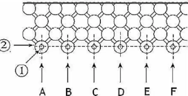
Figure 4 - Regular network of the reactor NETmix® in an association chambers and channels. ... 14 Figure 5 - Representation of the feed system NETmix® reactor. (1) Feeding channels; (2) inlet
chambers; (A-F) reagents inlet. (Adopted from Faria et. al 2008). ... 15
Figure 6 - Metabolic activity/cell proliferation profile of MG63 osteoblast-like cells cultured in the
presence of different nano-HA concentrations, at days 2, 4 and 7. Nano-HA was added 24h after cell seeding. Cell metabolic activity was determined using the MTT assay. The in absence of nano-HA were used as control. Results of MTT assays were expressed as means ± standard deviation (SD). Asterisks (*) indicate a significant difference (p < 0.05) from control (absence of nano-HA). ... 25
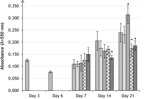
Figure 7 - Metabolic activity/cell proliferation profile of hMSCs cultured in the presence of
different nano-HA concentrations at days 7, 14 and 21, assessed by the MTT assay. Nano-HA was added at day 6 Results were expressed as means ± standard deviation (SD). Asterisks (*) indicate a significant difference (p < 0.05) from control (absence of nano-HA)... 26
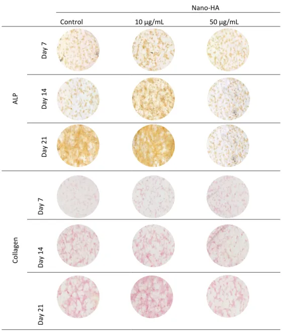
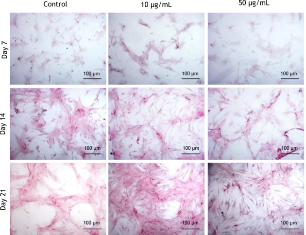
Figure 8 - Low magnification view of hMSCs cultured in the absence (control) and in the presence of
10 and 50 µg/mL nano-HA, stained for ALP and Collagen, at days 7, 14 and 21. ALP is shown by a brown to black stain, and collagen by a red stain. Magnification: 2X. ... 27
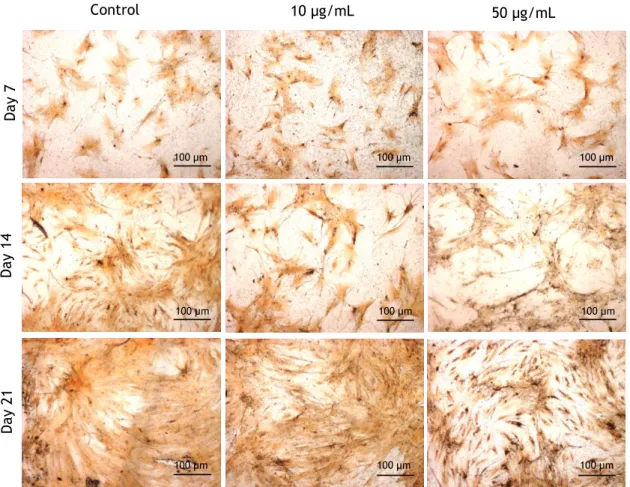
Figure 9 - Images of ALP histochemical staining of hMSCs cultured in the absence (control) and in
the presence of 10 and 50 µg/mL nano-HA, at days 7, 14 and 21. The presence of ALP is shown by a brown to black stain. Magnification: 200 X. ... 28
Figure 10 - Images of Collagen histochemical staining of hMSCs cultured in the absence (control)
and in the presence of 10 and 50 µg/mL nano-HA, at days 7, 14 and 21The presence of collagen is shown by red to pink stain. Magnification: 200X. ... 29
Figure 11 - SEM images of hMSCs cultured in the absence (control) and in the presence of 10 and 50
µg/mL nano-HA, at days 7, 14 and 21. Magnifications: 1000x. ... 30
Figure 12 - High magnification SEM images of hMSCs cultured in the presence of nano-HA. (A)
Cultures exposed to 10 µg/mL, 14 days; (B) cultures exposed to 50 µg/mL, 21 days. Magnification: 5 000 x. ... 30
Figure 13 - SEM images (left side) and EDS spectra of selected areas (right side). A: detailed view of
the interaction of hMSCs with the nano-HA (10 µg/mL, day 7), showing particles agglomerates over the culture substratum (#), over the cell surface (+) and, apparently, inside the cell (*). B: high magnification image of a nano-HA agglomerate showing the individual nanoparticles. Z1, Z2 and Z3: EDS spectra of the marked areas in the SEM images. A (backscattered image): Bar = 20 µm; B: Bar = 2 µm. ... 31
Figure 14 - Confocal laser scanning microscopy images of hMSCs cultured in the absence (control)
and in the presence of different nano-HA concentrations (1 to 100 µg/mL), at days 7 and 14 (a). High magnification images of the control cultures and the cultures exposed to 10 µg/mL
xii
nano-HA, at day 14 (b). Cultures were stained for actin cytoskeletal organization (green) and nuclei (red). Scale bar = 50 µm.. ... 32
Figure 15 - Transmission electron microscopy images of hMSCs cultured in the absence (control)
and in the presence of nano-HA, at day 7, i.e. 1 day after the addition of nano-HA. A: control cell; B and C: interaction of nano-HA with the cell membrane; D: internalized particles in a cell exposed to 10 µg/mL nano-HA; E: internalized particles in a cell exposed to 50 µg/mL nano-HA; F: apoptotic cell found in the cultures exposed to 50 µg/mL nano-HA. ... 33
Figure 16 - Apoptosis profile of hMSCs cultured in the absence (control) and in the presence of
nano-HA (1 to 100 µg/mL), by the evaluation of the amount of caspase-3, at days 7, 14 and 21. Results were expressed as means ± standard deviation (SD). Asterisks (*) indicate a significant difference (p < 0.05) from control (absence of nano-HA). ... 34
Figure 17 - RT-PCR analyses of hMSCs cultured in the absence (control) and in the presence of
nano-HA 10 and 50 µg/mL, at days 7, 14 and 21. Bands of the RT-PCR products (A) were subjected to a measurement of the area and integrated density (ID) by ImageJ 1.41 software. The ID values of gene expression were normalized to the corresponding GAPDH ID value. The expression profile of hMSCs was evaluated for Runx-2 (B), Col-1 (C), ALP (D), BMP-2 and OPG (F). Results were expressed as means ± standard deviation (SD). Asterisks (*) indicate a significant difference (p < 0.05) from control and number sign (#) indicate a significant difference between nano-HA 10 and 50 µg/mL. ... 35
xiii
List of Abbreviations
ALP Alkaline phosphatase BMPs Bone morphogenic proteins BMU Basic multicellular units BSP Bone sialoprotein CaP Calcium phosphate CDNAs Complementary DNAs
CLSM Confocal Laser Scanning Microscopy Col I Type I collagen
DHA Calcium deficient hydroxyapatite DMSO Dimethyl sulphoxide
dNTPS Deoxynucleotide triphosphates ECM Extracellular matrix
FEUP Faculdade de Engenharia da Universidade do Porto GAPDH Glyceraldehyde-3-phosphate dehydrogenase
HA Hydroxyapatite
hMSCs Human mesenchymal stem cells ID Integrated density
Micro-HA Microphase hydroxyapatite MRI Magnetic resonance imaging MSCs Mesenchymal stem cells Nano-HA Nanoscale hydroxyapatite
OC Osteoclacin
OP Osteopontin
OPG Osteoprotegerin
PBS Phosphate-buffered saline PF Primers sequence forward PFA Paraformaldehyde
PR Primers sequence forward reverse
RANKL Receptor activator of nuclear factor kappa B ligand RT-PCR Reverse transcription-polymerase chain reaction Runx-2 Runt related protein 2
SEM Scanning Electron Microscope TEM Transmission electron microscopy Wnts Wingless-ints pathway
1
Chapter 1
Introduction
In bone regeneration, the success of using materials with nanosized features, namely hydroxyapatite (HA), comparatively to microstructure materials, is considerable. These nanomaterials can be used as nanocomposites containing dispersed nanosized calcium phosphate (CaP) and, also, as simple nanoscale grains [1]. The reason for the success of nanomaterials in bone applications is because they display unique physical and chemical properties. Decreasing material size into the nanoscale, it dramatically increases surface area, surface roughness and surface area to volume ratios [1-3]. Thus, these materials represent an increasingly important material in the development of novel materials which can be used in numerous applications.
When a material is implanted in the bone tissue, the osteointegration process is influenced by the properties of the material surface (as topography and chemistry), that have the ability to modulate the molecular and cellular events at the implant interface. Nanomaterials with surface properties similar to the bone are undoubtedly appropriate for the bone regeneration and, therefore, are expected to improve orthopaedic/dental implant efficacy [4].
Nanophase ceramics, especially nano-hydroxyapatite (nano-HA), are widely used for bone regeneration and/or replacement in many biomedical applications due to their documented ability to promote mineralization [2]. Synthetic HA is a CaP with the chemical formulae Ca10(PO4)6(OH)2, which is similar to the principal inorganic constituent of bone and teeth [3,5].
Synthetic nano-HA can be produced using two routes, i.e. dry and wet methodologies. Fluidinova has a new technology to produce nano-HA, the NETmix® technology. By this technology, the Fluidinova developed and patented an industrial process based on the wet chemical reaction for controlling, at the molecular level, the calcium and phosphate reaction to produce HA nanoparticles [6], thus creating a novel product - nanoXIM•Hap102®. This product provides promising features for bone regeneration, as bone tissue engineering strategies might involve the use of synthetic nano-HA and precursor cells with the ability to differentiate into the osteogenic lineage.
Chapter 1
2
1.1 - Thesis Objectives and Layout
For bone regeneration has been increasing much investigation for materials to replace bone defect over last decades. This thesis aims to study biological profile of the commercially available preparation nanoXIM•HAp102® regarding relevant cellular and molecular events involved in the proliferation/differentiation behaviour of human Mesenchymal Stem Cells (hMSCs).
This synthetic HA, in a paste form, is the focus of this work, as the HA nanoparticles present in this paste are a very promising material for bone regeneration strategies [6]. In this context, the main objective of the work described in this thesis is to acquire information on the interaction of these nano-HA particles with human mesenchymal stem cells, which is essential to their application in bone tissue engineering. Therefore, it is intended to obtain integrated information on the uptake and intracellular fate of HA nanoparticles by hMSCs and the elicited cell response regarding proliferation and differentiation events.
The layout of the present work is divided as follows. In Chapter 2 the state-of-the-art, on the prospective use of nano-HA in bone regeneration and, also, to overview the most recent developments in this field. Moreover, it reports the relevant cellular processes occurring in bone tissue during normal metabolism and regeneration events, as well as general background on mesenchymal stem cells, which can be used to study the suitability of a material for bone regeneration approaches. In summary, the contents addressed have a fundamental aim to provide background for a rational intervention in this work.
The experimental methodology is described in Chapter 3. Results are presented in Chapter 4, and discussed in Chapter 5. Finally, the general conclusions drawn from this work are presented in Chapter 6.
3
Chapter 2
Literature Review
2.1 - Tissue engineering
Tissue engineering is an emerging field of interdisciplinary science and research that encompass several scientific areas such as medicine, biochemistry and materials science. It generally involves the use of materials and cells with goal of trying to understand tissue function and restore of damage tissues. In summary, it aims at developing new approaches for encouraging tissue growth and repair. [7-10]
Nowadays, there are several bone pathological conditions that might require the use of biomaterials to restore bone structure and function, such as bone fracture, osteoporosis, and bone cancer, among others. However, traditional implant materials may be associated with several complications, namely failures originating from implant loosening, inflammation, infection, osteolysis and wear debris. Bone tissue engineering has a major role in bone repair approaches, particularly by using nanotechnology, which becomes obvious when examining nature. [2]
With the entry of the nanotechnology in the field of regenerative medicine, there are many developments of nanostructured biomaterials, which have the ability for guiding cellular behaviour by presenting specific morphological and biological cues [1].
Accordingly in bone tissue engineering, one of the main interests is the development of novel nanomaterials with appropriate mechanical properties and, also, biomimetic in terms of their nanostructure [1-3].
Therefore, bone tissue engineering develops new approaches inspired by the hierarchical nanostructure of bone. This approach intends to obtain adequate synthetic nanomaterials (such as calcium material bioceramics) that should ideally be biocompatible, osteocondutive, osteoinductive, osteogenic, and biodegradable, leading to osteointegration [1].
Chapter 2
4
2.2 - Bone
2.2.1 - Bone function
Bone is a living conjunctive tissue in permanent growth, since throughout life it is constantly removed and replaced [11]. It has the mechanical function of providing attachment for muscles, facilitating the locomotion process, and it provides structural support for the other systems and organs [11-13]. Furthermore, bone is considered as a reservoir of calcium, phosphate and other inorganic ions [14].
2.2.2 - Bone composition
Bone is a composite material, constituted by bone cells dispersed in a bone matrix, which has an organic and a mineral component. The organic part of this matrix is mainly composed of fibrillar collagen type I (~90%) and noncallagenous proteins (10%) such as osteonectin, osteocalcin, osteopontin, bone sialoprotein, proteoglycans, glycoproteins, enzymes (e.g. alkaline phosphatase) and cytokines [1, 15]. The mineral phase of mature bone tissue is composed by calcium and phosphate. This phase has many similarities to the synthetic hydroxyapatite (HA), which has the chemical structure Ca10(PO4)6(OH)2 [3]. The bone mineral phase also contains other ions, such as carbonate, citrate, sodium, magnesium, fluoride, hydroxyl, potassium and others that can be found in smaller amounts [15, 16].
2.2.3 - Bone Structure
The morphological structure of the bone can be classified as cancellous (spongy or trabecular bone) and as cortical (compact or dense bone) [11]. Both types of bone exist in different proportions in several locations of de skeleton.
The compact bone is dense and it is located on the external parts of the bone. Its main function is to provide strength, it is relatively acellular and it has low metabolic activity [17]. Cortical bone is organized in cylindrical units, Haversian systems also known as osteons, which forms the diaphysis of long bones [15]. The Haversian systems are surrounded by a concentric layer of rings or lamellae [13, 15]. In the inner of the lamellae there are tiny spaces called lacunae containing bone cells, the osteocytes [13]. These units are considered the structural units of this type of bone. Osteons are typically circular or oval in cross section, 20 to 110 µm in diameter, and contain central canals with blood vessels to provide nutrition, lymph vessels and occasionally nerves [11, 13, 17]. The intercommunicating pore systems, constituted by canaliculi, lacunae and Volksmann’s canals which connect with Haversian canals, allow the transport of metabolic substances [15, 17].
Cancellous bone is porous and it is sited in the interior of bone. In general, cancellous bone is more closely associated with metabolic processes [11, 15]. It is found in the epiphysis of bone and this type of bone is lamellar in structure. The microstructure of cancellous bone shows interconnecting rods or occasionally plates of bone called trabecular [11, 13]. Cancellous bone is arranged as open cell porous networks that allow the transport of metabolic substances [15].
Literature Review
5
The periosteum is the membrane that covers bone external surface, not including the articulating joints. It consists of an outer fibrous layer containing collagen fibres and fibroblasts and an inner layer that contains the osteogenic cells [12, 13, 18]. The inner surface of the bone is covered by a thin membrane called the endosteum. This membrane is found facing the medullary cavity. The endosteum contains a single layer of cells and a small amount of connective tissue [18]. Both lining membranes, periosteum and endosteum, contain osteoblasts and their progenitor cells [13].
2.2.4 - Bone cells
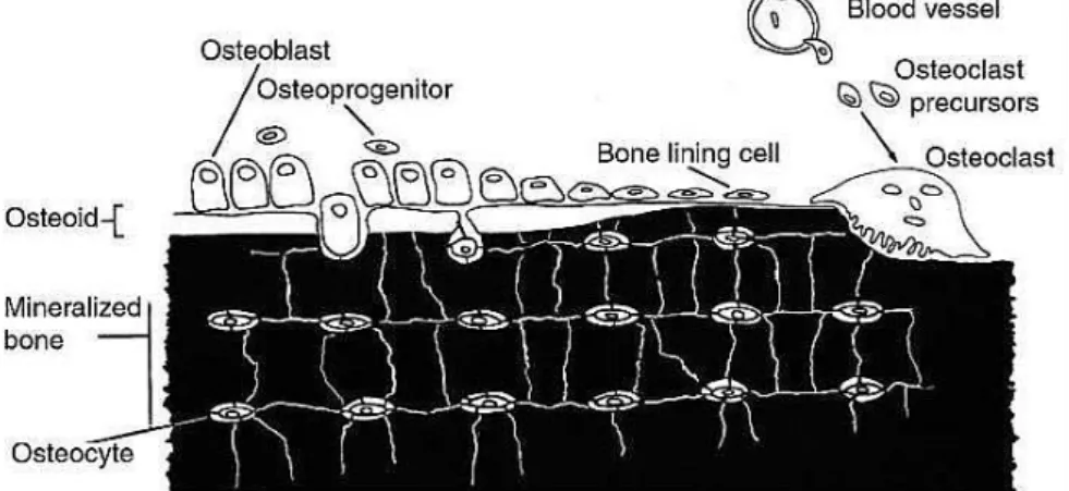
Bone tissue is dynamic organ undergoing constant remodelling. This is possible to the important role of bone cells. These cells are responsible for producing, maintaining and modifying the bone tissue in order to maintain its mechanical and structural properties [11]. There are four types of cells in bone tissue: osteoblasts, bone lining cells, osteocytes, and osteoclasts (figure 1) [11, 18]. Osteoblasts, osteoclasts and bone lining cells, that are located on the bone surfaces, and osteocytes, that can only be found in the interior of the mineralized extracellular matrix (ECM) [11].
Figure 1 - The origins and locations of bone cells (Adopted from Downeyet et. al 2006).
2.2.4.1 - Osteoblast
Osteoblasts are mononuclear and differentiated cells, with low mobility and their function is to synthesize the bone matrix and regulate the mineralization of the osteoid [15, 19]. Therefore, the osteoblasts are responsible for bone building because they play an active role in the synthesis of the proteins and polysaccharides of the bone matrix [20]. When active, osteoblasts have a characteristic morphology, since they have a marked cytoplasmic basophilia, round nuclei rich in ribonucleic acid located at the base of the cell opposite to the bone surface, contain large quantities of rough endoplasmic reticula, and the Golgi apparatus is a well visible, features typical of a protein producing cell [21-23].
Osteoblasts, during their lifetime, have several fates. They can follow one of three pathways: (1) remain active osteoblasts, (2) become surrounded by matrix and become osteocytes, or (3) transform into the relatively inactive bone lining cells [21, 24]. Briefly, osteoblasts may disappear by transformation into either bone-lining cells or osteocytes, or even by apoptosis mechanisms [20].
Chapter 2
6
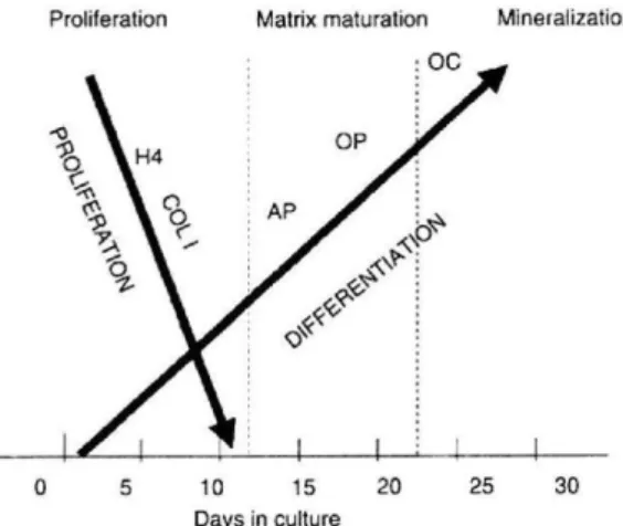
The osteoblast cells originated from undifferentiated mesenchymal stem cells (MSCs) that have the potential to become osteoblasts. MSCs are found in bone canals, endosteum, periosteum and marrow [11, 21, 25]. The undifferentiated cells remain in this state until they are stimulated to proliferate and differentiate into the osteoblast, thus MSCs behave as a pre-osteoblast, and they can migrate from neighbouring tissues or through the vascular system up to a target site [11, 21, 22, 26]. The osteogenic cells, MSCs, can be induced to differentiate into osteoblasts producing a bone like matrix by the synthetic glucocorticoid, dexamethasone, or by the bone morphogenic proteins (BMPs) [27]. Differentiation of MSCs into osteoblast cells is controlled by the regulated expression of genes that define three main periods of osteoblast phenotype development (proliferation, extracellular matrix (ECM) development and maturation, and mineralization). During proliferation, several ECM proteins, such as histone H4 and type I collagen (Col I) can be detected in this stage. Expression of alkaline phosphatase (ALP) is an earlier marker of the matrix maturation phase, while the expression of osteopontin (OP) and osteocalcin (OC) is found in the late maturation or early mineralization phases [23, 27].
Figure 2 - The relationship between cell growth and differentiation-related gene expression during
development in culture of the osteoblast phenotype: histone H4, COL I, ALP, OP, OC. The three principal stages of the osteoblast developmental are shown: proliferation, ECM development and maturation, and mineralization. (Adopted from Lian et. al 1995).
2.2.4.2 - Bone lining cells
Bone lining cells are inactive osteoblasts with flat appearance that cover most of the surface area of the normal bone [11, 21]. These cells are considered to be in communication with osteocytes and are found linked to each other, through cytoplasmic extensions or gap junctions [21].
The functions of the bone lining cells include the protection of the bone from the extracellular fluids, maintaining the bone own environment, the response to mechanical forces on the skeleton [21, 24] and, also, to several hormones, playing a role in the bone remodelling events [11, 20-22]. In addition, they are involved in mineral homeostasis, namely in the exchange of calcium and phosphorus ions into and out of the bone [11, 20].
Literature Review
7
2.2.4.3 - Osteocytes
The osteocytes are mature bone cells, and are the main cells in bone tissue (>90%) [11, 18, 21, 22, 24]. These bone cells are spidery in shape with a plump cell body and reside in a lacuna, and the slender cytoplasm processes that radiate from osteocytes are found in canaliculi [11, 21]. In general, the osteocytes are surrounded by bone matrix, thus they have small nuclei, scanty cytoplasm with mitochondria and a small Golgi zone indicating their low activity compared to the osteoblasts [11, 22]. An osteocyte may derive from the osteoblast that became embedded in its own osteoid [11].
The function of this cell is to maintain the bone tissue, being involved in the exchange of nutrients and wastes with the blood. Like osteoblasts, osteocytes do not undergo cell division. [18]~
2.2.4.4 – Osteoclasts
The osteoclasts are multinucleated giant cells differentiated from the monocyte/macrophage lineage, wherein are formed to carry out the unique function of bone resorption under both normal and pathological conditions. [11, 13, 21]. Once the osteoclasts are derived from a monocyte stem-cell lineage, they are equipped with phagocytic-like mechanisms similar to circulating macrophages. So, following osteoclast attachment to the bone surface, bone degradations begins by direct chemical and enzymatic attack [12].
Morphologically, the osteoclast can have several forms because they have a high mobility, moving from various sites and along the bone surface [21]. In general, these cells are found at the surface of bone and they may be much larger than other bone cells, because they might contain up to 50 nuclei [11, 21].
2.2.5 - Bone remodelling, healing and repair
Bone is constantly being remodelled by the highly regulated bone resorption and forming actions of the osteoclasts and osteoblasts, respectively [11, 13]. Thus, the bone is found throughout life in remodelling, i.e. bone has a continuous shaping. As the old bone is removed, new bone is produced to replace it, and calcium ions are released for other tissues [11, 18].
The processes involved in the bone replacement, bone reabsorption and bone formation, are regarded as independent events, but according to Frost [28] that described the sequential activities of bone replacement, they are closely linked spatially, in an anatomical and temporal sequence, to ensure a balance in the removal of mineralized matrix and the bone replacement [11, 29]. The anatomical structure is named basic multicellular unit (BMU), and this structure describes the sequential activities of osteoclasts and osteoblasts during remodelling [29].
In the process of bone reabsorption, the osteoclasts adhere strongly to the bone surface at the endosteum or periosteum, and releases protein-digesting lysosomal enzymes and several acids into the sealed surface. Thereby, the collagen fibres and other organic substances are digested by enzymes, while the acids dissolve the bone minerals. The degradation products of the bone, mainly calcium and phosphorus, go to the bloodstream by
Chapter 2
8
exocytosis. Thus, bone remodelling occurs when the small area removed by the osteoclasts is rebuilt by the osteoblasts [18].
The remodelling rate for compact bone is about 4% per year, and for spongy bone it is about 20% per year [18, 30]. However, the bone remodelling can also take place at different rates in different regions of the body [30].
The bone remodelling process allows the removal and the repair of bone microdamage, the adaptation to mechanical stress and the maintenance of its strength and integrity [18, 31]. Furthermore, the skeletal remodelling maintains the mineral ion homeostasis. Remodelling is regulated and stimulated by a number of hormones, such as vitamin D, parathyroid hormone, calcitonin and estrogen [24].
Injury in the bone, such as a fracture or the implant replacement, results in the loss of continuity of bone tissue, and it is associated to the damage of the blood vessels. Consequently, haemorrhage occurs and there is the formation of a blood clot, or hematoma [33]. Healing of bone tissue provides the capacity to repair the tissue continuity, form and function with a mechanism scar free [34, 35]. Bone tissue has a potential to heal itself, resulting in the regeneration of the anatomy of the bone and complete function.
The bone repair process is very complex, because it involves the coordination and sequence of many biological events [35, 36]. Bone healing can be divided into four phases: blood clotting, inflammatory phase, reparative phase and remodelling phase [34-36].
Once the bone tissue injury leads to damage to surrounding tissues, the bone healing is initiated by the activation of the coagulation cascade and by the formation of the blood clot [35, 36]. The blood clot allows the cellular migration and proliferation; furthermore, it functions as a primary source of growth factors [34]. Thus, inflammatory cells are recruited, such as neutrophils, macrophages, monocytes, lymphocytes, and cells of other lineages, such as fibroblasts and endothelial cells. Then, recruitment of the cells is followed by an acute inflammatory response resulting in the edema and cytokine and growth factor release [36].
The healing process continues by the reparative phase with resorption of the blood clot and its replacement by the deposition and formation of granulation tissue [34]. In this phase, there is vascular growth and migration of osteogenic cells. The granulation tissue is responsible for providing blood supply and for the recruitment of undifferentiated mesenchymal stem cells. At the lesion site, these cells proliferate and differentiate because they responded to growth factors released by the injured tissues and from the clot [3]. Finally, after 6 to 8 weeks, bone tissue is completely remodelled in the original shape, structure, function and mechanical properties [34, 35].
2.3 - Bone marrow and stromal cells
Bone marrow is a very complex organ, and it is composed by stromal tissue, hematopoietic cords and sinusoidal capillaries [36, 37]. The stromal tissue supports haematopoiesis and is made up of a network of extracellular matrix and cells [36, 38, 39]. Furthermore, stromal tissue has well known osteogenic potential [40].
Bone marrow stromal cells are widely used in tissue engineering [40, 41]. These cells are a heterogeneous population of non-hematopoietic cells of mesenchymal origin and fibroblastic cells [36, 40, 41]. Other cells are present in this population, such as monocytes, macrophages and endothelial cells [36, 40]. Following the culture of bone marrow stromal cells, the non-adherent cells are removed after medium change and cell passage [41].
Literature Review
9
Included in the adherent cell population, the mesenchymal stem cells (MSCs) are multipotent cells widely used in tissue engineering.
2.3.1 - Mesenchymal Stem Cells
The MSCs have the capacity of self-renewal and they are defined as a type of adult stem cells which contribute, in some circumstance, to the regeneration of mesenchymal tissues, once they can produce progeny that differentiate across the three primary germ layers (ectoderm, mesoderm and endoderm) [36, 42, 43]. Human MSC are easy to isolate from small aspirate of bone marrow via their adherence ability and they can be easily expanded in vitro [39].
MSCs that can give rise to mature cells, and in defined in vitro conditions, these cells readily differentiate to multiple connective tissue lineages, including osteoblasts, chondrocytes, and adipocytes [39, 43].
The morphology of the MSCs is characterized by a small cell body with a few cell processes that are long and thin. Furthermore, MSCs have a nucleus with prominent nucleolus which is surrounded by finely dispersed chromatin particles, giving the nucleus a clear appearance [20].
These cells are widely used in tissue engineering, because the selection of the culture conditions and the subsequent manipulations allow defining cell differentiation.
2.3.2 - Osteogenic differentiation potential of MSCs
The in vitro expansion of MSCs has been extensively studied by tissue engineering owing to their potential to differentiate into osteogenic tissue. The osteoblastic behaviour of MSCs in culture is normally achieved by culturing in an appropriate standard cell culture medium (i.e., α-minimum essential medium with 10% foetal bovine serum) supplemented with β-glicerophosphate, ascorbic acid and dexamethasone [27, 44-46]. Under these conditions, the MSCs in culture acquire an osteoblastic morphology and differentiate along the osteogenic phenotype, being able to express typical osteoblastic genes and to form a collagenous mineralized matrix [45, 46].
2.3.2.1 - Gene expression of MSCs during osteogenic differentiation
The MSCs have a potential to generate many cellular lineages such as osteogenic, adipogenic or chondrogenic cell lineage [47, 48]. The differentiation of MSCs into a cell lineage is dependent on the activated signalling pathways and several transcription factors [13, 47, 49, 50].
The transcriptional factors runt related protein 2 (Runx-2), osterix and β-catenin are essential to regulate the differentiation of MSCs into osteoblast lineage [47].
Osteoblast commitment and differentiation are guaranteed by Runx-2, it is responsible for inducing the osteogenic phenotype at an early stage and also to inhibit the differentiation of MSCs into adipocytes and chondrocytes [13, 47, 49, 51]. Runx-2 is known to activate and up-regulate the expression of major bone matrix protein genes of osteoblast differentiation, such as type I collagen (Col I), osteopontin (OP), bone sialoprotein (BSP) and osteocalcin (OC)
Chapter 2
10
[47, 49, 52]. Therefore, this transcriptional factor allows cells acquiring an osteoblast phenotype, although keeping the osteoblast cells in an immature stage, the preosteoblast [47, 49]. Preosteoblast proliferation and differentiation follows distinct phases: preosteoblast, mature osteoblast, and osteocyte, which are regulated by transcription factors and express specific phenotypic genes [53].
Other factors can regulate the activity of specific transcription factors maintaining an osteoblastic fate. They include bone morphogenetic proteins (BMPs) that are recognized for their osteoinductive proprieties, and regulate the differentiation of MSCs into osteoblasts components of bone [49]. Furthermore BMP-2 and wingless-ints (Wnts) pathways together have a critical role in promoting Runx-2 expression to promote osteoblast differentiation [13, 51].
The osteoblastic phenotype is characterized by the synthesis of specific bone proteins that define a genic expression pattern. The gene markers expression of osteoblast allows defining the distinct stages of osteoblast phenotype development: proliferation, matrix maturation and mineralization. In each stage there are characteristic changes in gene expression [49, 54, 55].
As already mentioned, in an early stage during the proliferative phase, there is the expression of genes that support the proliferation, such as Col I and fibronectin [23, 27, 49, 56]. However, in this phase, BMP-2 and BMP-5 have a significant role in increasing ALP activity and OC synthesis [49]. So, the accumulation of these matrix proteins allows, in part, the end of cell proliferation. The differentiation stage is recognized by early cell differentiation that is characterised by the transcription and expression of the ALP [56]. Thereby, these markers are widely used to evaluate the osteogenic differentiation. Ultimately, the ECM becomes into the mineralization phase in which osteoblastic cell express osteopontin (OP) and osteocalcin (OC). These protein markers are found in the late maturation or early mineralization phases [23, 27, 49].
Osteoblasts produce osteoprotegerin (OPG) which is a soluble decoy receptor for the receptor activator of nuclear factor kappa B ligand (RANKL), and it is known that the major effect of OPG is to inhibit osteoclastogenesis [50, 57]. However, its role in osteoblast differentiation is still unclear. More recently, Yu et al. (2011) showed that OPG expression is associated with preosteoblast maturation and promotes matrix maturation [57].
In a study on osteoblast behaviour or osteogenic differentiation of MSCs it is very important to analyse the gene expression profile of the cells. The interest in analysing the RNA is explained by a cell’s ability to adjust its mRNA copy numbers in response to environmental changes, being a crucial element in the complex regulation of gene expression. Moreover, at the mRNA level, it is known that a wide spectrum of pathological processes is associated with changes in gene expression [58].
The reverse transcription-polymerase chain reaction (RT-PCR) is a powerful tool for gene expression analysis. This technique is a PCR method that uses reverse transcriptase enzymes for making DNA from RNA [59]. The RT-PCR combines synthesis of the cDNA with its amplification allowing the cloning of genes from its mRNA. Thereby, this technique allows the analysis of the gene expression [59]. The result obtained from RT-PCR analysis allows to know whether a specific gene is being express into a cell [59].
The PCR method is characterized by the ability of DNA polymerase enzyme to synthetize a new strand of complementary DNA. The PCR reaction requires: a biological sample that contains DNA or RNA; a DNA polymerase enzyme; some salts for the DNA polymerase to
Literature Review
11
function; deoxynucleotide triphosphates (dNTPS) which provide essential nucleotides for new DNA strands; and finally oligonucleotides primers, which are responsible for initiating the chain reaction, being extremely specific to the precise genetic sequence of interest [59-61]. After PCR reaction, the reaction components are placed into a thermocycler, where they undergo basic PCR steps [59]. The PCR procedure involves repeated cycles of three steps, namely heat denaturation, annealing, and primer extension [59, 62].
In the first step, the thermocycler drives the reaction components up to denaturation temperature, typically 92 to 94°C. The denaturation allows that the hydrogen bonds of the double DNA helix are broken, and thereby the DNA double helix is reduced into single strands [59, 62].
In the next step, the temperature of the mixture decreased to a predetermined annealing temperature, usually between 50 and 60°C. In this step of PCR, the oligonucleotide primers can hybridise specifically to their complementary sequences, meaning that the primers can anneal to the DNA target sequence [59, 62].
Finally, the replication of the DNA strands occurs. After the annealing step, the temperature is raised to 72°C, and a DNA polymerase extends the primers by incorporating deoxynucleotides to form a new complementary strand of DNA [59, 62].
The cycle denaturation-annealing-extension is repeated over and over, usually 30 times, and new strands themselves as template for the DNA primers and the process lead to an exponential amplification of the DNA bounded by the primers [59, 61, 62]. These cycles allow an estimated enrichment of the selected sequence of 105 to 106 [61].
After the termination of the reaction cycles, the reaction products are separated by agarose gel electrophoresis, and finally they can be visualized directly by staining with ethidium bromide and examination of the gel under ultraviolet light [61].
An important aspect to take into consideration when analysing PCR results is that several parameters need to be controlled to obtain reliable quantitative expression measures. To this, it is often used an approach for the normalization of several parameters with an internal control gene. A gene valid to use as a reference gene requires an expression that do not vary in the tissues or cells under certain experimental conditions [48, 63]. One example of the most usually described normalization gene is glyceraldehyde-3-phosphate dehydrogenase (GAPDH). This gene is present in most cell types and it is considered a simple “housekeeping” protein that has been shown to be involved in many cellular processes in addition to glycolysis, where its function is catalysing the reversible oxidative phosphorylation of glyceraldeyade-3-phosphate [48, 63, 64].
2.4 - Nanomaterials in bone regeneration
Nanomaterials can be defined as the materials with dimensions less than 100 nm [1, 2, 65]. These materials include nanoparticles, nanoclusters, nanocrystals, nanotubes, nanofibres, nanowires, nanorods, nanofilms, etc. [2]
Nanomaterials are promising candidates in bone regeneration, since nanostructure provides a closer approximation to native bone architecture, compared to micron materials [2, 66].The decreasing material size leads to modification of the nanofeatured surfaces, increased surface area, surface roughness and surface to volume ratio, which creates superior physiochemical properties to the cellular behaviour [2, 66]. For example, increase of surface
Chapter 2
12
energy and wettability, seen in nanostructured surfaces, allows to an increased protein adsorption and cell adhesion [66, 67].
Recently, more efforts are being made to repair and reconstruct bone damage using biomimetic bone tissue. To accomplish this, it is necessary to select biomaterials that are present in bone (e.g. HA and collagen), and it is also possible to incorporate growth factors (e.g. BMPs) and/or cells for bone repair and regeneration [66].
Based on the structure of the natural HA, nano-HA has been synthesized for various tissue engineering applications, such as for bone replacement and for the delivery of relevant drugs in the management of several bone diseases [68]. Furthermore, many studies revealed that biomimetic nano-HA enhances the proliferation and activity of bone cells in the regeneration process compared to the microstructured ceramic [67, 69-72]. Webster et al. (2000) showed that cell differentiation markers, such as alkaline phosphatase (ALP) synthesis and calcium deposition, were significantly greater on nanophase HA than on conventional ceramic [73].
2.4.1 - Hydroxyapatite
Bioceramics materials, namely calcium phosphate (CaP)-based materials, are widely used for bone regeneration and/or replacement in many biomedical applications. Synthetic HA is a CaP with the chemical formulae Ca10(PO4)6(OH)2, and a Ca/P ratio of 1.67, which is similar to the principal inorganic constituent of bone and teeth [3,5] . However, CaP can also exist with a Ca/P ratio lower than 1.67, referred as calcium-deficient hydroxyapatite (DHA), denoted by Ca10-x (HPO4)x(PO4)6-x(OH)2-x [5, 73, 74]. HA (Ca/P=1.67) has been extensively investigated because it is a thermodynamically stable CaP in the physical environment [3] and shows excellent biological characteristics, due to its natural biocompatibility and potential osteoconductive, osteoinductive, osteogenic and biodegradable properties [1, 69].
2.4.1.1 - Nanostructured and properties
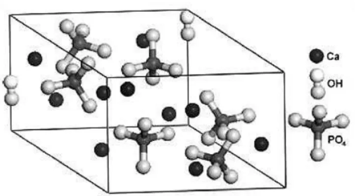
HA is a polycrystalline CaP, with a definite crystallographic structure. The hexagonal crystalline structure of HA allows exact atomic positions, and this structure have a space group P63/m. This space is characterized by a six-fold c-axis perpendicular to three equivalent a-axes at 120◦ angles to each other. Calcium cations (Ca2+) and phosphate anions (PO43-) are arranged around columns of monovalent hydroxyl anions (OH-) [3, 76, 77]. Figure 3 shows the unit of HA, composed by Ca, PO4 and OH groups closely packed together. Also, it can be noted that PO4 group provides the skeleton framework and gives the structure and its stability [78].
HA is a mineral with small nanosized dimensions and low crystallinity, properties that provide distinct features of the biological apatites. The synthetic HA can be synthesized using many processes. Crystallographic and chemical studies have confirmed that synthetic HA is similar to the natural HA found in bone and teeth [1, 77]. Therefore, the synthetic HA is widely used for bone regeneration and/or replacement in many biomedical applications.
Besides the similarities of the synthetic HA and natural HA, there are more advantages for its application in tissue engineering. Thus, synthetic HA shows biocompatibility, slow biodegradability in situ, and good osteoconductive and osteoinductive capabilities [1, 69, 79-81].
Literature Review
13
Figure 3 - Unit of hydroxyapatite hexagonally crystalline structure. (Adopted from White et. al 2009). The nanophase HA powders show improved properties when compared with microphased powders, such as surface grain size, pore size and wettability, which may control protein interactions and thus guide cellular responses. Therefore, the nano-HA has a better bioactivity [3, 81], hence it allows a good osteoblast adhesion, proliferation, differentiation , osteointegration, and deposition of calcium-containing minerals on its surface, leading to an improvement of the composition of regenerated tissue within a short period [67,69-72,82].
In summary, according to the literature, nano-HA appears to have clear advantages in bone regeneration applications [83].
2.4.1.2 - Synthesis Methods
The nano-HA is an important material in bone regeneration, therefore there is a strong interest in its synthesis in an extra pure and well defined HA nanocrystals [20].
Several techniques have been developed to prepare nanosized HA, most of them involving wet and dry methods.
The dry methods for nano-HA synthesis are characterized by the preparation of the powder via application of high temperature, high-energy mechanical and controlled pressure. In this process, calcium and phosphorous compounds are used as reactants. These powders are mixed in acetone, calcinated in vacuum and, lastly, heat-treated with the supply of water vapour as the source of the hydroxyl group [1, 85]. The reactants used in the dry process can be several calcium and phosphorous compounds such as, dicalcium phosphate anhydrous (CaHPO4), dicalcium phosphate dihydrate (CaHPO4.2H2O), monocalcium phosphate monohydrate (Ca(H2PO4)2.H2O) , calcium pyrophosphate (Ca2P2O7), calcium carbonate (CaCO3), calcium oxide (CaO) and calcium hydroxide (Ca(OH)2), among others [85].
The wet chemical precipitation method (i.e. wet chemical precipitation, sol-gel, emulsion, biomimetics, among others) is widely used because it is significantly cheaper and easier than other methods of HA formation [1, 3, 74] and the probability of contamination during processing is very low [85]. Thus, this technique is more popular for synthesis nano-HA since, no specialized equipment is required and large quantities of material can be produced of nanostructured HA from aqueous solution [1, 3, 74].
The reaction of Yagai and Aoki (1) can be followed in order to obtain suspensions of HA nanoparticles. A solution of H3PO4 is dropped into a basic suspension of Ca (OH)2 [1, 69, 86].
Chapter 2
14
Other calcium compounds may be used to obtain suspensions of nanoparticles. For example, calcium nitrate tetrahydrate (Ca(NO3)2.4H2O can be added to ammonium dihydrogen phosphate (NH4H2PO4)[1, 69].
In the wet chemical precipitation method, it is important that the reactants have the correct molar ratio of calcium and phosphorous and, also, to adjust and maintain the appropriate pH of the solution [87].
Synthesis of nano-HA by wet chemical precipitation allows controlling the particle shape, size and specific surface area, by monitoring the reactant addition rate, reaction temperature, pH, and the presence of additives [1, 69]. In literature, the crystallinity of HA is largely increases by reaction temperature [1, 69, 88].
2.4.1.3 - NETmix
®Technology
The NETmix® is a novel technology (development at FEUP) that enables the production of nanomaterials, such as nano-HA, with a high reproducibility, mainly in terms of size distribution [4]. This technology is now being commercially applied by Fluidinova to the synthesis of highly pure hydroxyapatite nanoparticles, nanoXIM-HAp® [5, 15, 89].
The NETmix® is a patented static mixer, on continuous mode operation [89] for mixing of liquids and/or gases; it allows the control of fluid mixture in an optimized and reproducible way. [90] Furthermore, this technology shows reproducible and satisfactory results, especially with regard to distribution of particle sizes [91].
Its principal component is a network of chambers interconnected by channels, that is, the network combines, in an organized manner, chambers and channels [92]. This interconnection create zones that promote complete mixing and of complete segregation, as shown in figure 4 [5].
The implementation of the static mixer brought improvements in comparison with other static systems. For example, its structure is considerably simple; it allows the introduction of temperature, pressure and concentration sensors; it provides an easy implantation of different pre-mixing reactants injection schemes; it has the ability to control the mean residence time of the reactants and the mixing intensity and scale. Thus, this reactor is particularly suitable for reactions in which mixing quality and intensity are critical. [92]
Figure 4 - Regular network of the reactor NETmix® in an association chambers and channels. (Adopted
from Silva et. al 2008).
The NETmix® regulates micromixing; this phenomenon is associated to the homogenization of the mixture at the smallest scale [92, 93]. The properties of the HA produced by NETmix®, such as purity, crystallinity, particle size distribution, crystallites size and morphology, can be determine by the micromixing characteristics [74].
Literature Review
15
In the laboratory, the prototype of this technology, consisting of a network of spherical chambers and cylindrical channels, was developed and used for the synthesis of nano-HA with high reproducibility, mainly in terms of size distribution [5]. The NETmix® reactor, for the synthesis of nano-HA, is based on the wet chemical precipitation method at room temperature [89].
Therefore, to produce nano-HA is necessary to feed the reactor with a calcium solution, a phosphorous solution and an alkaline solution and, optionally, one solvent or dispersing agent [74]. The initial reactants are injected in inlet chambers through the back and front feeding channels (figure 5).
Figure 5 - Representation of the feed system NETmix® reactor. (1) Feeding channels; (2) inlet
chambers; (A-F) reagents inlet. (Adopted from Faria et. al 2008).
The products synthesized by this technology can be collected in the form of suspension at the exit or the intermediate position of the reactor. Then, the suspension can be subjected to a separation process in order to concentrate the particles. Moreover, the concentrated particles can be washed for the elimination of the supernatant, and they can be exposed to drying processes. Thereafter, the particles may be subjected to several stages of grinding and thermal treatments. [89]
From its versatile technology, NETmix® Fluidinova is responsible by launching several products with a single phase and high purity, such as the nanoXIM•HAp102®. This product is a highly pure nanocrystalline hydroxyapatite in paste form at 15% (weight/volume) nano-HA concentration in pure water.
In summary, the NETmix® technology results from the application of a novel concept for producing HA nanoparticles, based on wet chemical precipitation method at room temperature. This patented industrial process is able of controlling, at the molecular level, the calcium and phosphate reaction to produce HA nanoparticles [6, 74, 94].
2.4.1.4 - Applications
Synthetic nanosized HA is the most stable CaP and it has been used for a variety of biomedical applications, such as in dentistry, matrices for drug release control and surgery for hard tissue repair [1, 3, 77, 95]. The previous studies have been so far mostly used owing to its chemical similarity to the mineral component of calcified tissue [3, 5, 95]. In addition, other properties have attracted interest, such as their high surface area, their ability to become dispersed in aqueous solutions and their higher reactivity and adsorption, over the same material of micrometric size [2].
Nano-HA is a bioactive ceramic that has been preferred for hard tissue repair and as coatings for metal prosthesis over autografts and allografts. For some time now, it appears
Chapter 2
16
that synthetic HA increases the response regarding bone bonding. Accordingly, the nano-HA is used as an implant coating to stimulate bone growth around the implant. [3, 96]
Many studies have demonstrated that the mechanical strength and fracture toughness of HA can be improved by the use of nanoscale powders; the large surface area to volume ratios of the particles contributed for a better densification [76]. Accordingly, nano-HA can be used to produce scaffolds for tissue engineering, and when nano-HA is compressed, for example into a cylinder, it has attracted interest for bone replacement [3, 97].
The nano-HA particles with high surface area and un-agglomerated are of interest for injectable or controlled setting bone cements, high strength porous or non-porous synthetic bone grafts and the reinforcing phase in nanocomposites that attempt to mimic the complex structure and superior mechanical proprieties of bone [98]. Furthermore, the nano-HA has been widely used in dentistry. Huang et al., [99] for example, showed that nano-HA had the potential to remineralize initial enamel caries lesions.
Recently, many researchers have interest in using the magnetic nano-HA, since it is possible to couple HA with magnetic particles such as cobalt (Co), iron (Fe) and magnetite (Fe3O4) [3, 77]. This magnetic coupling aims at particle delivery by external control of a magnetic field. The magnetic HA composites are adapted for biological applications such as cell separation, drug delivery, contrast agents for magnetic resonance imaging (MRI), and heat mediators for hyperthermia [3, 100]. Moreover, other studies suggested that nano-HA coated magnetic particles could be used to reverse osteoporosis [101-103], once osteoblast density and osteoblast differentiation markers are significantly increased in the present HA-coated-Fe3O4 [102].
2.5 - Interactions cells – Nanostructured materials
Nanostructured materials for bone applications have the ability to improve surface reactivity, because surface area-to-volume ratios increase with decreasing particle size [1, 104]. According to literature, the surface properties (such as surface area, charge, and topography) are related to the grain size of a material, thus the cell response to the nanoparticles is significantly different that the one observed during the cell interaction with the bulk materials [66, 68, 104, 105].
In particular, for cell interaction it is beneficial if the surface roughness of HA is in the nanoscale range but the reasons for this improvement are not completely understood. [3] However, it is believed that protein adsorption and bioactivity on particles with nanometer features is different from that on conventional materials. Interaction between cells and nanostructured materials also depends on receptors on the cell surface and the protein layer adsorbed onto the material surface [3]. It is reported that the adsorption and conformation (or bioactivity) of proteins that mediate specific osteoblast adhesion (such as fibronectin and vitronectin) are enhanced on nanophase material [67, 84, 73].
Several studies on the cell response to HA nanoparticles have shown that HA particles are internalized through endocytosis, and the cell response appears to depend on the combined interaction of multiple nanoparticles characteristics [5, 68, 106-108]. An important aspect in the modulation of the cell behaviour is that the HA nanoparticles may be digested in the lysosomes, leading to an increase of the levels of Ca2+ in the cytoplasm. Suitable concentration of Ca2+ is favourable for the proliferation and osteoblast differentiation [109].
Literature Review
17
2.5.1 - Apoptosis
Investigation of the cell-material interaction is mainly focused on biocompatibility. However, nowadays much attention is given to the possibility of apoptosis induced by biomaterial, once apoptosis could be caused by inducing factors, and many disease therapies are closely related to inducing apoptosis.
Apoptosis is recognized by an important mode of programmed cell death, which involves the genetically determined elimination of cells. It is a homeostatic mechanism responsible for the maintenance of normal cellular homeostasis [110, 111]. Moreover, this process can occur as a defence mechanism against cellular damage caused by disease or noxious agents. [110, 111] In other words, this phenomenon can be activated or inhibited by several stimuli, both physiological and pathological [111].
It is important to note that there are other forms of cell death, such as, necrosis, and other forms may be discovered. Moreover, there are cells that die with atypical characteristics, which are not categorized as apoptosis or necrosis [112].
Apoptosis contrasted with necrosis. Necrosis is considered to be a toxic process characterized by loss of cell membrane integrity leading to the release of the cytoplasmic contents, which causes the recruitment of inflammatory cells. Once apoptotic cells do not release the cytoplasmic contents and apoptotic cells or cell fragments are quickly phagocytized they have a little or no immune response [112].
Apoptosis can be considered a coordinated and often energy dependent process that involves caspase activity, DNA fragmentation and a complete cascade of events [110, 1110, 113].
Caspases are a family of cysteine aspartate-specific proteases that have a critical role in biochemical events associated with apoptosis [114, 115]. In particular, the activation of caspase-3 protease has been described as an “effector” caspase associated with the initiation of the “death cascade” and is therefore an important marker of the cells when they are into apoptotic signalling pathway [116].
Nowadays, there are many methods to determine apoptosis in cells through the evaluation of the activity of proteins involved in apoptosis. Note that when there is the activation of caspase-3 there is substrate specificity for the amino sequence aspartate-glutamate-valine-aspartate. Thereby, the substrate can be used to continuously monitor the activity of caspase-3. Upon enzymatic cleavage, the nonfluorescent substrate is converted in a two-step process to a fluorescent compound. So, this is the base for caspase-3 activity assay that detect apoptotic events [110,112]. This assay requires cell lysis, which aims to release the enzyme into solution, followed by detection with a fluorescent labelled substrate with the appropriate excitation and emission settings [110,112]. Detection of caspases represents a rapid and consistent method for quantification of apoptotic cells [110,112].
Another form to analyse cell apoptosis is through morphological characteristics observation of the cells by standard transmission electron microscopy (TEM). This technique allows to define characteristics of an apoptotic cell, such as intact cellular membranes, dark and dense cytoplasm and nucleus, blebs at the cell surface, large clear vacuoles, disorganized cytoplasmic organization and nuclear fragmentation [110-112]. Thereby, TEM is used to confirm apoptosis once the categorization of apoptotic cells is irrefutable in case the cell contains certain morphological characteristics of an apoptotic cell [111, 112].