Assessment of genetic mutation frequency induced by oxidative stress in
Trypanosoma cruzi
Carolina Furtado Torres-Silva
1*, Bruno Marçal Repolês
1*, Hugo Oliveira Ornelas
1, Andréa Mara Macedo
1,
Glória Regina Franco
1, Sérgio Danilo Junho Pena
1, Erich Birelli Tahara
1and Carlos Renato Machado
11
Departamento de Bioquímica e Imunologia, Universidade Federal de Minas Gerais, Belo Horizonte, MG,
Brazil.
Abstract
Trypanosoma cruzi is the etiological agent of Chagas disease, a public health challenge due to its morbidity and mor-tality rates, which affects around 6-7 million people worldwide. Symptoms, response to chemotherapy, and the course of Chagas disease are greatly influenced byT. cruzi‘s intra-specific variability. Thus, DNA mutations in this parasite possibly play a key role in the wide range of clinical manifestations and in drug sensitivity. Indeed, the envi-ronmental conditions of oxidative stress faced byT. cruzi during its life cycle can generate genetic mutations. How-ever, the lack of an established experimental design to assess mutation rates inT. cruzi precludes the study of conditions and mechanisms that potentially produce genomic variability in this parasite. We developed an assay that employs a reporter gene that, once mutated in specific positions, convert G418-sensitive into G418-insenstitiveT. cruzi. We were able to determine the frequency of DNA mutations in T. cruzi exposed and non-exposed to oxidative insults assessing the number of colony-forming units in solid selective media after plating a defined number of cells. We verified thatT. cruzi‘s spontaneous mutation frequency was comparable to those found in other eukaryotes, and that exposure to hydrogen peroxide promoted a two-fold increase inT. cruzi‘s mutation frequency. We hypothesize that genetic mutations inT. cruzi can arise from oxidative insults faced by this parasite during its life cycle.
Keywords:T. cruzi, DNA, mutation frequency, H2O2.
Received: September 05, 2017; Accepted: November 29, 2017.
Introduction
Trypanosoma cruziis the etiological agent of Chagas
disease, a complex zoonosis that affects more than seventy
genera of mammalian hosts (Zingaleset al., 2012; Baptista
et al., 2014). According to the World Health Organization
(WHO), around 6-7 million people are affected by this dis-ease in 21 countries, most of them in Latin America [WHO Chagas disease (American trypanosomiasis) fact sheet, 2017]. Also noteworthy is the fact that nowadays this dis-ease is spreading to non-endemic regions due to human mi-gration (Schmunis, 2007).
The life cycle ofT. cruziis complex and involves two
hosts: an invertebrate and a mammalian. Humans are con-sidered accidental hosts, in which the classic vectorial in-fection generally occurs at night when the blood-sucking triatomines defecate during feeding (Frasch, 2000). Once the feces droplets expelled by the triatomine reach the bloodstream or get in contact with eyes, nose or mouth
mu-cosa, the infection is then perpetrated (Prata, 2001).
Hu-mans may also be infected withT. cruzi through blood
transfusion, organ transplantation, from mother to infant during pregnancy, laboratory accidents, as well as through ingestion of food contaminated with triatomine feces
(Shi-kanai-Yasudaet al., 1991; de Noya and González, 2015).
Following the infection by T. cruzi, a short acute
phase characterized by high parasitemia takes place, along
with unspecific symptoms (Macedoet al., 2004). During its
chronic phase, Chagas disease presents a large spectrum of symptoms and low parasitemia. Interestingly, 30% of in-fected humans will develop cardiomyopathy, digestive im-plications or both (Rassi Jr and Marin-Neto, 2010), and a small percentage of them may still develop neurological symptoms (Prata, 2001). Although the mechanisms and factors influencing this clinical unpredictability have not been fully elucidated, the variability in the course of Cha-gas disease seems to be related to a number of factors such as parasite strain, host age, reinfection, and genetic factors of both host and parasite (Prata, 2001).
Since 2009,T. cruzistrains have been divided into six
discrete taxonomic units, namelyT. cruziI – VI, based on
its intra-specific genetic variability (Zingaleset al., 2009;
Baptista et al., 2014). Unquestionably, diverse tissue
DOI: http://dx.doi.org/10.1590/1678-4685-GMB-2017-0281
Send correspondence to Carlos Renato Machado. Departamento de Bioquímica e Imunologia, Universidade Federal de Minas Ge-rais, Belo Horizonte, MG, Brazil. E-mail: crmachad1967@gmail.com
*These authors contributed equally to this work
tropisms, response against immune system, and respon-siveness to chemotherapy have been frequently observed in Chagas disease (Revolloet al., 1998; Andradeet al., 2010).
In fact, genetic factors are able to strictly regulate infection capacity of parasites, as there is a correlation between ge-netic diversity and rate of success in escaping the host
im-mune response (Frasch, 2000; Burgoset al., 2013).
It has long been known that several microorganisms display intrinsic, spontaneous mutability events that lead to intra-specific genetic diversity (Steinberget al., 1971;
Tad-deiet al., 1997; Rosche and Foster, 2000). The generation
of spontaneous mutation is a very complex subject since several intrinsic and extrinsic factors might be involved in the process – like the environment in which the organism is found (Maticet al., 1997), location of mutation-prone sites
in the genome (Patrushev and Minkevich, 2008), and the behavior of the DNA repair system (Hoeijmakers, 2001). However, a number of studies have already shed light on the mechanisms and importance of spontaneous mutation rate in bacteria (Choiet al., 2011; Fordet al., 2013), yeast
(Magni and von Borstel, 1962; Glassneret al., 1998;
Ben-sasson, 2011), and in other non-disease causing eukaryotes (Provanet al., 1999; Shikazonoet al., 2003). Also, it has
al-ready been shown that certainT. cruzihaplogroups display
mutations in microssatelite alleles after being cultured in
media supplemented with hydrogen peroxide (H2O2)
(Augusto-Pintoet al., 2003).
Therefore, the study of the mechanisms related to the generation of genetic mutations and diversity inT. cruziis imperative since they may play a role in how this parasite deals with genotoxic stress and drug response; in fact, ex-perimental analysis of the antigenic diversity generation
re-mains a challenge since few works tried to investigateT.
cruzi‘s mutation rate. In this work, we developed a model that allows the detection of mutational events through the
selection ofT. cruziresistant to the aminoglycoside G418.
We found that the mutation frequency in this parasite is similar to other eukaryotic cells, being substantially
in-creased by challenging T. cruzi with exogenous H2O2.
SinceT. cruzihas to cope with oxidative stress situations
during its complex life cycle (Piacenzaet al., 2009;
Ma-chado-Silvaet al., 2016), we hypothesize that immunologic
evasion and chemotherapy resistance in Chagas disease could be associated to the generation of genetic variability inT. cruzienhanced by oxidative stress conditions.
Material and Methods
Plasmid construction and bacterial transformation
Wild-type Neo (NeoWT) and its mutant variants –
NeoD90, NeoD180, NeoD270, Neostop, and NeostopT
®G – were
amplified by PCR from the pROCK_Neo vector (da Rocha
et al., 2004), using the primers indicated in Table 1. All
re-sultant amplicons (Table 1) were digested withXhoI and
XbaI and then ligated to pMAL-c2G (New England Biolabs
Inc., Massachusetts, USA) previously digested with the
same endonucleases. Electrocompetent Escherichia coli
DH5a(Gonzaleset al., 2013) were transformed with
liga-tion products and plated onto 2xYT medium [1.6% tryptone, 1.0% yeast extract, 0.5% NaCl (pH 7.0)]
supple-mented with 100 mg/mL ampicillin. Bacterial positive
clones were screened using the colony PCR method (Bergkessel and Guthrie, 2013) and further isolated.
Bacterial kanamycin resistance assay
DH5apositive clones for all Neo constructs (Table 1) were grown in 2xYT liquid medium supplemented with
100mg/mL ampicillin, under orbital agitation (180 rpm) at
37 °C for 16 h. Bacterial cells were then subject of a serial dilution (suspensions with final concentrations of 10-4, 10-6,
10-8, and 10-10cells/mL), and 2.5
mL of each suspension
were added onto plates containing 2xYT solid medium
(liq-uid 2xYT plus 2.0% agar) supplemented with 100mg/mL
ampicillin and 0.1 mM isopropyl b
-D-1-thiogalacto-pyranoside (IPTG) in the presence or absence of either 10
mg/mL kanamycin or 10mg/mL neomycin. Plates were
in-cubated at 37 °C for 18 h at the end of which they were photo-documented.
T. cruzitransfection, selection, and genotyping of transfected clones
Epimastigotes of T. cruzi clone CL Brener were
grown in liver infusion tryptose medium [0.9% liver
infu-sion broth, 0.5% tryptose, 0.1% NaCl, 0.8% Na2HPO4,
0.04% KCl, 0.2% hemin, 10% fetal bovine serum; 200
mg/mL streptomycin; 200mg/mL penicillin (LIT); pH 7.2],
at 28 °C. Cells were transfected by electroporation, as
de-scribed elsewhere (da Rocha et al., 2004), using the
pROCK_Hygro-Neostopconstruct generated as described in
Results, Item 2, and then selected in liquid LIT medium
supplemented with 200mg/mL hygromycin B – cells were
transferred to fresh hygromycin B-added LIT weekly, for 4-5 weeks. Then, exponentially-grown transfected cells were plated onto blood-agar medium [48.4% LIT, 48.4% brain-heart infusion and 2.5% defibrinated blood (Gomes
et al., 1991)] supplemented with 200mg/mL hygromycin B
Table 1- Primers used for reporter construction.
Primer # Name Sequence
for selection of transfected clones. Colony forming units (CFU) were then picked and cultured in hygromycin-added liquid LIT medium, and after 7 days were subjected to
genomic DNA extraction as follows: 108from each
T. cruzi
culture was centrifuged at 5000 xgfor 5 min and pelleted
cells were resuspended in 100mL Milli-Q water and
incu-bated at 95 °C for 10 min. After another centrifugation, the supernatants were collected and genotyping was conducted by PCR using primers 5 and 8, listed in Table 1.
Determination ofT. cruzigrowth rate and survival
A defined number (5x10⁶/mL) of transfected
T. cruzi
cells (T. cruziNeostop) were cultured for 2 days in fresh
hygromycin B-added LIT, until they reached logarithmic growth phase, with cellular concentration around
2x10⁷/mL. After repeating this procedure three times,
T. cruziNeostopcells had their growth rate monitored for 7 or 42
days. After that, transfected cells were transferred to hygro-mycin B-added LIT supplemented with either 200 or 400
mg/mL G418 and cultured for 2 days. The number of viable
cells was determined using a hemocytometry chamber by the use of erythrosine as a vital stain for differentiation be-tween live and dead cells. All experiments were performed in biological triplicates and results are reported in mean±
standard deviation. Statistical analyses (one-way ANOVA) were performed using GraphPad Prism v6.0 (GraphPad Software, Inc.).
T. cruzigenomic DNA extraction
T. cruzigenomic DNA was extracted through cellular
lysis, deproteination and precipitation, as described in
Andradeet al.(1999). Briefly, a defined number of
expo-nentially-grown T. cruzi cells (108) were washed three
times with PBS and incubated in 200mL of lysis solution
[(0.5% SDS, 100 mM EDTA, and 10 mM Tris-HCl (pH
8.0)] with 20mg/mL RNase, for 1 h, at 37 °C. Then, 100
mg/mL proteinase K was added to the lysate, which was
in-cubated at 50 °C for 3 h. Deproteination was conducted by
the addition of 200mL saturated phenol followed by gentle
homogenization and centrifugation; the organic phase was then dispose – the same procedures were repeated for the
addition of 200mL of phenol/chloroform 1:1 (v/v) and 200
mL of chloroform. DNA precipitation was carried out using
absolute isopropanol at -80 °C overnight. The isopro-panolic suspension of DNA was then centrifuged at 16,000
xg, for 10 min, and pelleted DNA was washed twice with
ethanol 70% before being dried and resuspended in sterile MilliQ water.
T. cruzigenomic DNA sequencing
Genomic DNA fromT. cruziwas sequenced through
the Sanger method using a MegaBACE 1000 DNA Se-quencing System (GE Healthcare). For each reaction, DYEnamic ET Dye Terminator MegaBACE kit and the
specific set of primers were used. Sequences were analyzed
by the Phred-Phrap algorithm (Ewinget al., 1998) and
ex-amined with MultAlin for multiple sequence alignment (Corpet, 1988).
Mutation frequency assay
A defined number of T. cruziNeostop epimastigotes
(107) was cultured for 42 days in hygromycin-added LIT in
the presence or absence of 50mM H2O2. Cells were then
washed and resuspended in PBS, and counted as described in Materials and Methods, item 4. A volume of suspension
containing 108cells were plated onto hygromycin B-added
solid blood-agar, either in the presence or absence of G418. After 8 weeks, CFUs were counted, and mutation fre-quency was determined by dividing the number of CFUs observed on the plate per the number of cells/mL present in the liquid LIT culture from which epimastigotes were col-lected.
Results
Development of the Neostop
reporter
We developed a methodology to assess DNA
muta-tion rates inT. cruzibased on a system that carries a Neo
gene variant unable to encode an amino 3’-glycosyl phos-photransferase [APH(3’)-II] that displays its biological ac-tivity, unless a genomic mutational event takes place and restores this ability. APH(3’)-II is an enzyme responsible for microbial resistance against aminoglycosides such as
neomycin, kanamycin, and G418 (Hächleret al., 1996).
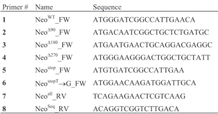
First, we sought to determine which segments from APH(3’)-II were essential to its activity. For such, we gen-erated three Neo gene shorter variants lacking their first 90, 180 and 270 nucleotides, using primers 2 – 4, indicated in Table 1. Each Neo gene variant were ligated into pMAL c-2G (which harbors thelacpromoter; Walkeret al., 2010),
giving rise to NeoD90-pMAL, NeoD180-pMAL and NeoD270
-pMAL constructs (Figure 1A). We next transformedE. coli
DH5awith all aforementioned constructs, as well as with
the wild type Neo construct (NeoWT-pMAL) (Figure 1A),
and bacterial transformants were selected from 2xYT plates
supplemented with 100 mg/mL ampicillin. DH5a
trans-formants were cultivated overnight in liquid ampicillin-added 2xYT, and then plated onto ampicillin-ampicillin-added solid 2xYT supplemented with 0.1 mM IPTG, in the presence or
absence of 10mg/mL kanamycin. We then verified that,
un-like NeoWT, none of the three obtained Neo gene variants
(NeoD90, NeoD180, and NeoD270) were able to confer DH5 a
resistance against kanamycin (Figure 1A, B). We therefore concluded that the first 30 amino acids of the N-terminal portion of APH(3’)-II are essential to its biological activity. Once we determined that the Neo gene is required to promote resistance against aminoglycosides, we decided to
gene start codon using primers 5 and 7 listed in Table 1, cre-ating the Neostopvariant, in which a G – its fourth base – is
substituted by a T, generating the stop codon TGA (Figure 2A). This premature stop codon prevents the formation of APH(3’)-II, completely abrogating the growth capacity of
DH5ain the presence of kanamycin (Figure 1B). We next
manually performed anin silicoprediction of possible
mu-tations that would restore the translation of the N-terminal portion of APH(3’)-II, and thus provide resistance against aminoglycosides. Interestingly, from all predicted muta-tional events (Figure 2B), two of them – G®T at position 5,
and T®G at position 15 – are classic mutations generated
by cellular exposure to H2O2(Shibutaniet al., 1991).
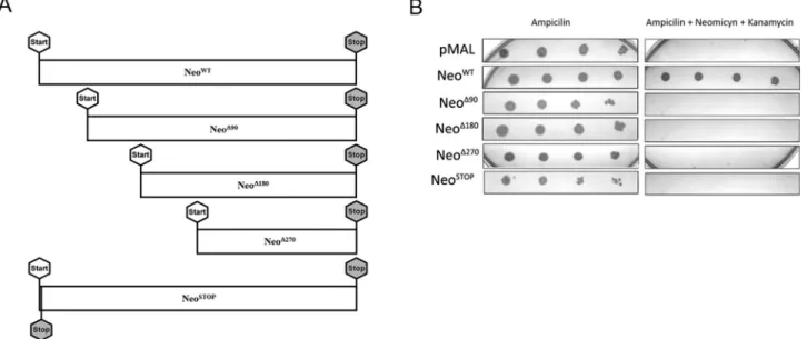
Long-term cultivation induces mutational events inT. cruzi
After (i) observing that DH5atransformed with the
Neostop-pMAL construct did not exhibit growth in 2xYT
supplemented with kanamycin (Figure 1B), and (ii) that ox-idation could lead to mutational events that might restore the translation of APH(3’)-II from the Neostopvariant
(Fig-ure 2B), we sought to transfect T. cruziwith the Neostop
gene variant. As expected, we were unable to observe, through erythrosine vital stain assay, visible growth of clones #1 and #5 ofT. cruziNeostopcultured in
hygromycin-added liquid LIT supplemented with G418 (Figure 3A). We then investigate if long-term cultivation –i.e., 42 days – of T. cruziNeostopwas capable of generating G418-insensitive clones for such, clones #1 and #5 were subject to the same experimental design described above, being cultured for 42 days, instead. Surprisingly, upon the increase of the cultiva-tion period, we were able to verify the presence of
G418-resistantT. cruzi cells from NeoStop clones #1 and #5 in
hygromycin-added liquid LIT supplemented with 200 or 400 mM G418 (Figure 3B).
Figure 1- The N-terminal region of Neo is required to promote resistance against kanamycin. Wild-type Neo gene (NeoWT) and its variants (NeoD90,
NeoD180, NeoD270and NeoSTOP) were obtained as described in Materials and Methods, item 1, and kanamycin- and neomycin-resistance assay was
con-ducted as described in Materials and Methods, item 2. (A) Diagram depicting wild-type Neo gene and deletions of N-terminal segments, which give rise to Neo gene variants. (B) NeoD90, NeoD180, NeoD270and NeoSTOPwere unable to confer to DH5aresistance against kanamycin. pMAL: empty vector.
Figure 2- Construction of the Neostopreporter and manually predicted mutations within its first seventy nucleotides. (A) The Neostopreporter was
con-structed substituting a guanine for a thymine at position 4 (as indicated by the arrow), generating the stop codon TGA right after the start codon ATG, as described inMaterials and Methods, item 1. (B) Manually predicted spontaneous and oxidation-induced mutations within the first seventy nucleotides of the Neostopreporter are indicated by underlined and bold-type letters, respectively. Insertion of a guanine, cytosine, and adenine at position 26 (indicated
Oxidative stress increases mutational events inT. cruzi
Given the fact that long-term cultivation allows the observation of mutational events inT. cruzi, we decided to
take advantage of the established protocol for isolation ofT. cruziclones using solid blood-agar to determine the
num-ber of CFUs of G418-insensitiveT. cruziNeostop generated
from a defined number of plated cells – this would allow us
to determine the frequency of mutation ofT. cruzi. Then,
1x10⁸cells from NeoStopclones #1 and #5, previously
cul-tured in hygromycin B-added liquid LIT for 42 days, in the
presence or absence of 50 mM H2O2, were plated onto
hygromycin B-added solid blood-agar, and the number of CFUs were determined, as described in Material and Methods, item 7. We verified thatT. cruziNeostopcultured in
the presence of H2O2, showed a mutation frequency of
1.56x10⁻⁷, while parasites cultured in control conditions,
i.e., in the absence of H2O2, exhibited a mutation frequency
of 0.71x10⁻⁷. This observation indicated that there is a
two-fold increase in mutation frequency when T. cruzi
faces situations of environmental oxidative stress. Besides, the experimental design was sensitive enough to allow us to
identify the basal frequency of genomic mutations ofT.
cruziNeostop, i.e., the frequency of mutational events
ob-served in parasites that were not exposed to H2O2during
this assay. This basal frequency – lower than the one ob-served in the presence of H2O2– may indicate the rate of
oxidation-independent mutational events that probably take place spontaneously inT. cruzi.
Screening genetic mutations from G418-resistantT. cruziNeostop
To determine the identity of the mutations present in G418-resistantT. cruziNeostopclones generated after 42 days
of culture in the presence or absence of H2O2(Material and
Methods, item 7), we next selected seven of them (#1-2 and
#5-2, from cultures conducted in the absence of H2O2;
#18-2, #34-2, #36-2, #40-2 and #43-2, from cultures car-ried out in the presence of H2O2) aiming to isolate, extract,
and sequence their genomic DNA by the Sanger method. Through this screening we verified that (i)T. cruziNeostop
clones #1-2, #5-2, #36-2, and #43-2 presented mutations that abrogate the TGA stop codon previously inserted in Neostop[#1-2: G
®A transversion, probably promoted by
replicative stress; #5-2: G®C transversion; #36-2 and
#43-2: G®T transversions, generated by a 8-oxoguanine
(8-oxoG) formed by the oxidation of a guanine from the genomic DNA]; and that (ii) clones #18-2, #34-2 and #40-2
showed a T®G transversion – probably caused by 8-oxoG
formation by the oxidation of a guanine from the nucleotide pool at positon 15, allowing the creation of an in-frame start codon at position 13 (Figure 4A). It is noteworthy that all G418-resistantT. cruziNeostopclones picked from the 42-day
cellular culture under oxidative stress conditions (#18-2, #34-2, #36-2, #40-2 and #43-2) presented classic trans-versions that arise from the exposure to reactive oxygen species (Figure 4A). Also, clones #1-2 and #5-2, selected from non-oxidative cellular cultures, despite presenting mutations that abrogate the inserted stop-codon, lacked the classic mutation signature promoted by conditions of oxi-dative stress.
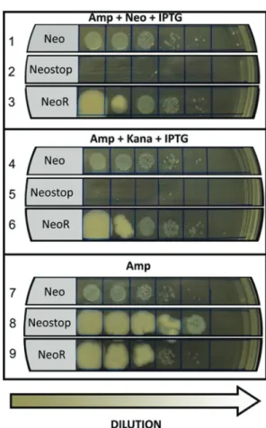
The NeostopT
®G reporter confers kanamycin resistance to DH5a
We next designed a forward primer carrying a gua-nine in its 4thposition (#6, Table 1) to artificially obtain the
Neostop gene variant mimicking the oxidation-induced
T®G mutation, which creates a downstream start codon, as
found in Neostop clones #18-2, #34-2 and #40-2 (Figure
4A). The resultant amplicon (NeostopT®G) was ligated into
pMAL c-2G plasmid, generating the NeostopT
®G-pMAL
construct, which was used to transform DH5a, whose
transformants were selected from ampicillin-supplemented
Figure 3- Long term incubation leads to selection ofT. cruziNeostoprevertant clones.T. cruzineostoptransfection, selection, genotyping, and growth rate and
2xYT plates. After isolation, the NeostopT
®G-pMAL
con-struct was used to obtain DH5atransformants from solid
ampicillin-added 2xYT plates. Once selected, one clone from these bacterial transformants was cultured overnight in liquid ampicillin-added 2xYT, and then plated onto ampicillin-added solid 2xYT supplemented with 0.1 mM
IPTG, in the presence or absence of 10mg/mL kanamycin.
We were then able to verify that DH5a harboring
NeostopT
®G became G418-resistant (Figure 4B),
confirm-ing that the aminoglycoside resistance observed in theT.
cruziNeostop clones #18-2, #34-2 and #40-2 is in fact
pro-moted by the T®G mutation, a nucleotide transversion
classically induced by oxidants (Shibutaniet al., 1991).
Discussion
Genetic diversity is an important factor that is directly related to adaptation and survival ofT. cruziin its hosts; in
fact, DNA metabolism and mutagenesis may allow this par-asite to increase the chances to adapt to different
environ-ments during its complex life cycle (Machado-Silvaet al.,
2016). In this sense, the study of mechanisms that govern
this phenomenon is crucial for the understanding of howT.
cruzievade the immune system and show resistance against
drugs, and for the development of new therapeutic strate-gies. However, currently, other than a restricted number of
studies employingin silicoapproaches to study
mutagene-sis and variability inT. cruzi(Azuajeet al., 2007a,b), there
is scarce information regarding the exact cellular events that may generate intra-specific genomic variability and few biological assays that allow the determination and de-tection of mutation rates in this parasite.
The Neo gene encodes APH(3’)-II, a phosphotrans-ferase that contains 267 amino acids, and is responsible for conferring microbial resistance against aminoglycosides
(Hächler et al., 1996). APH(3’)-II displays an ATP
bid-ing-site and can transfer theg-phosphoryl group from an
ATP molecule to the aminoglycoside, converting the latter to its phosphorylated, inactive form (Eustice and Wilhelm, 1984; Shawet al., 1993; Thompsonet al., 2002). We
gener-ated a number of mutations in the Neo gene, which gave rise to shorter APH(3’)-II variant forms (Table, 1, Figure
1A) that were ineffective in conferring DH5a resistance
against kanamycin (Figure 1B). Then, once we determined
Figure 4- NeostopT®G transversion can rescue aminoglycoside resistance to DH5a. (A) Sequencing analysis of G418-resistant clones shows that expo-sure to H2O2leads to classic transversions arisen from oxidative damage (bold-type letters). Oxidative-unrelated mutations were also found (underlined
letters). (B) To verify if T®G at position 15 could restore aminoglycoside resistance in DH5awe generated this transversion through the use of the
primer NeostopT®G_FW (#6, Table 1) – which generates a start codon into the Neostop– to obtain the NeostopT®G reporter, that confers kanamycin- and
that the N-terminal segment of Neo was required to provide resistance against kanamycin and G418, we introduced a premature stop-codon right after Neo’s ATG through a G®T mutation at position 4 (Figure 2A), creating a variant (Neostop) that would re-establish resistance against amino-glycosides if the mutated codon underwent a mutational
event. This was observed when T. cruziNeostop was
culti-vated for 42 days in hygromycin B-added liquid LIT (Fig-ure 3).
During its life cycle,T. cruziundergoes an obligatory
intracellular amastigote stage in which the immune system promotes the release of reactive oxygen and nitrogen spe-cies to halt the infection (Piacenzaet al., 2009); thus,
repli-cation of amastigotes under a scenario of oxidative stress can promote a condition from which mutated cells can
ulti-mately increase the pool of mutatedT. cruzi, which could
lead to intra-specific genetic diversity. Although epimas-tigotes and amasepimas-tigotes are subjected to different extents of
oxidative stress, data from the literature (Aguiar et al.,
2013), as well as unpublished observations from our group,
suggest that both aforementionedT. cruzi life forms are
equally affected by oxidative stress and share the same re-sponses against this biological condition. Therefore, the observation that epimastigotes treated with H2O2display a
2-fold increase in mutational events (Results, item 3) sug-gests that oxidative stress promoted by the host may play a direct role in genetic variability ofT. cruziamastigotes. In
fact, for several other organisms, including E. coli,
Helicobacter pylori, Salmonella typhimurium, Bacillus subtilis, Pseudomonas, Clostridium, Saccharomyces cerevisiae, andCandida albicans, increased mutation rates
are often correlated with increased survival and infection
rates in adverse conditions (Wang et al., 2001; Foster,
2000; Linzet al., 2014). In this manner, the increase in the
number of G418-resistantT. cruziNeostopclones after
long-term oxidative insult (Figure 3) suggests that this type of stress could stimulate intra-specific genetic variability.
It is well-established that oxidative stress promotes a range of modifications in nucleic acids, such as double-strand breaks and nitrogenous base modification (Friedberg
et al., 2006). Interestingly, the generation of 8-oxoG, one of
the most frequent lesions derived from oxidative stress, has a high mutagenic potential, since the oxidized guanine, if localized in the genomic DNA, promotes a mismatched
pairing with adenine resulting in G®T or C®A
trans-versions. In addition, the generation of 8-oxoG in the
nucle-otide pool also promotes a T®G transversion,
conse-quently leading to nucleotide mismatches (Dizdaroglu et
al., 2002; van Loonet al., 2010). In fact, the severity of
ef-fects that can arise from the formation of 8-oxoG became evident when the GO system – a pathway specialized in preventing mutagenicity promoted by 8-oxoG, comprised of three enzymes, namely MYH (MutY homologue), MTH (MutT homologue), and OGG1 (FPG homologue) – was
first described (Michaelset al., 1992; Michaels and Miller,
1992; Davidet al., 2007).
InT. cruzi, long-term exposure to H2O2induced DNA
mutations related to the generation of 8-oxoG, as clones #18-2, #34-2 and #40-2 showed mutations that are likely consequence of a guanine oxidation (Figure 4A). Likewise, clones #36-2 and #43-2 also presented formation of 8-oxoG mutations, since guanine in DNA undergoes a mispairment with adenine during replication (Figure 4A). The mis-pairing observed in clones #1-2 and #5-2 – which were not exposed to H2O2– are possibly products of an impaired
rep-lication process induced by a wobble conformation, al-though the DNA template and protein conformation are not disturbed (Johnson and Beese, 2004). These mismatches allow the formation of a structure closer to Watson-Crick base pair than that one observed in G:A and A:G mis-matches. Altogether, these verifications indicate that
muta-tions observed in T. cruzi cells exposed to H2O2 are
products of generation or misincorporation of 8-oxoG in the DNA, since those mutations are deleterious and do not easily arise in normal environments, considering the abnor-malities they cause to the polymerase structure (Johnson and Beese, 2004). Alterations in DNA metabolism can also increase genetic mutation frequency (Castillo-Acostaet al.,
2012). Organisms like yeast seem to preferentially insert cytosine opposing apurinic/apyrimidinic sites, and this
mechanism could lead to the increase of AT®GC
trans-versions (Thomaset al., 1997).
As suggested forT. cruzi,the presence of mutations,
to some extent, are possibly related to the survival of some
other organisms. In fact,Trypanosoma bruceistrain relies
on variant surface glycoproteins (VSG) switching to escape from the host immune system, a process in which recombi-nation plays a crucial role (Hartley and McCulloch, 2008; Horn and McCulloch, 2010). Deletion of deoxyuridin 5’-triphosphate pyrophosphatase (dUTPase) can cause a 9-fold increase in spontaneous mutation, and the appearance of double strand breaks inT. brucei, which could lead to a recombination process, increasing VSG switching (Cas-tillo-Acostaet al., 2012).
In this work, through a novel assay to assess
muta-tional events inT. cruzi, we demonstrated that oxidative
stress increases the mutation frequency in this parasite.We hypothesize that the 2-fold increase in mutation frequency after exposure to H2O2– which mimics the reactive oxygen
Acknowledgments
We thank Neuza Antunes Rodrigues for excellent technical support. This work was supported by the follow-ing Brazilian research fundfollow-ing institutions: CNPq (Con-selho Nacional para o Desenvolvimento Científico e Tecnológico) and FAPEMIG (Fundação de Amparo a Pes-quisa de Minas Gerais).
References
Aguiar PH, Furtado C, Repolês BM, Ribeiro GA, Mendes IC, Peloso EF, Gadelha FR, Macedo AM, Franco GR, Pena SD et al.(2013) Oxidative stress and DNA lesions: the role of 8-oxoguanine lesions inTrypanosoma cruzi cell viability. PLoS Negl Trop Dis 7:e2279.
Andrade LO, Machado CR, Chiari E, Pena SD and Macedo AM (1999) Differential Tissue Distribution of Diverse Clones of Trypanosoma Cruzi in Infected Mice. Mol Biochem Para-sitol 100:163-72.
Andrade LO, Galvão LM, Meirelles MdeN, Chiari E, Pena SD and Macedo AM (2010) Differential tissue tropism of Trypanosoma cruzi strains: an in vitro study. Mem Inst Oswaldo Cruz 105:834-837.
Augusto-Pinto L, Teixeira SM, Pena SD and Machado CR (2003) Single-nucleotide polymorphisms of theTrypanosoma cruzi MSH2 gene support the existence of three phylogenetic lin-eages presenting differences in mismatch-repair efficiency. Genetics 164:117-126.
Azuaje F, Ramirez JL and Da Silveira JF (2007a) An exploration of the genetic robustness landscape of surface protein fami-lies in the human protozoan parasiteTrypanosoma cruzi. IEEE Trans Nanobiosci 6:223-228.
Azuaje FJ, Ramirez JL and da Silveira, JF (2007b)In silico, bio-logically-inspired modelling of genomic variation genera-tion in surface proteins ofTrypanosoma cruzi. Kinetoplastid Biol Dis 6:6.
Baptista RP, D’Ávila DA, Segatto M, do Valle ÍF, Franco GR, Valadares HM, Gontijo ED, Galvão LM, Pena SD, Chiari E et al.(2014) Evidence of substantial recombination among Trypanosoma cruzi II strains from Minas Gerais. Infect Genet Evol 22:183-191.
Bensasson D (2011) Evidence for a high mutation rate at rapidly evolving yeast centromeres. BMC Evol Biol 11:211. Bergkessel M and Guthrie C (2013) Colony PCR. Methods
Enzymol 529:299-309.
Burgos JM, Risso MG, Brenière SF, Barnabé C, Campetella O and Leguizamón MS (2013) Differential distribution of genes encoding the virulence factor trans-sialidase along Trypanosoma cruzi discrete typing units. PLoS One 8:e58967.
Castillo-Acosta VM, Aguilar-Pereyra F, Bart JM, Navarro M, Ruiz-Pérez LM, Vidal AE and González-Pacanowska D (2012) Increased uracil insertion in DNA is cytotoxic and in-creases the frequency of mutation, double strand break for-mation and VSG switching inTrypanosoma brucei. DNA Repair 11:986-995.
Choi SS, Chivers PT and Berg DE (2011) Point mutations in Helicobacter pylori’s fur regulatory gene that alter resis-tance to metronidazole, a prodrug activated by chemical re-duction. PloS One 6:e18236.
Corpet F (1988) Multiple sequence alignment with hierarchical clustering. Nucleic Acids Res 16:10881-10890.
da Rocha WD, Silva RA, Bartholomeu DC, Pires SF, Freitas JM, Macedo AM, Vazquez MP, Levin MJ and Teixeira SM (2004) Expression of exogenous genes in Trypanosoma cruzi: improving vectors and electroporation protocols. Parasitol Res 92:113-120.
David SS, Shea VLO and Kundu S (2007) Base excision repair of oxidative DNA damage. Nature 447:941-950.
de Noya BA and González ON (2015) An ecological overview on the factors that drives toTrypanosoma cruzioral transmis-sion. Acta Trop 151:94-102.
Dizdaroglu M, Jaruga P, Birincioglu M and Rodriguez H (2002) Free radical-induced damage to DNA: Mechanisms and measurement. Free Radic Biol Med 32:1102-1115. Eustice DC and Wilhelm JM (1984) Mechanisms of action of
aminoglycoside antibiotics in eucaryotic protein synthesis. Antimicrob Agents Chemother 26:53-60.
Ewing B, Hillier L, Wendl MC and Green P (1998) Base-calling of automated sequencer traces using phred. I. Accuracy as-sessment. Genome Res 8:175-185.
Ford CB, Shah RR, Maeda MK, Gagneux S, Murray MB, Cohen T, Johnston JC, Gardy J, Lipsitch M and Fortune SM (2013) Mycobacterium tuberculosismutation rate estimates from different lineages predict substantial differences in the emer-gence of drug-resistant tuberculosis. Nat Genet 45:784-790. Frasch AC (2000) Functional diversity in the trans-sialidase and
mucin families in Trypanosoma cruzi. Parasitol Today 16:282-286.
Friedberg EC, Walker GC, Siede W, Wood RD, Schultz RA and Ellenburger T (2006) DNA repair and mutagenesis. 2nd edi-tion. ASM Pres, Washington, 1118 p.
Foster PL (2000) Adaptive mutation: implications for evolution. Bioessays 22:1067-1074.
Glassner BJ, Rasmussen LJ, Najarian MT, Posnick LM and Sam-son LD (1998) Generation of a strong mutator phenotype in yeast by imbalanced base excision repair. Proc Natl Acad Sci U S A 95:9997-10002.
Gomes ML, Araujo SM and Chiari E (1991)Trypanosoma cruzi: Growth of clones on solid medium using culture and blood forms. Mem Inst Oswaldo Cruz 86:131-132.
Gonzales MF, Brooks T, Pukatzki SU and Provenzano D (2013) Rapid protocol for preparation of electrocompetent Esche-richia coliandVibrio cholerae. J Vis Exp 8:80.
Hächler H, Santanam P and Kayser FH (1996) Sequence and char-acterization of a novel chromosomal aminoglycoside phos-photransferase gene, aph (3’)-IIb, in Pseudomonas aeruginosa. Antimicrob Agents Chemother 40:1254-1256. Hartley CL and McCulloch R (2008) Trypanosoma brucei
BRCA2 acts in antigenic variation and has undergone a re-cent expansion in BRC repeat number that is important dur-ing homologous recombination. Mol Microbiol 68:1237-1251.
Hoeijmakers JHJ (2001) Genome maintenance mechanisms for preventing cancer. Nature 411:366-374.
Horn D and McCulloch R (2010) Molecular mechanisms underly-ing the control of antigenic variation in African trypano-somes. Curr Opin Microbiol 13:700-705.
Linz B, Windsor HM, McGraw JJ, Hansen LM, Gajewski JP, Tomsho LP, Hake CM, Solnick JV, Schuster SC and Mar-shall BJ (2014) A mutation burst during the acute phase of Helicobacter pylori infection in humans and rhesus ma-caques. Nat Commun 13:4165.
Macedo AM, Machado CR, Oliveira RP and Pena SD (2004) Trypanosoma cruzi: Genetic structure of populations and relevance of genetic variability to the pathogenesis of cha-gas disease. Mem Inst Oswaldo Cruz 99:1-12.
Machado-Silva A, Cerqueira PG, Grazielle-Silva V, Gadelha FR, Peloso EF, Teixeira SM and Machado CR (2016) How Trypanosoma cruzideals with oxidative stress: Antioxidant defence and DNA repair pathways. Mutat Res Rev Mutat Res 767:8-22.
Magni GE and von Borstel RC (1962) Different rates of spontane-ous mutation during mitosis and meiosis in yeast. Genetics 47:1097-1108.
Matic I, Radman M, Taddei F, Picard B, Doit C, Bingen E, Denamur E, Elion J (1997) Highly Variable Mutation Rates in Commensal and Pathogenic Escherichia Coli. Science 277:1833-34.
Michaels ML and Miller JH (1992) The GO system protects or-ganisms from the mutagenic effect of the spontaneous lesion 8-hydroxyguanine (7,8-dihydro-8-oxoguanine). J Bacteriol 174:6321-6325.
Michaels ML, Cruz C, Grollman AP and Miller JH (1992) Evi-dence that MutY and MutM combine to prevent mutations by an oxidatively damaged form of guanine in DNA. Proc Natl Acad Sci U S A 89:7022-7025.
Patrushev LI and Minkevich IG (2008) The problem of the eukaryotic genome size. Biochemistry 73:1519-1552. Piacenza L, Alvarez MN, Peluffo G and Radi R (2009) Fighting
the oxidative assault: TheTrypanosoma cruzijourney to in-fection. Curr Opin Microbiol 12:415-421.
Prata A (2001) Clinical and epidemiological aspects of Chagas disease. Lancet Infect Dis 1:92-100.
Provan J, Soranzo N, Wilson NJ, Goldstein DB and Powell W (1999) A low mutation rate for chloroplast microsatellites. Genetics 153:943-947.
Rassi Jr A and Marin-Neto JÁ (2010) Chagas disease. Lancet 375:1388-1402.
Revollo S, Oury B, Laurent JP, Barnabé C, Quesney V, Carrière V, Noël S and Tibayrenc M. (1998)Trypanosoma cruzi: Im-pact of clonal evolution of the parasite on its biological and medical properties. Exp Parasitol 89:30-39.
Rosche WA and Foster PL (2000) Determining mutation rates in bacterial populations. Methods 20:4-17.
Schmunis GA (2007) Epidemiology of Chagas disease in non-endemic countries: the role of international migration. Mem Inst Oswaldo Cruz 102 Suppl 1:75-85.
Shaw KJ, Rather PN, Hare RS and Miller GH (1993) Molecular genetics of aminoglycoside resistance genes and familial re-lationships of the aminoglycoside-modifying enzymes. Microbiol Rev 57:138-163.
Shibutani S, Takeshita M and Grollman AP (1991) Insertion of specific bases during DNA synthesis past the oxidation-damaged base 8-oxodG. Nature 349:431-434.
Shikanai-Yasuda MA, Marcondes CB, Guedes LA, Siqueira GS, Barone AA, Dias JC, Amato Neto V, Tolezano JE, Peres BA, Arruda Jr ERet al.(1991) Possible oral transmission of acute Chagas’ disease in Brazil. Rev Inst Med Trop Sao Paulo 33:351-357.
Shikazono N, Yokota Y, Kitamura S, Suzuki C, Watanabe H, Tano S and Tanaka A (2003) Mutation rate and novel tt mu-tants ofArabidopsis thalianainduced by carbon ions. Ge-netics 163:1449-1455.
Steinberg CM, Diuision B and Ridge O (1971) Inheritance of spontaneous mutability in yeast. Genetics 69:17-27. Taddei F, Vulic M, Radman M and Matic I (1997) Genetic
vari-ability and adaptation to stress. EXS 83:271-290.
Thomas D, Scot AD, Barbey R, Padula M and Boiteux S (1997) Inactivation of OGG1 increases the incidence of G.C®T.A transversions in Saccharomyces cerevisiae: Evidence for endogenous oxidative damage to DNA in eukaryotic cells. Mol Gen Genet 254:171-178.
Thompson PR, Boehr DD, Berghuis AM and Wright GD (2002) Mechanism of aminoglycoside antibiotic kinase APH(3’)-IIIa: role of the nucleotide positioning loop. Biochemistry 41:7001-7007.
van Loon B, Markkanen E and Hübscher U (2010) Oxygen as a friend and enemy: How to combat the mutational potential of 8-oxo-guanine. DNA Repair 9:604-616.
Wang G, Wilson TJ, Jiang Q and Taylor DE (2001) Spontaneous mutations that confer antibiotic resistance inHelicobacter pylori. Antimicrob Agents Chemother 45:727-733. Zingales B, Andrade SG, Briones MR, Campbell DA, Chiari E,
Fernandes O, Guhl F, Lages-Silva E, Macedo AM, Machado CRet al.(2009) A new consensus forTrypanosoma cruzi intraspecific nomenclature: Second revision meeting recom-mends TcI to TcVI. Mem Inst Oswaldo Cruz 104:1051-1054.
Zingales B, Miles MA, Campbell DA, Tibayrenc M, Macedo AM, Teixeira MM, Schijman AG, Llewellyn MS, Lages-Silva E, Machado CRet al.(2012) The revisedTrypanosoma cruzi subspecific nomenclature: rationale, epidemiological rele-vance and research applications. Infect Genet Evol 12:240-253.
Internet Resources
WHO Chagas disease (American trypanosomiasis) fact sheet, http://www.who.int/mediacentre/factsheets/fs340/en/ (No-vember 13, 2017).
Associate Editor: Carlos F.M. Menck