Analytical Methods
Bioassay-guided isolation of proanthocyanidins with antioxidant activity
from peanut (Arachis hypogaea) skin by combination of chromatography
techniques
Tatiane L.C. Oldoni
a,⇑, Priscilla S. Melo
b, Adna P. Massarioli
b, Ivani A.M. Moreno
b,
Rosângela M.N. Bezerra
c, Pedro L. Rosalen
d, Gil V.J. da Silva
e, Andréa M. Nascimento
f,
Severino M. Alencar
baDepartment of Chemistry, Federal Technological University of Paraná (UTFPR), 85501-970 Pato Branco, PR, Brazil
bDepartment of Agri-Food Industry, Food and Nutrition, ‘‘Luiz de Queiroz’’ College of Agriculture, University of Sao Paulo (USP), 13418-900 Piracicaba, SP, Brazil cFaculty of Applied Sciences, Piracicaba Dental School, University of Campinas (UNICAMP), 13484-350 Piracicaba, SP, Brazil
dDepartment of Physiological Sciences, University of Campinas (UNICAMP), 13414-903 Piracicaba, SP, Brazil eDepartment of Chemistry, FFCLRP, University of São Paulo (USP), 14040-901 RibeirãoPreto, SP, Brazil
fInstitute of Biological and Exact Sciences, Federal University of Ouro Preto (UFOP), 35400-000 OuroPreto, MG, Brazil
a r t i c l e
i n f o
Article history:
Received 24 June 2014
Received in revised form 10 May 2015 Accepted 4 July 2015
Available online 6 July 2015
Chemical compounds studied in this article:
(+)-Catechin (PubChem CID: 9064)
p-Coumaric acid (PubChem CID: 637542) ()-Epicatechin (PubChem CID: 72276) Epigallocatechin (PubChem CID: 72277) Ferulic acid (PubChem CID: 445858) Gallic acid (PubChem CID: 370) Quercetin (PubChem CID: 5280343) Proanthocyanidin A1 (PubChem CID: 9872976)
Proanthocyanidin A2 (PubChem CID: 124025)
Keywords:
Purification By-product Chromatography Antioxidant activity Phenolic compounds
a b s t r a c t
Purification and bioassay-guided fractionation were employed to isolate proanthocyanidins with antiox-idant activity from peanut skin (Arachis hypogaeaRunner 886). The crude extract was prepared with acetone (60% v/v) and purified using chromatographic methods, including a semipreparative HPLC technique. As a result, two proanthocyanidins were isolated and identified using NMR, epicatechin-(2 b?O?7, 4b?8)-catechin (proanthocyanidin A1) and epicatechin-(b?2 O?7, 4b?8)-epicatechin (proanthocyanidin A2). Despite the structural similarity, differences were observed in their antioxidant activity. Proanthocyanidin A1 proved to be more active, with EC50value for DPPH radical scavenging
of 18.25lg/mL and reduction of Fe3+–TPTZ complex of 7.59 mmol/g, higher than that of synthetic
antiox-idant BHT. This compound evaluated by ABTS+was similar to that of natural quercetin. Therefore, peanut
skin is an important source of bioactive compounds that may be used as a mild antioxidant for food preservation.
Ó2015 Elsevier Ltd. All rights reserved.
1. Introduction
Proanthocyanidins have nutritional and biological interest because they are considered potent antioxidants (Manach, Mazur, & Scalbert, 2005) due to the presence of a B-ring catechol
group (dihydroxylated B-ring) capable of readily donating hydrogen (electron) to stabilize a radical species (Williams, Spencer, & Rice-Evans, 2004). Furthermore, because of the various o-dihydroxy groups present in structures with high molecular weight, procyanidins have a high ability to complex metal ions, such as Fe (III), Cu (II) and Al (III), as well as proteins.
Proanthocyanidins are well-known for their potential benefits to human health and for presenting valuable biological activities,
http://dx.doi.org/10.1016/j.foodchem.2015.07.004 0308-8146/Ó2015 Elsevier Ltd. All rights reserved.
⇑Corresponding author at: CP 591, 85501-970 Pato Branco, PR, Brazil.
E-mail address:[email protected](T.L.C. Oldoni).
Contents lists available atScienceDirect
Food Chemistry
such as antimicrobial, antioxidant, anticancer, antifungal, antialler-gic, anti-inflammatory, and vasodilatory actions (Pearson, Schmitz, Lazarus, & Keen, 2001; Pizzolitto et al., 2013; Zhang, Liu, Han, & Wei, 2013).
While the kernel of the peanut is a prized commodity, the skin is a low value by-product of peanut processing with a commercial value of approximately US$ 12–20 per tones (Sobolev & Cole, 2004). Skins have been used for animal feed or burned for energy. However, recent research has shown that skin has a high content of phenolic compounds as flavonoids, phenolic acids, procyanidins dimmers and oligomers (Ballard & Mallikarjunan, 2009; Ballard, Mallikarjunan, Zhou, & O’Keefe, 2010; Lou et al., 2004; Sarnoski, Johnson, Reed, Tanko, & O’Keefe, 2012; Tsujita, Shintani, & Sato, 2014; Yu, Ahmedna, & Goktepe, 2005; Yu, Ahmedna, Goktepe, & Dai, 2006) that makes it a product with potential benefits for human health and valuable biological activities (Awad, Chan, Downie, & Fink, 2000; Yu, Ahmedna, & Goktepe, 2010).
Lou et al. (2004)andSarnoski et al. (2012)isolated proantho-cyanidins from peanut skin and identified them. The compounds were tested for antioxidant activity. However, the authors did not use bioassay-guided fractionation, and the compounds were isolated at random. Thus, it is not possible to sure the bioactive compounds were, in fact, isolated.
The bioassay-guided fractionation technique has been employed by many researchers to study natural products (Baldé et al., 2010; Bargougui et al., 2014; Campos, Azevedo, Filho, Perez, & Braga, 2013; Ding, Ding, Zhang, & Luo, 2013; Oldoni et al., 2011; Teke et al., 2011). The main reason to choose this tech-nique is the rationalization process used to isolate biologically active substances from complex natural extracts.
Although few studies have been carried out with peanut skin, it is possible this low cost product is potentially rich in compounds with functional and biological activities, such as phenolic com-pounds. Therefore, the aim of the present study was to evaluate the antioxidant activity of crude extract and fractions of peanut skin and, subsequently, isolate proanthocyanidins using the bioassay-guided fractionation technique.
2. Materials and methods
2.1. Samples and standards
The peanut skin samples (Arachis hypogaeaRunner 886), sup-plied by CAP Agroindustrial (Dumont, SP, Brazil) in 2010 and 2011, were obtained as a by-product of the blanching process of peanuts. The skin samples were lyophilized, weighed, and stored at18°C until analysis.
Silica gel 60 and 2,2-diphenyl-1-picrylhydrazyl hydrate (DPPH) were purchased from Merck (Darmstadt, Germany); butylated hydroxytoluene (BHT), and butylated hydroxyanisole (BHA) standards from Synth (Diadema, SP, Brazil); Amberlite XAD-2Ò
resin, 2-20-azino-di-(3-ethylbenzthiazoline sulfonic acid) (ABTS),
2,4,6-tri(2-pyridyl)-s-triazine (TPTZ) analytical standards querce-tin, (+)-catechin, ()-epicatechin, gallic acid, ferulic acid,p -couma-ric acid and epigallocatechin from (Sigma Ald-couma-rich, St. Louis, MO, USA); and Sephadex LH-20 from Amersham Pharmacia (Uppsala, Sweden). All the solvents used for chromatography were of high performance liquid chromatography (HPLC) grade and all the other chemicals were of analytical-reagent grade.
2.2. Extraction and isolation bioguided of bioactive compounds
A representative sample of peanut skin (23 g) was extracted with a mixture of acetone: water (60:40), acidified to pH 1.5 with 0.1 mol/L HCl, in a thermostatized bath at 70°C for 30 min. The
extract was centrifuged at 5000g for 15 min, filtered, and concentrated in a rotary evaporator at 36°C until the acetone
had evaporated completely. The sample was lyophilized and the concentrated solid, i.e., the acetone extract of peanut skin (AEPS) was used for purification and subsequent analyses. The AEPS was further purified using Amberlite XAD-2Ò
resin, resulting in the methanolic fraction (Met-fr) and the aqueous fraction (Aqu-fr).
The active Met-fr was separated on a Sephadex LH-20 column (5 cm30 cm), using the technique of gel filtration (hydrox-ypropylated, cross-linked dextran), and eluted with methanol, yielding 123 subfractions. The subfractions obtained were moni-tored by thin layer chromatography (TLC) using the anisaldehyde reagent (4-methoxy-benzaldehyde), and acetic acid as a develop-ing reagent, followed by incubation at 100°C for 5 min.
Fluorescent substances were viewed under ultraviolet (UV) light at 254 nm and 366 nm (Alencar et al., 2007).
Subfractions that were similar in color and Rf were regrouped into 18 subfractions. These subfractions were tested and evaluated for their antioxidant activity and chemical profile using HPLC (Francisco & Resurreccion, 2009a). Subfraction 10 showed the highest activities using the DPPHand ABTS+ radical scavenging methods. Therefore, it was purified by semipreparative HPLC using an Agilent Prep-ODS (H) column (250 mm20 mm). The mobile phases were composed of water (solvent A) and acetonitrile (sol-vent B) at a constant flow rate of 6 mL/min. The gradient started with 20% solvent B to 30% B in 30 min, 55% B in 35 min, and 20% B in 40 min. The column was maintained at a constant temperature of 30°C. The peaks of eluted compounds were collected in a
frac-tion collector coupled to a liquid chromatography system, tested for antioxidant activity using the DPPHand ABTS+radical scav-enging methods and ferric reducing antioxidant power (FRAP), and also analyzed by nuclear magnetic resonance (NMR) for elucidation of their chemical structures.
2.3. Nuclear magnetic resonance (NMR)
NMR spectra, obtained in CD3OD using tetramethylsilane (TMS) as internal standard, were recorded in a Bruker DRX 500 spectrom-eter, operating at 500.13 MHz for1H and 125.76 MHz for13C. The data were obtained by 1D and 2D NMR experiments (1H–1H COSY, HMQC and HMBC). The chemical shifts are expressed in d
(parts per million) and the coupling constants (J) in Hz (Freimund, Sauter, Käppeli, & Dutler, 2003).
All the compounds, already described in the literature, were identified by comparison of their spectral data (nuclear magnetic resonance – NMR) with reported values.
Epicatechin-(2b?O?7, 4 b?8)-catechin (A1):1H and13C
NMR data were in agreement with the reported literature values (Lou et al., 2004).
Epicatechin-(b?2 O?7, 4 b?8)-epicatechin (A2): 1H and
13
C NMR data were in agreement with the reported literature values (Lou et al., 2004).
2.4. Antioxidant activity using the 2,2-diphenyl-1-picrylhydrazyl hydrate (DPPH) free radical scavenging method
DPPH free radical scavenging activity was measured as described by Moraes de Souza, Oldoni, Regitano-d’Arce and Alencar (2008). The reaction medium consisted of 0.5 mL of the extract or purified fractions, 3.0 mL of ethanol, and 0.3 mL of 0.5 mmol/L DPPHsolution in ethanol. The mixture was incubated in the absence of light at room temperature for 45 min. Subsequently, the absorbance was measured using a spectropho-tometer (UV mini-1240, Shimadzu Co., Kyoto, Japan) at 517 nm. The anti-radical activity was calculated using the following equation:
Antioxidant activityð%Þ ¼100 ½ðAaAbÞ 100=Ac
where:
Aa = absorbance of the sample; Ab = absorbance of the blank (prepared by replacing the volume of DPPHsolution with an equal volume of ethanol); Ac = absorbance of the control (prepared by replacing the volume of extract with an equal volume of ethanol).
To determine the antioxidant activity of the isolated com-pounds, the analysis of the DPPH scavenging activity was per-formed in a 96-well polystyrene microplate. Aliquots of 170
l
L of the solution, 60l
L of the DPPHradical solution in ethanol, and 50l
L of the isolated compounds were added to each microplate cavity. After incubation for 45 min at room temperature, the absor-bance was measured at 517 nm with a spectrophotometer. The antioxidant activity was determined as previously described.The concentrations of the samples (AEPS, purified fractions, and isolated compounds) responsible for a 50% decrease in the DPPH free radical initial activity (EC50,
l
g/mL) were calculated by linear regression of the antioxidant activity, measured at various concen-trations of each sample. All analyzes were carried out in triplicate.2.5. Antioxidant activity using the 2-20 -azino-di-(3-ethylbenzthiazoline sulfonic acid) (ABTS) method
The antioxidant activity by the ABTS method was performed according toRe et al. (1999). The ABTS+radical was formed by the reaction of 7 mmol/L ABTS with 140 mmol/L potassium persul-fate, incubated at 25°C in the dark for 12–16 h. The radical was diluted with ethanol to obtain the absorbance value of 0.700 ± 0.200 at 734 nm. Under dark conditions, 3.0 mL of the ABTS+ radical solution was added to 30
l
L of each dilution of AEPS, purified fractions, and isolated compounds and the absor-bance was read at 734 nm in a spectrophotometer after 6 min against ethanol as blank. Trolox was used as reference at concen-trations ranging from 100 to 2000l
mol/L and the results of theantioxidant activity were expressed as mmol of trolox equivalent antioxidant capacity (TEAC)/g of peanut skin. All analyzes were carried out in triplicate.
2.6. Antioxidant activity using the ferric reducing antioxidant power (FRAP) method
The determination of antioxidant activity using the FRAP method was performed as described byKukic´ et al. (2008). FRAP reagent was prepared at the moment of analysis by mixing 25 mL of 300 mmol/L acetate buffer (pH 3.6), 2.5 mL of 10 mmol/L 2,4,6-tri(2-pyridyl)-s-triazine (TPTZ) in 40 mmol/L HCl, and 2.5 mL of 20 mmol/L FeCl3 in aqueous solution. An aliquot of 100
l
L of each extract was added to 3 mL of FRAP reagent and incu-bated at 37°C in water bath for 30 min. The absorbance was mea-sured, aqueous solutions of ferrous sulfate (100–2000l
mol/L) were used for calibration, and the results were expressed as mmol of Fe2+/g of lyophilized sample. All analyzes were carried out in triplicate.2.7. Statistical analysis
A randomized design was used for all experiments. Data were analyzed by ANOVA using the Statistical Analysis System (SAS) software. Mean separation was performed using the Tukey’s test at the 95% confidence interval.
3. Results and discussion
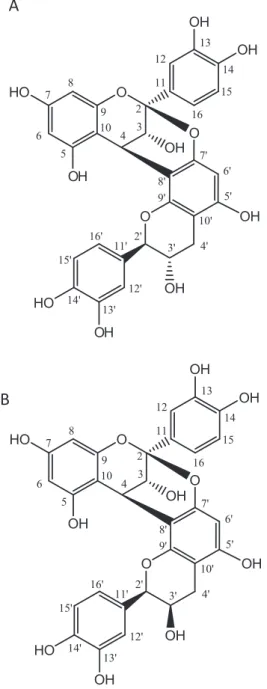
In this study, the antioxidant activities of AEPS, purified frac-tions, and isolated compounds obtained from the peanut skin were investigated. The activity-directed purification (Fig. 1) of the crude extract using several chromatographic separations resulted in the isolation of two compounds with high antioxidant activity, 10 mg of epicatechin-(2b?O?7, 4 b?8)-catechin (A1) and 2 mg of
epicatechin-(b?O?7, 4 b?8)-epicatechin (A2) (Fig. 2). The
Acetone extract of peanut skin (AEPS) (23 g)
Methanolic fraction (Met-fr) (1.05 g)
123 Subfractions – regrouped in 18
Subfraction 10 (160 mg) Extraction with acetone 60%
70 oC/30 min
Purification in Amberlite XAD2 resin
Aqueous fraction (Aqu-fr)
Column chromatography on Sephadex LH-20
Rechromatography on semi-preparative HPLC Lyophilized peanut skin (25 g)
Proanthocyanidin A1 (10 mg) Proanthocyanidin A2 (2 mg)
tests for detection of antioxidant activity were monitored throughout the fractionation and isolation steps.
The structures of the compounds were identified by their spec-troscopic data (1H NMR,13C NMR) measurements and comparison with published values (Lou et al., 2004).
3.1. Bioassay-guided for antioxidant activity
The first step in the process of isolation of active compounds was the purification with Amberlite XAD-2Ò
resin. The Met-fr and Aqu-fr were evaluated for their antioxidant activity using the DPPH and ABTS+ radical scavenging methods (Table 1) and chemical profile by HPLC-DAD (Fig. 3).
The results of antioxidant activity, expressed as radical scaveng-ing of DPPHand ABTS+, showed that Met-fr has higher activity than AEPS, while Aqu-fr displayed the lowest activity (Table 1).
These results indicate that the purification process using Amberlite XAD-2Ò
resin was efficient, because it resulted in an increase in antioxidant capacity of Met-fr compared with AEPS.
The results found herein for the antioxidant activity of peanut skin are higher than those reported by Francisco and Resurreccion (2009b)for peanut skin from different regions and processed under different temperatures, which ranged from 0.62 to 2.56 mmol of TEAC/g of peanut skin.
Although the Met-fr elicited a more potent antioxidant effect than the Aqu-fr, the HPLC-DAD profiles registered for the fractions were similar, with variation in the intensities of peaks (Fig. 3).
In view of the higher antioxidant activity, chemical profile and amount of the met-fr available, this was purified on a column of Sephadex LH-20. Elution of Met-fr allowed the collection of 123 subfractions, which were analyzed by TLC.
Among the subfractions evaluated, subfraction 10 showed the highest antioxidant activity and a HPLC-DAD positive profile (Fig. 3) for chemical isolation indicating a mixture with UV spectra compatible with phenolic compounds. The major constituents were collected providing compounds with high purity (proantho-cyanidins A1 and A2) (Fig. 3).
3.2. Antioxidant activity of isolated compounds
The results of antioxidant activity, expressed as EC50, indicated that the isolation and purification procedures were adequate. A decrease in EC50value was observed during the purification pro-cess (Table 2). The best activities were obtained for Met-fr, subfrac-tion 10, and proanthocyanidin A1, with EC50values of 16.10, 15.65, and 18.25
l
g/mL, respectively. Compared with the synthetic antioxidant BHA (EC50= 25l
g/mL) (Yuan, Bone, & Carrington,2005), the two compounds isolated in this study and their extracts are very promising, since they have a great potential for applica-tions in food and pharmaceutical industries.
To the best of our knowledge, this is the first time that bioactive proanthocyanidins have been isolated from peanut skin using the bioassay-guided fractionation technique, and that their antioxi-dant activity and structure elucidated.
AlthoughLou et al. (1999)isolated proanthocyanidins A1 and A2 from the water soluble fraction of peanut skin, the authors did not determine their antioxidant activity. Despite isolating other constituents from peanut skin and evaluating them for their antioxidant activity using the EC50test,Lou et al. (2004)did not use bioassay-guided isolation.Appeldoorn et al. (2009)also iso-lated procyanidins A-type dimers and B-type dimers from peanut skin and grape seeds, respectively. However, because the authors did not evaluate bioactivity, they could not determine whether these compounds were responsible for the antioxidant activity. The bioassay-guided fractionation technique ensures that the com-pounds responsible for biological activity are isolated.
The standards quercetin, catechin, and epicatechin were also tested in our study, and their antioxidant activities (radical ABTS method,Fig. 4) were compared with the AEPS, purified fractions and isolated compounds. The highest activity was found for proan-thocyanidin A1 (6.54 mmol TEAC/g), very similar to the standards quercetin, catechin, and epicatechin (7.52, 7.33, and 7.50 mmol TEAC/g, respectively). The mixture of proanthocyanidin A1 + proanthocyanidin A2 (1:1 mix in the same proportion) and proanthocyanidin A2 alone had the lowest values for antioxidant activity.
The isolated compounds proanthocyanidin A1 and A2 are diastereoisomers and the only difference between them is the stereochemistry of theAOH group at the 30position. Despite their
structural similarity, these compounds exhibited different physi-cal, chemiphysi-cal, and biological features. Proanthocyanidin A1 proved to be more bioactive than proanthocyanidin A2, and the
O
OH
OH
OH
OH
HO
O
O
HO
OH
OH
2 3 4 5 6 7 8 9 10 11 12 13 14 15 16OH
7' 6' 8' 5' 10' 2' 3' 4' 9' 11' 12' 13' 14' 15' 16'O
OH
OH
OH
OH
HO
O
O
HO
OH
OH
2 3 4 5 6 7 8 9 10 11 12 13 14 15 16OH
7' 6' 8' 5' 10' 2' 3' 4' 9' 11' 12' 13' 14' 15' 16'A
B
Fig. 2.Structures of the bioactive compounds isolated from peanut skin: (A) epicatechin-(2b?O?7, 4b?8)-catechin (proanthocyanidin A1), (B)
epicate-chin-(2b?O?7, 4b?8)-epicatechin (proanthocyanidin A2).
antioxidant activity of an equimolar mixture of both compounds was considerably less, which may be due to the reduced activity showed by the latter.
The isolated compounds were also evaluated for their antioxi-dant activity by the FRAP method. The activities obtained for the AEPS, Met-fr, subfraction 10, and proanthocyanidin A1 were 7.75, 7.33, 7.55, and 7.59 mmol Fe2+/g, respectively (Fig. 4). The values found for the standards, epicatechin and catechin, were 21.3 and 20.9 mmol Fe2+/g, respectively. Comparing the results found in this study using the FRAP method with the synthetic standard BHT, which presents antioxidant activity of 1.59 mmol Fe2+/g (Borneo,
León, Aguirre, Ribotta, & Cantero, 2009) and with natural antioxi-dants tested, the isolated compounds show great potential as antioxidants in the food and pharmaceutical industries.
Given that studies on antioxidant activity using the FRAP method are scarce, this discussion focuses on compounds isolated from other matrices. Using the FRAP method Cerezo, Cuevas, Winterhalter, Garcia-Parrilla, and Troncoso (2010)evaluated the antioxidant activity of compounds isolated from strawberry and found values ranging from 2.75 to 7.67 mmol Fe2+/g. The isolated Table 1
Antioxidant activity of the acetone extract of peanut skin (AEPS) and its methanolic (Met-fr) and aqueous (Aqu-fr) fractions using the ABTS and DPPH methods.a
Sample Antioxidant activity
ABTS (mmol TEAC/g) DPPH (%) AEPS 4.4b± 0.10 55.9a± 2.55
Met-fr 5.5a± 0.50 60.0a± 1.44
Aqu-fr 3.2c± 0.15 41.3b± 1.80
Each value is expressed as mean (triplicate) ± standard deviation (SD). The same letters in the column are not significantly different at the 0.05 level.
The samples were tested at the concentration of 50lg/mL.
Proanthocyanidin A1
Proanthocyanidin A2
Minutes
mAU
Proanthocyanidin A2 Proanthocyanidin A1
Proanthocyanidin A2 Minutes
mAU
A
B
Fig. 3.RP-HPLC profile registered for peanut skin fractions and isolated compounds. (A) Methanolic and aqueous fractions; (B) subfraction 10 and collection of proanthocyanidin A1 and proanthocyanidin A2 using semipreparative RP-HPLC.
Table 2
Antioxidant activity of the acetone extract of peanut skin (AEPS), its methanolic fraction (Met-fr), subfraction 10, and isolated compounds (proanthocyanidin A1 and proanthocyanidin A2) using the DPPH method, expressed as EC50.
Sample Antioxidant activity – DPPH EC50(lg/mL)
AEPS 20.62b± 0.05
Met-fr 16.10d± 0.21
Subfraction 10 15.65e± 0.07
Proanthocyanidin A1 18.25c± 0.01
Proanthocyanidin A2 31.10a± 0.05
compounds were identified as anthocyanins and the most active one was pelargonidin-3-glucoside.
4. Conclusions
From the present work, we can conclude that the antioxidant activity presented by AEPS indicates high potential. To the best of our knowledge, this is the first report describing the bioassay-guided isolation of active compounds from peanut skin. The process isolated compounds with higher activities than AEPS, proving that the isolation process was performed properly. The antioxidant activities of proanthocyanidin A1 and A2 were consid-erably higher than the synthetic antioxidants BHA and BHT, and activities near to potent natural antioxidants, such as quercetin and epicatechin. The proanthocyanidin A1 is a potent antioxidant with no synergistic effect with its A2 isomer.
On the whole, it is interesting to note that a by-product from Brazilian peanut skin is a valuable source of natural bioactive molecules and has properties that suggest applications in the food and pharmaceutical industries. We suggest that, in vivo
studies should be carried out to verify the effectiveness of these antioxidant compounds in biological systems.
Conflicts of interest
The authors declare no conflict of interest.
Acknowledgements
The authors are thankful to State of São Paulo Research Foundation (FAPESP – Brazil) (Proc. Nos. 200855492-7 and 200907944-9) for the financial support and to Direction of Research and Post Graduation (DIRPPG) of the Federal Technological University of Parana (UTFPR).
References
Alencar, S. M., Oldoni, T. L. C., Castro, M. L., Cabral, I. S. R., Costa-Neto, C. M., Cury, J. A., et al. (2007). Chemical composition and biological activity of a new type of Brazilian propolis: Red propolis.Journal of Ethnopharmacology, 113, 278–283. Appeldoorn, M. M., Sanders, M., Vincken, J., Cheynier, V., Le Guernevé, C., Hollman,
P. C. H., et al. (2009). Efficient isolation of major procyanidin A-type dimers from
Fig. 4.Antioxidant activity of the acetone extract of peanut skin (AEPS), its methanolic fraction (Met-fr), subfraction 10, isolated compounds from peanut skin and natural standards using the ABTS (A) and FRAP (B) methods (results expressed as mean value ± standard deviation,n= 3).
peanut skins and B-type dimers from grape seeds. Food Chemistry, 117, 713–720.
Awad, A. B., Chan, K. C., Downie, A. C., & Fink, C. S. (2000). Peanuts as a source of beta-sitosterol, a sterol with anticancer properties.Nutrition and Cancer, 36, 238–241.
Baldé, E. S., Megalizzi, V., Traoré, M. S., Cos, P., Maes, L., Decaestecker, C., et al. (2010).In vitroantiprotozoal, antimicrobial and antitumor activity ofPavetta crassipesK. Schum leaf extracts.Journal of Ethnopharmacology, 130, 529–535. Ballard, T., & Mallikarjunan, P. (2009). Optimizing the extraction of phenolic
antioxidants from peanut skins using response surface methodology.Journal of Agricultural and Food Chemistry, 57, 3064–3072.
Ballard, T. S., Mallikarjunan, P., Zhou, K., & O’Keefe, S. (2010). Microwave-assisted extraction of phenolic antioxidant compounds from peanut skins. Food Chemistry, 120, 1185–1192.
Bargougui, A., Champy, P., Triki, S., Bories, C., Le Pape, P., & Loiseau, P. M. (2014). Antileishmanial activity of Opuntia ficus-indica fractions. Biomedicine & Preventive Nutrition, 4, 101–104.
Borneo, R., León, A. E., Aguirre, A., Ribotta, P., & Cantero, J. J. (2009). Antioxidant capacity of medicinal plants from the Province of Córdoba (Argentina) and their
in vitrotesting in a model food system.Food Chemistry, 112, 664–670. Campos, J. J., Azevedo, A. D. O., Filho, J. D. D. S., Perez, A. C., & Braga, F. C. (2013).
Bioguided isolation of myricetin-3-O-b-galactopyranoside with antinociceptive activity from the aerial part of Davilla elliptica St.-Hil. Journal of Ethnopharmacology, 150, 270–274.
Cerezo, A. B., Cuevas, E., Winterhalter, P., Garcia-Parrilla, M. C., & Troncoso, A. M. (2010). Isolation, identification, and antioxidant activity of anthocyanin compounds inCamarosastrawberry.Food Chemistry, 123, 574–582.
Ding, L.-J., Ding, W., Zhang, Y.-Q., & Luo, J.-X. (2013). Bioguided fractionation and isolation of esculentoside P fromPhytolacca americanaL..Industrial Crops and Products, 44, 534–541.
Francisco, M. L. L. D., & Resurreccion, A. V. A. (2009a). Development of a reversed-phase high performance liquid chromatography (RP-HPLC) procedure for the simultaneous determination of phenolic compounds in peanut skin extracts.
Food Chemistry, 117, 356–363.
Francisco, M. L. L. D., & Resurreccion, A. V. A. (2009b). Total phenolics and antioxidant capacity of heat-treated peanut skins.Journal of Food Composition and Analysis, 22, 16–24.
Freimund, S., Sauter, M., Käppeli, O., & Dutler, H. (2003). A new non-degrading isolation process for 1,3-b-d-glucan of high purity from baker’s yeast
Saccharomyces cerevisiae.Carbohydrate Polymers, 54, 159–171.
Kukic´, J., Popovic´, V., Petrovic´, S., Mucaji, P., C´iric´, A., Stojkovic´, D., et al. (2008). Antioxidant and antimicrobial activity ofCynara cardunculus extracts. Food Chemistry, 107, 861–868.
Lou, H., Yamazaki, Y., Sasaki, T., Uchida, M., Tanaka, H., & Oka, S. (1999). A-type proanthocyanidins from peanut skins.Phytochemistry, 51, 297–308.
Lou, H., Yuan, H., Ma, B., Ren, D., Ji, M., & Oka, S. (2004). Polyphenols from peanut skins and their free radical-scavenging effects. Phytochemistry, 65(16), 2391–2399.
Manach, C., Mazur, A., & Scalbert, A. (2005). Polyphenols and prevention of cardiovascular diseases.Current Opinion in Lipidology, 16, 77–84.
Moraes de Souza, R. A., Oldoni, T. L. C., Regitano-d’Arce, M. A. B., & Alencar, S. M. (2008). Antioxidant activity and phenolic composition of herbal infusions consumed in Brazil.Ciencia Y Tecnologia Alimentaria, 6, 41–47.
Oldoni, T. L. C., Cabral, I. S. R., Regitano d’Arce, M. A. B. R., Rosalen, P. L., Ikegaki, M, Nascimento, A. M., & Alencar, S. M. (2011). Isolation and analysis of bioactive isoflavonoids and chalcone from a new type of Brazilian propolis.Separation and Purification Technology, 77, 208–213.
Pearson, D. A., Schmitz, H. H., Lazarus, S. A., & Keen, C. L. (2001). Inhibition of in vitro low-density lipoprotein oxidation by oligomeric procyanidins present in chocolate and cocoas.Methods in Enzymology, 335, 350–360.
Pizzolitto, R. P., Dambolena, J. S., Zunino, M. P., Larrauri, M., Grosso, N. R., Nepote, V., et al. (2013). Activity of natural compounds from peanut skins onFusarium verticillioidesgrowth and fumonisin B1 production.Industrial Crops and Products, 47, 286–290.
Re, R., Pellegrini, N., Proteggente, A., Pannala, A., Yang, M., & Rice-Evans, C. (1999). Antioxidant activity applying an improved ABTS radical cation decolorization assay.Free Radical Biology & Medicine, 26, 1231–1237.
Sarnoski, P. J., Johnson, J. V., Reed, K. A., Tanko, J. M., & O’Keefe, S. F. (2012). Separation and characterisation of proanthocyanidins in Virginia type peanut skins by LC–MSn.Food Chemistry, 131, 927–939.
Sobolev, V. S., & Cole, R. J. (2004). Note on utilisation of peanut seed testa.Journal of the Science of Food and Agriculture, 84, 105–111.
Teke, G. N., Kuiate, J.-R., Kueté, V., Teponno, R. B., Tapondjou, L. A., Tane, P., et al. (2011). Bio-guided isolation of potential antimicrobial and antioxidant agents from the stem bark ofTrilepisium madagascariense. South African Journal of Botany, 77, 319–327.
Tsujita, T., Shintani, T., & Sato, H. (2014). Preparation and characterisation of peanut seed skin polyphenols.Food Chemistry, 151, 15–20.
Williams, R. J., Spencer, J. P. E., & Rice-Evans, C. (2004). Serial review: Flavonoids and isoflavones (phytoestrogens): Absorption, metabolism, and bioactivity. Free Radical Biology & Medicine, 36, 838–849.
Yu, J., Ahmedna, M., & Goktepe, I. (2005). Effects of processing methods and extraction solvents on concentration and antioxidant activity of peanut skin phenolics.Food Chemistry, 90, 199–206.
Yu, J., Ahmedna, M., & Goktepe, I. (2010). Potential of peanut skin phenolic extract as antioxidative and antibacterial agent in cooked and raw ground beef.
International Journal of Food Science & Technology, 45, 1337–1344.
Yu, J., Ahmedna, M., Goktepe, I., & Dai, J. (2006). Peanut skin procyanidins: Composition and antioxidant activities as affected by processing.Journal of Food Composition and Analysis, 19, 364–371.
Yuan, Y. V., Bone, D. E., & Carrington, M. F. (2005). Antioxidant activity of dulse (Palmaria palmata) extract evaluatedin vitro.Food Chemistry, 91, 485–494. Zhang, H., Liu, M., Han, S., & Wei, Y. (2013). Optimizing the extraction of catechin